Acute Necrotizing Gastritis with Gangrene: A Case Report
Acute Necrotizing Gastritis with Gangrene: A Case Report
Acute Necrotizing Gastritis with Gangrene: A Case Report

You also want an ePaper? Increase the reach of your titles
YUMPU automatically turns print PDFs into web optimized ePapers that Google loves.
Gastric gangrene31<strong>Acute</strong> <strong>Necrotizing</strong> <strong>Gastritis</strong> <strong>with</strong> <strong>Gangrene</strong>: A <strong>Case</strong> <strong>Report</strong>RUEY-AN CHIANG, SHAN-LONG CHEN, YAO-CHUNG TSAIAn 80-year-old man presented <strong>with</strong> signs of peritonitis. Laparotomy revealed gangrene of the stomachand abdominal esophagus <strong>with</strong>out obvious cause. The patient underwent total gastrectomy <strong>with</strong> partialesophagectomy. Bacterial culture of the peritoneal fluid grew Morganella morganii and Proteus mirabilis.The patient died of irreversible sepsis on the second postoperative day.Key words: necrotizing gastritis, gangrene, stomachIntroduction<strong>Gangrene</strong> of the stomach is a rare and fulminatingcatastrophic event that is often fatal. Itscause has been attributed to embolization of atheroscleroticplaque, thrombosis of major arterialsupply, occlusion of gastric vessels by therapeuticallyinjected foreign bodies, psychogenic polyphagiaresulting in massive gastric dilatation, ingestionof corrosive materials, intrathoracic herniationof the stomach through the diaphragm, gastricvolvulus, and necrotizing gastritis caused byorganisms.We report on a man <strong>with</strong> stomach gangrenethat appeared to be caused by a severe necrotizinginfection <strong>with</strong> no known portal of entry.<strong>Case</strong> <strong>Report</strong>An 80-year-old, previously healthy man, wassent to our emergency room <strong>with</strong> the chief complaintof abdominal distension and pain for oneday. On examination, the patient was afebrile,tachycardic (110 beats/min), tachypneic (26 breaths/min), <strong>with</strong> a systolic blood pressure of 90 mmHg.His abdomen was distended, guarded and rigid,<strong>with</strong> diffuse rebound tenderness and absent bowelsounds. White blood cell count was 18,700/mm 3 ,chest and abdomen x-rays showed no gas underthe diaphragm and no air-fluid levels.With a clinical diagnosis of peritonitis, anexploratory laparotomy was performed after initialresuscitation. When the peritoneum was opened,about 1000 ml of brown hemorrhagic, foul-smellingfluid could be seen in the perinoneal cavity.The whole of the stomach and abdominal esophaguswere black in color, the walls appearingpaper-thin and friable. Pulsations of major gastricarteries were present. The liver, spleen, duodenum,gallbladder, pancreas, small bowel and colon allappeared normal. The diaphragmatic domes werenormal and there was no evidence of gastricvolvulus. En-bloc resection of the entire stomach,omentum and distal esophagus was performed;cervical esophagostomy and tube jejunostomy werecreated. Immediate reconstruction for alimentarycontinuity was not performed due to the criticalcondition of the patient. The patient died of septicReceived: July 15, 2005 Accepted for publication: November 16, 2005From the Department of Surgery, Mackay Memorial Hospital Taitung Branch, TaiwanAddress for reprints: Dr. Ruey-An Chiang, 1 Lane 303, Changsha Street, Taitung City, Taitung County 950, Taiwan, R.O.C.Department of Surgery, Mackay Memorial Hospital Taitung Branch, TaiwanTel: (089) 310150 Fax: (089) 321240E-mail: ryan@ttms.mmh.org.tw
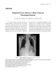
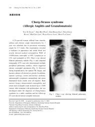
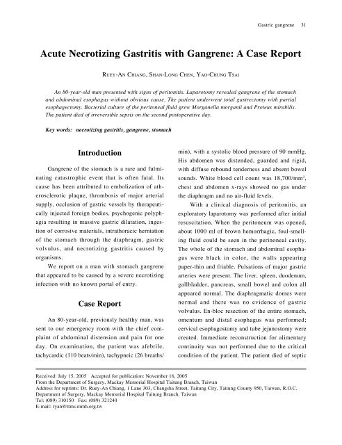
32J Emerg Crit Care Med. Vol. 17, No. 1, 2006shock on the second postoperative day.Gross examination of the specimens revealedgangrenous, black discoloration, thinning and absentrugal folds in the entire stomach and abdominalesophagus; the proximal esophagus and omentumappeared uninvolved. (Fig. 1). Microscopicexamination of the stomach showed destruction ofthe wall by a diffuse necrotic process extendingthrough all layers. An extensive purulent processhad completely destroyed the mucosa and hadinvaded the submucosa, <strong>with</strong> massive accumulationof inflammatory cells and necrosis of theunderlying muscularis. The esophagus revealeddetachment of the squamous epithelium and acuteinflammation of the mucosa and muscle layers.The omentum was unremarkable.Cultures of the peritoneal fluid taken at thetime of surgery grew Morganella morganii andProteus mirabilis that were sensitive to cefuroximeand gentamicin.DiscussionThe abundant and anastomotic nature of thestomach s vascular supply makes gangrene veryrare. In 1943, Babkin et al (1) . found that tying allthe gastric arteries did not cause gastric infarctionin the dog. The lack of infarction was explainedby the presence of multiple anastomoses betweenthe left gastric artery and branches of the phrenicand esophageal arteries. However, Harvey et al (2) .reported a case of multifocal gastric infarctionsecondary to atheromatous emboli originating in athoracic aortic aneurysm. In another report (3) , aFig. 1The specimen shows gangrene of the entire stomach andabdominal esophagus. The attached omentum is not involved.
Gastric gangrene33patient was described <strong>with</strong> extensive gastric necrosisafter therapeutic transcatheter embolizationof the left gastric artery <strong>with</strong> fragments of gelatinsponge for recurrent massive upper gastrointestinalhemorrhage. Ovnat et al (4) . reported three casesof acute obstruction of the celiac trunk. The firstpatient was diagnosed by abdominal computedtomography (CT) and angiography, and was treatedsuccessfully <strong>with</strong> thrombolytic therapy. Theother two patients underwent explorative laparotomyto establish the diagnosis: the most impressivefinding was that all of the gastric mucosa alongthe lesser curvature was necrotic but the appearanceof the stomach was only mildly ischemic.Both these patients required total gastrectomy andsplenectomy.In our case, the finding of gangrene of thestomach was an operative surprise. Pulsatile gastricarteries made vascular accident unlikely. Boththe domes of the diaphragm were normal, whichruled out a diaphragmatic hernia content strangulationas the cause of the gastric gangrene. Notwisting of the stomach was noted, and so volvuluswas ruled out. There was no history or evidenceof swallowing any corrosive substance.The peritoneal cavity fluid grew Morganellamorganii and Proteus mirabilis. Transmigration oforganisms into the peritoneal cavity across thegangrenous stomach was considered but, normally,the contents of the stomach are sterile. Therefore,we suspect that the stomach wall was involved asa result of some necrotizing infection.Abscess or spreading cellulitis of the stomachwall caused by microorganisms, known as phlegmonousgastritis, is a rare condition, <strong>with</strong> onlyabout 500 cases having been reported in the worldliterature. The pathogenesis is unclear, althoughpredisposing factors include chronic gastritis, increasedage, alcoholism, hypoacidity, protein-energymalnutrition and immunosuppression (5-7) . Phlegmonousgastritis may arise from a local or disseminatedhematogenous infection, and may involve aportion of the stomach (localized type) or theentire stomach (diffuse type). The most frequentcausative agents, in order of frequency, areStreptococcus, Staphylococcus, Escherichia coli,Haemophilus influenza, Proteus and Clostridia.Mixed bacterial infections have also been reported (8) .Patients <strong>with</strong> acute phlegmonous gastritis havesevere upper abdominal pain <strong>with</strong> associated fever,nausea and vomiting. The pain usually increasesin severity as the abscess enlarges, does notradiate and is non-colicky in nature. Physical findingsinclude fever, signs of peritoneal irritationand, occasionally, a palpable mass (5,8) . Diagnosismay be delayed due to the lack of typical signsand this, combined <strong>with</strong> the rapid progression toperitonitis, often results in a fatal outcome. Surgicalintervention <strong>with</strong> gastrectomy is thought to bethe most effective form of treatment. At laparotomy,the stomach is usually found to have intact extrinsicblood supply, <strong>with</strong> thrombosis of the microscopicintrinsic vascular plexus producing theappearance of extensive infarction. There is darkdiscoloration of the stomach wall and sloughing ofthe mucosa. The submucosa is the layer mostcharacteristically involved by contiguity but necrosisis rare and, if it occurs, is usually focal.<strong>Acute</strong> necrotizing gastritis is a variant ofphlegmonous gastritis, <strong>with</strong> organisms producingnecrosis and gangrene of the stomach wall ratherthan just an intramural abscess. Etiologically,Streptococci, fusiform and spirochetal organisms(commonly found in the mouth), or combinationsof various organisms have been reported.Diagnosis of acute necrotizing or gangrenousgastritis is usually made at laparotomy, althoughendoscopy, endosonography (9) and endoscopic snarebiopsy have also been used to reach a diagnosis.However, in a patient <strong>with</strong> frank signs of peritonitis,as in the present case, these are not feasible.Phlegmonous involvement of segments of the gas-
34J Emerg Crit Care Med. Vol. 17, No. 1, 2006trointestinal tract other than the stomach is extremelyrare: involvement of the esophagus (10) ,small intestine (11-13) and colon (12,13) have been reported.Our case had diffuse involvement of the stomachand the abdominal esophagus.During the last 50 years, there have beenreports of the successful treatment of patients <strong>with</strong>phlegmonous gastritis by medical therapy alone (14,15) .Overall, the mortality rate is 17% for patients <strong>with</strong>a medically treated localized disease and 60% forthe diffuse disease. Depending on the clinicalsituation, patients <strong>with</strong> the localized disease mayrespond to prompt and aggressive treatment. Polymicrobialinfection occurs in nearly a third of cases,so initial treatment should include broad-spectrumantibiotic coverage (i.e. ampicillin/sulbactam andciprofloxacin). Nevertheless, the combination ofantibiotic therapy and early gastric resection offersthe best prospect for patient survival. The mortalityrate of patients treated by surgical resection isfar lower than for those treated by medical therapyalone (20% vs. 50%) (16) .In conclusion, acute phlegmonous or necrotizinggastritis is a rare condition and clinicaldiagnosis may be difficult. Streptococcus is themost frequent organism identified but polymicrobialinfection has also been found. Early diagnosisand prompt treatment are essential to reduce themortality rate.References1. Babkin BP, Armour JA, Webster DR. Restorationof the functional capacity of the stomachwhen derived of its main arterial blood supply.Can Med Assoc J 1943;48:1-10.2. Harvey RL, Doberneck RC, Black WC. Infarctionof the stomach following atheromatousembolization. <strong>Report</strong> of a case and literaturereview. Gastroenterology1972;62:469-72.3. Bradley EL 3rd, Goldman ML. Gastric infarctionafter therapeutic embolization. Surgery1976;79:421-4.4. Ovnat A, Dukhno O, Pinsk I, Shaked G, LevyI. <strong>Acute</strong> obstruction of the celiac trunk. J ClinGastroenterol 2005;39:647.5. Haubrich WS, Schaffner F, Berk JE eds. Bockusgastroenterology, 5th ed. Philadelphia: W.B. Saunders Co, Philadelphia 1995.6. Zazzo JF, Troche G, Millat B, Aubert A,Bedossa P, Keros L. Phlegmonous gastritisassociated <strong>with</strong> HIV-1 seroconversion. Endoscopicand microscopic evolution. Dig Dis Sci1992;37:1454-9.7. Takeuchi M, Uno H, Matsuoka H, et al. <strong>Acute</strong>necrotizing gastritis associated <strong>with</strong> adult T-cell leukemia in the course of chemotherapy.Gan To Kagaku Ryoho 1995;22:289-92. Japanese8. Miller AI, Smith B, Rogers AI. Phlegmonousgastritis. Gastroenterology 1975;68:231-8.9. Iwakiri Y, Kabemura T, Yasuda D, et al. Acase of acute phlegmonous gastritis successfullytreated <strong>with</strong> antibiotics. J Clin Gastroenterol1999;28:175-7.10. Wakayama T, Watanabe H, Ishizaki Y, et al.A case of phlegmonous esophagitis associated<strong>with</strong> diffuse phlegmonous gastritis. Am J Gastroenterol1994;89:804-6.11. van Leeuwen ML, Tjiong HL, van BlankensteinM, Mulder AH, Bakker CM. Phlegmonousgastritis: an unusual presenting symptomof Sjogren’s syndrome. Gut 1993;34:1142-4.12. Blei ED, Abrahams C. Diffuse phlegmonousgastroenterocolitis in a patient <strong>with</strong> an infectedperitoneo-jugular venous shunt. Gastroenterology1983;84:636-9.13. Rosen Y, Won OK. Phlegmonous enterocolitis.Am J Dig Dis 1978;23:248-56.14. Schultz MJ, van der Hulst RW, Tytgat GN.<strong>Acute</strong> phlegmonous gastritis. Gastrointest En-
Gastric gangrene35dosc 1996;44:80-3.15. Hu DC, McGrath KM, Jowell PS, KillenbergPG. Phlegmonous gastritis: successful treatment<strong>with</strong> antibiotics and resolution documentedby EUS. Gastrointest Endosc 2000;52:793-5.16. Kim GY, Ward J, Henessey B, et al. Phlegmonousgastritis: case report and review. GastrointestEndosc 2005;61:168-74.
36J Emerg Crit Care Med. Vol. 17, No. 1, 2006 !"#$%" &'()*+ !"#$!%&' !"#$%&'()*+,-./0123456789:;? @ABCD+C !"#$%& '()"*+,-.#$/0123456+789:;? !"#$%&'()*+ !"#$%&'()*+,-./01234356789:;+97?@AB4C !"#$%&'()*+,'-./#01234$56789: ;?@AB !"#$%&'(&(#) 94 7 15 !"#$94 11 16 !"#$%&#'()*+ !"#$%&'()*+,-./303 1 !"#$%&'(%)*+, (089)310150 !(089)321240E-mail: ryan@ttms.mmh.org.tw