

dhjBa
dhjBa
dhjBa

Create successful ePaper yourself
Turn your PDF publications into a flip-book with our unique Google optimized e-Paper software.
REVIEW<br />
Technology Update<br />
Edited by Michael Colvard, MD, and Steven Charles, MD<br />
The Integrated<br />
Cataract Surgical Suite<br />
By linking diagnostic instruments with surgical ones, companies<br />
hope to improve surgical safety and effi cacy.<br />
Walter Bethke, Managing Editor<br />
The drive to integrate and connect<br />
a practice’s equipment is extending<br />
into the cataract suite, as well,<br />
with the recent release of integrated<br />
systems from both Alcon and Carl<br />
Zeiss Meditec. Users say the benefits<br />
can run from simply having all the patient<br />
data transferred to the operating<br />
microscope to having the microscope<br />
display digital overlays to aid in the<br />
positioning of toric lenses. Here’s a<br />
look at the two new systems and the<br />
features they bring to the practice.<br />
Alcon’s Applied Integration<br />
Alcon’s Cataract Refractive Suite<br />
connects an imaging/measurement<br />
device called the Verion with the Centurion<br />
phaco machine, the LenSx femtosecond<br />
cataract laser and the Luxor<br />
operating microscope.<br />
“The goal is to have all of the surgeon’s<br />
in-office data acquisition easily<br />
transferable to the OR suite,’” says<br />
Richard J. Mackool Jr., MD, of Astoria,<br />
N.Y., who uses the system. “In the<br />
past, we would do the testing, obtaining<br />
the IOL power and astigmatism<br />
results on paper, and then make decisions<br />
about our lens choice in the<br />
Richard Mackool, MD<br />
OR based on those measurements.<br />
With the Verion, the data is stored<br />
digitally on a USB memory stick that<br />
is taken to the OR and inserted into<br />
the system. The microscope retrieves<br />
the data, giving you a digital overlay<br />
that’s visible in the microscope oculars<br />
and indicates the correct axis at which<br />
to implant the toric IOL. Before this,<br />
we’d mark the eye with a pen with the<br />
patient sitting up to indicate the principal<br />
meridians, then use a toric axis<br />
marker to locate the desired meridian.<br />
With the Verion, these steps are<br />
eliminated. Instead, you have a digital,<br />
real-time overlay as a direct indicator<br />
of the preop data.”<br />
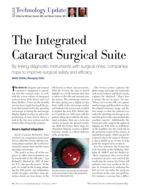
The Alcon Verion can provide an axis guide<br />
in the microscope to aid in the placement<br />
of a toric intraocular lens.<br />
The Verion system captures the<br />
globe image and maps out landmarks<br />
such as iris features and blood vessels,<br />
explains Dr. Mackool. “That’s how<br />
it orients the eye in space,” he says.<br />
“When you’re in the OR, you capture<br />
another image and the system overlays<br />
the original reference image and the<br />
new image, so that the patient’s eye<br />
under the operating microscope is oriented<br />
in space in the exact position the<br />
machine expects.” Additionally, the<br />
Verion gives the surgeon the option of<br />
aligning any IOL, toric or multifocal,<br />
on the pupillary axis, the visual axis or<br />
the geometric center of the cornea. A<br />
capsulorhexis guide, which can also be<br />
centered where the surgeon chooses,<br />
is also available as an overlay, to help<br />
guide the surgeon as he creates it.<br />
For lens selection, the Verion has<br />
such formulas as the Holladay, Holladay<br />
II, SRK/T and the Hoffer Q.<br />
“More important, after you’ve done<br />
a number of cases and entered the<br />
results into the system, the Verion will<br />
optimize your case results in the future<br />
by retrospectively analyzing the<br />
data so you can tailor the program<br />
you choose to the cases you perform.<br />
For instance, everyone has a differ-<br />
18 | Review of Ophthalmology | February 2014<br />
This article has no commercial sponsorship.