PLANT DEFENCES AGAINST PATHOGENS David Guest and John ...
PLANT DEFENCES AGAINST PATHOGENS David Guest and John ...
PLANT DEFENCES AGAINST PATHOGENS David Guest and John ...

You also want an ePaper? Increase the reach of your titles
YUMPU automatically turns print PDFs into web optimized ePapers that Google loves.
17<br />
<strong>PLANT</strong> <strong>DEFENCES</strong> <strong>AGAINST</strong> <strong>PATHOGENS</strong><br />
Gontents<br />
<strong>David</strong> <strong>Guest</strong> <strong>and</strong> <strong>John</strong> Brown<br />
17. 1 Host-parasite reLationshrps ...........<br />
263<br />
17.2 Passiue deJences ............<br />
266<br />
PhgsicaL barriers....<br />
266<br />
ChemicaL barriers<br />
268<br />
17.3 Pathogen recognition ..........<br />
269<br />
N on- s p e cijic eLicito r s<br />
270<br />
Gene- speciJic elicttors ..............<br />
271<br />
Suppresso r s <strong>and</strong> comp atibtLitg Jactor s<br />
271<br />
PhgsioLogicaL roLe oJ eLicitors<br />
272<br />
Eu olution oJ ho st-p ar as ite s p ecificitg<br />
273<br />
17.4 Rapid actiue deJences<br />
274<br />
Chang e s in memb r ane Junction<br />
274<br />
Ttte oxidatiue burst<br />
274<br />
CeLL tuaLL reiryforcement ...........<br />
274<br />
Hg per sensrtiue ceLL de ath<br />
276<br />
PhgtoaLexins ............<br />
278<br />
17.5 DeLaged actiue deJences<br />
281<br />
Pattngen containment <strong>and</strong> tuound repair<br />
281<br />
P atho g e ne s is - r eLqte d p r ote in s<br />
282<br />
Systemic acqutred re sistance<br />
283<br />
17 . 6 The dgnamtcs <strong>and</strong> coordination oJ deJence responses........ 284<br />
17.7 F\Lrther reading<br />
285<br />
17.1 Host-parasite relationships<br />
Earlier chapters have described the diverse <strong>and</strong> constant threat pathogens pose<br />
to plant health. Yet, surprisingly, disease is the exception rather than the rule in<br />
natural plant communities. Put another way, most pathogens are unable to<br />
attack most plants; they have a restricted host range. Assuming environmental<br />
conditions favour pathogen development, the resistance or susceptibility of a<br />
plant to a particular pathogen depends on two interrelated factors: (i) the<br />
substrate requirements of the pathogen <strong>and</strong> (ii) the response of the plant to the<br />
pathogen.<br />
In the previous chapter two broad groups of pathogens, necrotrophs <strong>and</strong><br />
biotrophs, were distinguished by their different substrate requirements (Table<br />
16.f). Necrotrophs are'thugs'in the sense that they kill plant cells before<br />
parasitising them. Host <strong>and</strong> parasite cells cannot coexist harmoniously. Thus, an<br />
incompatible cellular relationship between the parasite <strong>and</strong> host is essential for<br />
disease development. If the toxins used to kill host cells are not released at the<br />
right time, place or concentration, or if a particular host genotype is insensitive to<br />
the toxin, host cells will not die. The necrotroph will be unable to colonise or<br />
reproduce <strong>and</strong> the plant will be resistant. TWo types of necrotrophic pathogens<br />
exist: (i) those with a wide host range involving many plant species <strong>and</strong> (ii) those
264 Dauid. Gue st <strong>and</strong> <strong>John</strong> Brotun<br />
with a host range restricted to a few plant species or even to cultivars within a<br />
species. The key difference between these two types of necrotroph is the<br />
specificity of the toxin(s) produced. Necrotrophs with a broad host range secrete<br />
toxins that act on metabolic targets common to many plants. In contrast, the<br />
pathogenic ability of necrotrophs that release host-specific toxins is conditioned<br />
by the gene that encodes the ability to produce the toxin <strong>and</strong> by a gene in<br />
susceptible cultivars of the host that encodes sensitivity to that toxin. Hostspecific<br />
necrotrophs usually form a pathogenic race or pathotype structure where<br />
some races can attack some cultivars within a species but not others. If the gene<br />
that conditions sensitivity to a particular host-specific toxin is absent from a<br />
cultivar, that cultivar will be resistant to the disease caused by that pathogen.<br />
Biotrophs on the other h<strong>and</strong> are obligate parasites that obtain nutrients from<br />
living cells. Consequently, they must establish a compatible cellular relationship<br />
with their hosts. Biotrophs act as 'sneaks'. They typically infect through natural<br />
openings or by directly penetrating their host's surface. They mostly then grow<br />
between the cells of their host <strong>and</strong> only penetrate host cell walls (but not host cell<br />
membranes) to form food-absorbing haustoria. The pathogen develops without<br />
eliciting the host's defence responses or by spreading in advance of the plant's<br />
ability to activate its defence responses. The level of specialisation required to<br />
establish this type of relationship usually means that biotrophs have a restricted<br />
host range <strong>and</strong> a well-defined pathogenic race structure. If host cells die in<br />
advance of invasion by a biotrophic pathogen, the plant will be resistant because<br />
the pathogen is unable to establish a parasitic relationship.<br />
A second factor that influences whether a parasitic relationship will become<br />
established is the way that the plant under challenge responds. Some<br />
interactions between individual pathogen propagules <strong>and</strong> plant cells may lead to<br />
successful pathogen establishment, while others may not. In this chapter it will<br />
become evident that resistance or susceptibility of a whole plant <strong>and</strong> plant<br />
communities is the sum of many individual cellular interactions. Plants that are<br />
resistant restrict or retard the development <strong>and</strong> reproduction of an overwhelming<br />
majority of individual pathogen propagules that attack it. In this sense resistance<br />
is quantitative-resistant hosts prevent or slow the development <strong>and</strong><br />
reproduction of a higher proportion of pathogen propagules than susceptible<br />
hosts. For the purposes of plant breeding, the response of a plant to pathogen<br />
inoculation is often categorised as either'resistant'or'susceptible', although from<br />
a cellular perspective this distinction is not always so clear. Resistance <strong>and</strong><br />
susceptibility are more accurately portrayed as the extremes of a continuum<br />
upon which most host-parasite interactions sit. Resistance may be expressed in<br />
many ways, from the inhibition of propagule germination <strong>and</strong> penetration, the<br />
killing of pathogens before establishment, to the restriction or retardation of<br />
colony development <strong>and</strong> reproduction once the pathogen has established. For<br />
example, different genes for stem rust resistance in wheat act at different stages<br />
of the host-parasite interaction. Some cause the rapid death of the pathogen<br />
following attempted penetration, others allow initial infection, but prevent<br />
haustorial development <strong>and</strong> starve the pathogen, while the 'slow-rusting' genes<br />
allow parasitism <strong>and</strong> pathogen reproduction, but at a much slower rate than in<br />
susceptible cultivars. Each type of interaction provides useful resistance for plant<br />
breeders because they all delay the onset of epidemics <strong>and</strong> reduce yield losses.<br />
The early steps involved in the establishment of a host-pathogen relationship<br />
are delicate <strong>and</strong> sensitive to environmental factors, including the presence of<br />
other micro-organisms. The host-parasite-environment interaction is mediated by<br />
a complex interchange of signals. Plants respond to pathogen attack by erecting a<br />
highly coordinated series of molecular, cellular <strong>and</strong> tissue-based defence barriers.
17. PLant deJences against pathogens<br />
265<br />
All plants have the capacity to activate these defences. However if they are<br />
activated too little, too late, or in the wrong place, they will fail to restrict the<br />
pathogen <strong>and</strong> the plant will be susceptible. Pathogens respond by escaping or<br />
suppressing plant defence responses or by rendering these responses impotent,<br />
for example by detoxiffing plant antibiotics.<br />
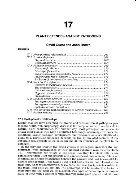
The interaction of pathogen nutrient requirements <strong>and</strong> host responses leads to<br />
five possible outcomes if environmental conditions favour infection (Fig. l7.l).<br />
. No relationship is established when the plant <strong>and</strong> the pathogen ignore each<br />
other. For example, a spore of a fungus may germinate, but because the host<br />
does not provide essential requirements for pathogen development, the<br />
resulting hypha fails to penetrate or establish a parasitic relationship. The<br />
fungus dies when its energy reserves are exhausted. The plant does not react<br />
in any way <strong>and</strong> is resistant by default. It is a non-host.<br />
No relationship<br />
between olant<br />
<strong>and</strong> pat n<br />
Mutual adjustment<br />
between the plant<br />
<strong>and</strong> the patho<br />
PATHOGEN<br />
Virus<br />
Bacterium<br />
Fungus<br />
Nematode<br />
Mutual antagonism<br />
between the olant<br />
<strong>and</strong> the<br />
Pathogen is<br />
antagonistic to the<br />
plant<br />
Figiure 17.1 Five possible relationships between plants <strong>and</strong> potential pathogens.<br />
. A plant is antagonistic to the pathogen when it secretes inhibitory<br />
compounds into its environment that prevents pathogen development. For<br />
example, the stubble of some brassicas releases 'biofumigants' into the soil<br />
that prevent the hatching of nematode eggs <strong>and</strong> inhibit the growth of some<br />
root-infecting fungi. Asparagus <strong>and</strong> marigolds {Tagetes spp.) secrete<br />
substances into the rhizosphere that are toxic to nematodes <strong>and</strong> provide<br />
useful protection against nematodes when interplanted with nematodesusceptible<br />
plants like tomato. Many plants secrete phenolic compounds onto<br />
their leaf surfaces that not only discourage herbivore feeding, but also inhibit<br />
many micro-organisms, including potential pathogens. In this relationship, the<br />
pathogen fails to develop <strong>and</strong> has no observable effect on the metabolism of<br />
the host plant. In some cases, such as in the quiescent infection of ripening<br />
avocado fruit with Coll.etotricltum gLoeosportoides, plant antagonists only<br />
temporarily inhibit pathogen development. Spores germinate to form<br />
appressoria, but their development is arrested by fungistatic substances in the<br />
peel. After harvest, these substances are enzymically degraded <strong>and</strong> the<br />
appressorium germinates to form infection hyphae. Eventually, anthracnose<br />
lesions develop. This type of interaction involving a quiescent stage in
266 Dauid <strong>Guest</strong> <strong>and</strong> <strong>John</strong> Broun<br />
pathogen development is common among the stem end rot pathogens of<br />
avocado <strong>and</strong> mango (e.g. DothiorelLa domtnicana, Lastodtplodta theobromae,<br />
Phomopsis spp. <strong>and</strong> ColLetotrichum gloeosporioide s).<br />
. The pathogen is antagonistic to the plant when it secretes compounds that<br />
damage the plant. For example, Periconia circinata, infects the roots of<br />
sorghum, but only those strains of the fungus that produce the host-specific<br />
toxin, periconin, induce symptoms of milo disease, but only in cultivars that<br />
are sensitive to this toxin. Similarly, some strains of Al.ternaria aLternata<br />
release host-specific toxins that kill cells of susceptible host species <strong>and</strong><br />
cultivars. For example, a strain of the fungus that is pathogenic on tomato<br />
produces AAl-toxin, to which tomato is uniquely sensitive. Strains producing<br />
AAM-toxin attack apples, AAK-todn producing strains affect Japanese pears,<br />
AAC toxin-producing strains affect citrus <strong>and</strong> so on. The tomato, apple <strong>and</strong><br />
Japanese pear strains are not pathogenic to citrus because citrus is only<br />
sensitive to the AAC-toxin. Cochliobolus uictoriae produces the toxin victorin<br />
that causes severe seedling blight on susceptible cultivars of oats, but has<br />
little effect on resistant cultivars or on other plant species. Resistance is the<br />
result of insensitivity to the toxin produced by the pathogen. If this<br />
insensitivity is common to all cultivars within a plant species, that species is<br />
said to be a non-host.<br />
o Mutual antagonism between plant <strong>and</strong> pathogen results in the inhibition or<br />
death of both the host tissue <strong>and</strong> pathogen. For example, an incompatible<br />
interaction between the stem rust pathogen, Puccinia gramtnis f. sp. tritici <strong>and</strong><br />
resistant cultivars of wheat causes the death of both host <strong>and</strong> pathogen cells.<br />
. Mutual adjustment leads to a compatible cellular relationship between the<br />
host <strong>and</strong> pathogen. Symbiotic relationships between mycorrhizal fungi <strong>and</strong><br />
plant roots <strong>and</strong> between nitrogen-fixing prokaryotes <strong>and</strong> plant roots, are<br />
examples of mutually beneficial interactions. Endophytic fungi <strong>and</strong> bacteria<br />
colonise the intercellular spaces of plant tissue, apparently without damaging<br />
their host cells. Many stem end rot pathogens have an endophytic phase in<br />
leaves <strong>and</strong> twigs before they infect fruits. Biotrophic pathogens, like the<br />
mildews <strong>and</strong> rusts, grow <strong>and</strong> reproduce on living host tissue. However, the<br />
diversion of nutrients to the invading pathogen adversely affects the growth of<br />
the host, even though host cells are not killed.<br />
In this chapter, plant defence mechanisms will be discussed in the order they<br />
are usually confronted by pathogens. Broadly speaking, passive defence<br />
mechanisms are those that are present before contact with the pathogen, while<br />
active defence mechanisms are activated only after pathogen recognition (Fig.<br />
L7.2).In reality this distinction is not always clear, as many pre-existing defences<br />
are modified after infection.<br />
17.2 Passive defences<br />
To gain access to the nutrients or replication machinery available within the host<br />
cell, pathogens must first breach the natural barriers presented by healthy<br />
plants. These barriers may be physical (the cuticle, cell wall, stomatal aperture or<br />
lenticel) or chemical (including inhibitory compounds or the absence of<br />
stimulatory compounds needed for pathogen development). Saprophytes lack the<br />
ability to penetrate these natural barriers.<br />
Physical barriers<br />
The importance of the cuticle as a barrier to penetration has been demonstrated<br />
by the dependence of many pathogens on adhesion <strong>and</strong> the subsequent release of
17. Plant deJences against pathogens 267<br />
cutin-degrading enzymes at the time of penetration. AJthough cutin-degrading<br />
enzymes are also secreted by many saprophytic fungi <strong>and</strong> bacteria, their primary<br />
activity is to allow access to cellulose in plant cell walls as a nutritional substrate.<br />
Different forms of cutin-degrading enzymes are used by pathogens to puncture<br />
the cell wall (Chapter 16). The activity of this type of cutinolytic enzyme in<br />
isolates of Fllsarrum sol.qnt f. sp. pisi is directly related to their aggressiveness on<br />
pea stems, indicating that pathogens unable to dissolve the cuticle at the point of<br />
penetration are excluded.<br />
Passive defences<br />
Physical barriers<br />
l:'m".<br />
lcell wall<br />
lstomata<br />
llenticels<br />
l-n utrie nt dep rivat i o n<br />
Chemical barriers I fil*"."ucipins<br />
| :lant defensins<br />
Active defences<br />
Rapid<br />
Delayed<br />
l-rnembrane function<br />
loxidative burst<br />
I cell wall reinforcement<br />
I hypersensitive cell death<br />
| 4ftoatexin<br />
accumulation<br />
I pathogenesis-related proteins<br />
I systemic acquired resistance<br />
lpathogen containment<br />
Figtrre 17.2 Some defence mechanisms in plants.<br />
Cuticle <strong>and</strong> cell wall thickness may influence resistance to certain pathogens.<br />
Some types of 'adult plant resistance' could be associated with a reduced ability<br />
of pathogens to enter through thicker, tougher cell walls. Some pathogens such<br />
as Puccinia graminis only infect young barberr5r leaves with thin cuticles <strong>and</strong> the<br />
germ tubes emerging from basidiospores do not penetrate thicker cuticles on<br />
mature leaves. Similarly, the ability of Taphrina dejorrnans to infect only young,<br />
newly unfolded leaves has been attributed to the inability of germ tubes to<br />
penetrate the thicker cuticles of older leaves. The presence of secondary cell walls<br />
in sclerenchyma, xylem or older plant tissue often retards pathogen development,<br />
leading, for example, to angular leaf spots where pathogen spread is restricted by<br />
leaf veins. Thick cuticles may physically prevent the eruption of sporophores <strong>and</strong><br />
release of spores. However, most experimental evidence suggests that toughened<br />
cuticles <strong>and</strong> cell walls are just one of the many factors that contribute to<br />
resistance.<br />
Waxy cuticles <strong>and</strong> vertically oriented leaves may prevent the formation of<br />
moisture films on leaf surfaces. Dry leaf surfaces intriUit infection by pathogens<br />
such as bacteria, nematodes <strong>and</strong> fungal zoospores that require a film of water for<br />
motility. Fungal spores might also be inhibited because most require moisture for<br />
germination. This must be balanced with the fact that vertically oriented leaves
268 Dauid. <strong>Guest</strong> <strong>and</strong> <strong>John</strong> Brown<br />
are more prone to impaction by wind-borne pathogen propagules <strong>and</strong> are likely to<br />
face higher inoculum levels compared with those that are horizontally oriented.<br />
Many pathogens enter through wounds, natural openings or are introduced by<br />
vectors. In these cases it is difficult to see how natural barriers such as the<br />
cuticle <strong>and</strong> cell wall could be involved in resistance. Some researchers have<br />
proposed that plants that have stomatal apertures that are the w'rong shape or<br />
size for pathogen infection structures to enter or that have stomata that close at<br />
the time of day that pathogen spores normally germinate, may be more resistant<br />
to pathogen attack. The black pod pathogen, Phgtophthora palmiuora, enters<br />
cocoa pods through stomata. Cocoa genotypes that produce pods with few,<br />
relatively smaller stomata, allow fewer lesions to establish than genotypes with<br />
more numerous, larger stomata. Not surprisingly, as the pathogen enters through<br />
stomatal pores, there is no correlation between cuticle thickness or pod case<br />
hardness <strong>and</strong> resistance to black pod. The bacterium that causes citrus canker,<br />
Xanthomonqs campestris pv. citri, enters grapefruit through open stomata.<br />
M<strong>and</strong>arins are resistant because their stomata are too small to allow entry of the<br />
bacterium. Similarly, lenticels that suberise rapidly so that their size is reduced<br />
may physically exclude pathogens such as StreptomAces scabies, the cause of<br />
common scab of potato.<br />
Chemical barriers<br />
Exudates on the surfaces of plants or compounds in plant cells may stimulate or<br />
inhibit the development of pathogens. Sometimes, plants resist infection because<br />
they do not provide the pathogen with its required nutrients. Resting spores of<br />
pathogens such as Spongospora subterranea (powdery scab of potato), Urocgstls<br />
agropgri (flag or leaf smut of wheat) <strong>and</strong> Plasmodtophora brassicae (club root of<br />
crucifers) <strong>and</strong> eggs of the potato cyst nematode, Globodera rostochiensis, require<br />
specific substances to stimulate germination or hatching. These are provided in<br />
secretions from certain plants, including potential hosts. Plants that fail to<br />
secrete these stimulators are resistant by default.<br />
Other plant secretions may simply not support the pre-penetration growth of<br />
the pathogen. Experimental depletion of iron availability using binding agents<br />
(siderophores) inhibits the growth of certain fruit-rotting bacteria. Host cultivars<br />
that secrete lower than normal levels of iron onto their surface may deprive<br />
pathogens of essential nutrients, inhibiting their growth. Similarly, micro,<br />
organisms that sequester available iron on leaf surfaces have potential as<br />
biocontrol agents (Chapter 27).<br />
Plants sometimes produce compounds during normal growth that inhibit the<br />
development of pathogens. Phytoanticipins may be excreted into the external<br />
environment (e.g. rhizosphere or phylloplane), accumulate in dead cells or they<br />
may be sequestered in vacuoles in an inactive form. The dead cells of brown<br />
onion skins contain the quinones catechol <strong>and</strong> protocatechuic acid, which inhibit<br />
germination of spores of the smudge pathogen, CoLLetotrichum circinans, <strong>and</strong> the<br />
neck rot pathogen, Botrytis cinerea. white onions do not produce these<br />
compounds <strong>and</strong> are susceptible to smudge. AspergiLLus niger is insensitive to<br />
these inhibitors <strong>and</strong> attacks both white <strong>and</strong> brown onions. Avocado rootstocks<br />
resistant to root rot caused by Phgtophthora ctnnamomr secrete borbinol, an<br />
antimicrobial phenolic compound, into the rhizosphere. The secretion of<br />
nematode-inhibiting substances into the rhizosphere surrounding asparagus <strong>and</strong><br />
marigold roots has already been mentioned. Symptoms of anthracnose of<br />
avocado, caused by CoLLetotrichum gLoeosporiordes, only develop on ripe fruit. The<br />
peel of unripe avocado fruit contains antifungal lipids called dienes that prevent
17. Ptant deJences against pathogens 269<br />
appressorial germination. As these dienes are gradually metabolised during fruit<br />
ripening to less toxic compounds, quiescent appressoria germinate <strong>and</strong><br />
susceptibility to anthracnose increases. In anthracnose-resistant cultivars, diene<br />
breakdown is blocked following infection, so that antifungal levels are sustained<br />
for longer periods. The resistance of immature apples <strong>and</strong> pears to scab, caused<br />
by Venturia inaequalis <strong>and</strong> V. pirina respectively, correlates with the presence of<br />
the phenolic compounds chlorogenic acid, phloridzin, arbutin <strong>and</strong> iso-chlorogenic<br />
acid in the outer layers of the fruit. These compounds also contribute to the bitter<br />
taste of unripe apples <strong>and</strong> pears <strong>and</strong>, as the fruit ripens <strong>and</strong> sweetens, it also<br />
becomes more susceptible to scab.<br />
One group of phytoanticipins, the saponins, are plant glycosides with<br />
surfactant (wetting agent) properties. Saponins bind sterols in pathogen cell<br />
membranes, destroying membrane integrity <strong>and</strong> function. In this way saponins<br />
are toxic to organisms containing sterols in their membranes (e.g. plants <strong>and</strong><br />
fungi, but not Oomycota). Inactive saponin precursor molecules appear to be<br />
stored in vacuoles of intact plant cells, but hydrolase enzymes released following<br />
wounding or infection convert these precursors to active, antimicrobial forms.<br />
Several lines of evidence suggest that saponins are involved in disease resistance<br />
<strong>and</strong> host range determination. It appears that the ability of some pathogens to<br />
detoxiff specific saponins matches their host range. For example, a strain of the<br />
take-all pathogen that attacks oats as well as wheat <strong>and</strong> barley<br />
(GaeumannomAces graminis var. auenae), releases the enzyme avenacinase.<br />
Avenacinase detoxifies the triterpenoid saponin, avenacin, found in epidermal<br />
cells of the roots of oat plants. Mutants in which the gene for avenacinase<br />
production has been deleted are sensitive to avenacin in vitro <strong>and</strong> are not<br />
pathogenic on oats, but remain pathogenic to wheat <strong>and</strong> barley.<br />
GaeumannomAces graminisvar. tritict lacks avenacinase <strong>and</strong> attacks wheat <strong>and</strong><br />
barley, but not oat species containing avenacin. An oat species that does not<br />
produce avenacin, Auena LongigLumis, is susceptible to GaeumannomAces<br />
graminis var. tritici. Another saponin, tomatine, contributes to the resistance of<br />
tomato leaves to Botrytis cinerea.<br />
Some plant peptides also inhibit the development of fungi, bacteria, viruses<br />
<strong>and</strong> insects. They act as proteinase <strong>and</strong> polygalacturonase-inhibitors, as<br />
ribosome inhibitors or lectins. These inhibitors interfere with pathogen nutrition<br />
<strong>and</strong> retard their development, thus contributing to disease resistance. Because of<br />
their similarity to peptides called defensins found in insects <strong>and</strong> mammals, they<br />
have been termed plant defensins. Secreted defensins provide an important<br />
defence against damping-off pathogens. While only O.5o/o of the total protein<br />
found in ungerminated radish seeds is defensin, it makes up 3O% of the proteins<br />
released from germinating seeds. It provides an antimicrobial micro-environment<br />
around the emerging radicle. Defensins may constitute up to lOo/o of the total<br />
proteins in cereal, legume <strong>and</strong> solanaceous seeds. Similar studies have shown<br />
defensins are also present in the outer cell layers of other plant organs such as<br />
flowers, leaves <strong>and</strong> tubers. While many defensins accumulate during normal<br />
plant development, others are induced, or their accumulation is enhanced, after<br />
wounding. Defensins, because of their anti-feeding activity against insects,<br />
provide a defence against insect-transmitted viruses.<br />
17.3 Pathogen recognition<br />
The ability of plants to respond to challenge by potential pathogens implies that<br />
plants recognise these potential pathogens as 'non-self. While mammals use<br />
antigen-antibody interactions to recognise non-self, plants recognise a vast array
270 Dauid <strong>Guest</strong> <strong>and</strong> <strong>John</strong> Brotun<br />
of signals originating from micro-organisms <strong>and</strong> the environment to elicit defence<br />
responses.<br />
N o n-s pec if i c e I icito rs<br />
Many signals of abiotic <strong>and</strong> biotic origin induce defence responses in a range of<br />
cultivars <strong>and</strong> host species that bear little relationship to pathogen host ranges.<br />
The magnitude of the response depends on the amount of elicitor present. Abiotic<br />
elicitors, including heavy metal ions, [fV light <strong>and</strong> some metabolic inhibitors,<br />
precipitate physiological stress responses, some of which contribute to resistance.<br />
Their effect is generally transitory <strong>and</strong> non-specific. The significance in hostparasite<br />
interactions of abiotic elicitors is not always obvious as they are rarely<br />
present at the infection court. However solar UV radiation may elicit stress<br />
responses in exposed plant tissues, providing an additional barrier for invading<br />
pathogens. On the other h<strong>and</strong>, environmental stresses usually increase the<br />
susceptibility of plants to necrotrophic pathogens.<br />
Cell wall fragments released from fungi <strong>and</strong> bacteria elicit defence responses<br />
in plants. Cell wall fragments from Phgtophthora megasperrna f. sp. glgcinea are<br />
potent elicitors of defence responses in soybeans. The smallest active fragment is<br />
a heptabetaglucan (seven glucose units) that is found in cell walls of many<br />
pathogenic <strong>and</strong> non-pathogenic races <strong>and</strong> species of oomycetes. Recently, a<br />
receptor was identified in the plasma membrane of soybean cells. This, together<br />
with its potency, suggests a role for heptabetaglucan <strong>and</strong> related oligosaccharins,<br />
in pathogen recognition.<br />
Hydrolytic enzymes of plant or pathogen origin also catalyse the release of<br />
plant cell wall fragments (endogenous elicitors) that elicit defence responses.<br />
For example, polygalacturonase enzymes released by fruit decay fungi <strong>and</strong><br />
bacteria dissolve the middle lamella of plant tissues. While this facilitates<br />
pathogen colonisation, it also causes the release of pectic fragments,<br />
oligosaccharides consisting of nine to thirteen polygalacturonate units, that are<br />
potent elicitors.<br />
A number of peptides <strong>and</strong> glycoproteins that elicit defence responses in<br />
plants have been isolated from culture filtrates of bacterial <strong>and</strong> fungal pathogens.<br />
A 46 kD glycoprotein extracted from culture filtrates of the black shank pathogen,<br />
Phgtopttthoranicotianae var. ntcottanqe <strong>and</strong> from tobacco leaves infected with this<br />
pathogen, is a potent elicitor. There is some evidence that Ppn 468, a 46 kD<br />
glycoprotein, has endoxylanase activity, suggesting that it may also elicit through<br />
the release of cell wall fragments. A 42 kD glycoprotein with glucanase activity<br />
has been isolated frorn Phgtophthora mega.sperma f. sp. gtgcinea. The active<br />
fragment of this glycoprotein is a thirteen-amino acid peptide that binds to a<br />
receptor on the host plasma membrane. These elicitors are found in both<br />
avirulent <strong>and</strong> virulent isolates, suggesting that their activity does not determine<br />
resistance.<br />
A family of lOkD peptides called 'elicitins' has been isolated from culture<br />
filtrates of Phgtophthora spp. <strong>and</strong> a number of related oomycetes. There are two<br />
groups of elicitins (i) the acidic a -elicitins such as parasiticein produced by<br />
P. ntcottanae var. parasitica. <strong>and</strong> capsicein produced by P. capstci <strong>and</strong> (ii) the<br />
basic B-elicitins such as cryptogein, produced by P. cryptogea, melonin produced<br />
by P. meLonis <strong>and</strong> cinnamomin produced by P. cinnamomi. All elicit systemic<br />
necrosis in tobacco. Elicitins are translocated when applied to the plant, but they<br />
have yet to be found at the infection court. They are not known to have any<br />
metabolic function in the fungi that produce them. Highly aggressive isolates of<br />
P. nicotianae var. nicotianae do not release an elicitin <strong>and</strong> do not elicit host
17. Plc:nt deJences against pathogens 27r<br />
defence responses. However, less aggressive isolates <strong>and</strong> isolates from hosts other<br />
than tobacco, release parasiticein. This evidence indicates that elicitin release<br />
may limit the host range of certain oomycetes. The black shank pathogen is a<br />
biotroph in the early stages of infection <strong>and</strong> aggressive mutants with low elicitin<br />
levels may have been selected during co-evolution with its host, tobacco.<br />
Polyunsaturated fatty acids like arachidonic <strong>and</strong> eicosapentaenoic acid from<br />
cell membranes of Phgtophthora infestans elicit defence responses in potato<br />
slices. Although they have lower elicitor activity in other plants when applied on<br />
their own, these fatty acids enhance the elicitor activity of glucans when applied<br />
in combinations. This, <strong>and</strong> other evidence, indicates that the complex responses<br />
of some infected plants may depend on the recognition of a combination of<br />
elicitors.<br />
Gene-spec iti c el i citors<br />
Gene-specific elicitors are those conditioned by avirulence genes in the pathogen.<br />
Their activity precisely matches the gene-for-gene hypothesis. Only recently has<br />
the application of molecular techniques allowed the characterisation of a few<br />
gene-specific elicitors, although their presence has been inferred for many years.<br />
A series of race-specific peptide products of the avirulence genes of FbtuiaJutua, a<br />
biotrophic pathogen of tomato, has been identified. These peptides were first<br />
isolated from intercellular fluids of infected leaves <strong>and</strong> have since been found<br />
around the infection site.<br />
A heat labile exudate from germinating basidiospores of incompatible races of<br />
cowpea rust (Uromgces uignae) elicits defence responses only in cowpeas with the<br />
corresponding resistance gene. Similarly, a 6.4 kD peptide from the barley leaf<br />
scald pathogen, Rhgnchosportum secalis, specifically elicits resistance in cultivars<br />
with the corresponding resistance gene. Host receptors for these peptides have<br />
yet to be identified.<br />
A number of avirulence genes have been identified in plant pathogenic<br />
bacteria, although their gene products are yet to be characterised. Avirulence<br />
(anr) genes determine host range (species/pathovar <strong>and</strong> cultivar/race<br />
interactions) according to the gene-for-gene hypothesis. However, studies with<br />
genetically-transformed bacteria show that qur genes only appear to function in<br />
the presence of another set of genes, the hrp (hypersensitive response <strong>and</strong><br />
pathogenicity) gene cluster. Hrp genes are found among a wide range of<br />
pathogenic <strong>and</strong> non-pathogenic Gram-negative bacteria. They function as<br />
pathogenicity genes in the absence of the aur gene <strong>and</strong> hypersensitive responseeliciting<br />
genes in their presence. One of these hrp genes encodes a heat stable<br />
protein, harpin, that is involved in membrane transport. Clusters of harpin<br />
subunits apparently line a pore allowing secretion of aur gene products. Hrp gene<br />
products are also involved in the secretion of the extracellular polysacchari.des<br />
that disguise the pathogen from host recognition, thus functioning in both<br />
virulence <strong>and</strong> avirulence.<br />
Suppressors <strong>and</strong> compati bility factors<br />
It has been proposed that compatibility factors operate at two levels. All biotrophs<br />
must establish basic compatibility with their hosts. Virulent races might also<br />
produce specific compatibility factors that delay, avoid or negate recognition by<br />
normally resistant cultivars of a host species. Experiments using a range of hostparasite<br />
interactions have demonstrated that co-inoculation of a host with<br />
compatible <strong>and</strong> incompatible strains of a pathogen allows the normally avirulent<br />
strain to infect, colonise <strong>and</strong> reproduce (Fig. 17.3). These results suggest that the
272 Dauid <strong>Guest</strong> <strong>and</strong> <strong>John</strong> Brotun<br />
virulent isolate somehow suppresses the resistance mechanisms of the host.<br />
However, if the virulent strain is inoculated some hours after the avirulent strain,<br />
the host is resistant to both, indicating that suppressors are unable to switch off<br />
resistance responses once they are activated. Water-soluble molecules found on<br />
the surface of virulent, but not avirulent, isolates of. Phytophthora iryfestans<br />
suppress defence responses in potato tuber slices. Glycopeptides produced by<br />
Ascochgta rabiei <strong>and</strong> MgcosphaereLla pisi suppress defence responses in their<br />
respective hosts, chickpea <strong>and</strong> pea. Such interactions may be common in nature.<br />
yellow isolate gave<br />
a susceptible<br />
infection type<br />
white isolate gave a<br />
resistant infection<br />
type<br />
when leaves were inoculated<br />
simultaneouslv. both<br />
isolates produced<br />
susceptible infection types<br />
resistant<br />
infection<br />
type<br />
Figure 17.3 Induced susceptibility to stripe rust of wheat induced by simultaneous<br />
inoculation of plants with compatible <strong>and</strong> incompatible isolates of Puccinta<br />
stngformis. (After Brown <strong>and</strong> Sharp, 1970.)<br />
Physiological role of elicitors<br />
To underst<strong>and</strong> fully how the discriminatory expression of active defence<br />
responses determines resistance or susceptibility, we must underst<strong>and</strong> the basis<br />
of specificity. In other words, why do incompatible pathogens trigger plant<br />
defence responses, while compatible pathogens do not?<br />
The simplest prediction from the gene-for-gene hypothesis would be that<br />
avirulence <strong>and</strong> resistance gene products recognise each other, triggering a racespecific<br />
response. Only recently have molecules been identified that elicit plant<br />
defence responses according to the gene-for-gene hypothesis. These molecules are<br />
peptides encoded by avirulence genes, <strong>and</strong> some, perhaps all, bind to receptor<br />
peptides encoded by host resistance genes. Of the half-dozen or so resistance<br />
genes sequenced, most have some homologr to genes encoding proteins involved<br />
in protein-protein interactions in cells, such as protein kinases <strong>and</strong><br />
polygalacturonase-inhibiting proteins. Some are membrane-bound, while others<br />
are cytoplasmic. Activation of these proteins following the recognition of<br />
avirulence gene products triggers a cellular alarm mechanism, involving signal<br />
transduction pathways that lead to a massive shift in gene transcription <strong>and</strong>
17. Plant deJences against pathogens 273<br />
plant cell metabolism. As well, local <strong>and</strong> systemic signals are released that prime<br />
the plant against further infection.<br />
Specific recognition takes place against a background of non-specific events<br />
triggered by the multitude of molecules produced by pathogens that are<br />
recognised by plants as non-self. The lack of race-specificity of these elicitors<br />
makes their role in disease resistance unclear. Furthermore, the relevance of<br />
many non-specific elicitors to recognition in host-parasite interactions is<br />
questionable as they have been isolated from cultures of the pathogen rather<br />
than from the infection court. For example, culture filtrates of MoniLiniafructicoLa<br />
contain a small peptide, monilicolin A, that elicits defence responses in pea pod<br />
cavities. However, M. Jructicola is not a pathogen of pea <strong>and</strong> monilicolin A is<br />
inactive on natural hosts of the pathogen such as peach.<br />
Non-specific elicitors present at the infection court may simply function to<br />
ampliff the defence response. Cell wall fragments, released from both the host<br />
<strong>and</strong> pathogen in increasing quantities as colonisation is attempted, activate<br />
responses that are amplified in a positive feedback loop. Combined signals from<br />
the pathogen <strong>and</strong> host could help the plant differentiate between damage <strong>and</strong><br />
infection.<br />
Evolution of host-parasite specificity<br />
Clearly, pathogens produce a diverse range of molecules able to elicit host<br />
defence mechanisms. Only a few of these elicitors define the pathotype-cultivar,<br />
or even species, specificity characteristic of the hosts they were extracted from. If<br />
one assumes that disease resistance in plants is due to an active response, this is<br />
not surprising. There would have been a hear,y selection pressure on individual<br />
plants with the ability to recognise <strong>and</strong> resist pathogens with the potential either<br />
to kill them or to reduce their fitness. Thus, any molecule released by a potential<br />
pathogen could function as an elicitor, whether or not that molecule has anything<br />
to do with virulence. Examples might include cell-wall fragments, membrane<br />
lipids or extracellular enzymes, none of which is specific to avirulent races of a<br />
pathogen. On the other h<strong>and</strong>, gene-for-gene or pathotype-specific resistance is<br />
determined by the interaction between products of pathogen avirulence genes,<br />
gene-specific elicitors, <strong>and</strong> products of host resistance genes.<br />
What do elicitors do? Where do they bind? Where do they act? The defence<br />
responses of plants are very rapid. Host gene expression begins within minutes or<br />
even seconds of exposure to elicitors or pathogens. Elicitors may act directly on<br />
host genes as regulators. However, the diversity of elicitors that activate a<br />
common suite of responses suggests that second messengers are involved <strong>and</strong><br />
that elicitors induce a range of responses through complementary action. This<br />
notion is supported by the recent identification of resistance gene products that<br />
appear to be membrane-bound proteins involved in signal transduction, by the<br />
involvement of active oxygen as a second messenger <strong>and</strong> by the identification of<br />
salicylic acid as a common mediator of systemic defence responses.<br />
An alternative explanation for the evolution of host-parasite specificity<br />
proposes that r<strong>and</strong>om mutations might confer the ability to produce host-specific<br />
toxins. The dependence of formae speciales of ALternaria alternata on host-specific<br />
toxins for virulence on their respective hosts described in the previous section<br />
supports this explanation. These toxins allow an otherwise saprophytic organism<br />
to necrotrophically colonise a previously unavailable host. These mutants, being<br />
able to occupy a new ecological niche, have an evolutionary advantage in the<br />
presence of their hosts. Here, virulence, rather than host resistance, is the active<br />
phenomenon that was selected under evolutionary pressure.
274 Dauid. <strong>Guest</strong> <strong>and</strong> <strong>John</strong> Brown<br />
17.4 Rapid active defences<br />
Plant responses to infection are complex <strong>and</strong> there is no universal model or<br />
sequence of events that accurately describes the dynamics of resistance in the<br />
few interactions studied, let alone the vast majority of undescribed interactions.<br />
Almost every host-parasite interaction is unique in the details of the activation,<br />
localisation, timing <strong>and</strong> magnitude of each component of the defence response.<br />
As previously stated, resistance is rarely absolute <strong>and</strong> whether a plant ends up<br />
being resistant or susceptible depends on the sum of many individual responses.<br />
Changes in membrane function<br />
Most studies on the earliest stages of the host-parasite interaction conclude that<br />
the host membrane is involved in pathogen recognition <strong>and</strong> signal transduction.<br />
Membrane permeability changes rapidly following the exposure of plant cell<br />
suspension cultures to fungal <strong>and</strong> bacterial elicitors, usually leading to a loss of<br />
cellular electrolytes such as K+ <strong>and</strong> an uptake of H+. At the same time, there is<br />
often an influx of Ca2+, a key intracellular signal in plants that is involved in the<br />
activation of enzymes <strong>and</strong> gene expression. The experimental blocking of Ca2+<br />
transpor[ across membranes in inoculated bean cells also inhibits gene activation<br />
<strong>and</strong> subsequent defence responses.<br />
The oxidative burst<br />
Membranes are also the sites where the oxidative burst occurs. The term<br />
'oxidative burst' was first used to describe a rapid increase in respiration<br />
observed in neutrophils involved in the immune response of mammals. This<br />
increased level of respiration is now known to be due to the generation of reactive<br />
oxygen species, especially hydrogen peroxide <strong>and</strong> the superoxide anion (Oz-),<br />
through the addition of electrons to 02 catalysed by the membrane-bound<br />
errzyme, NADPH oxidoreductase. Reactive o>(ygen species are also produced by<br />
errors in electron transport during respiratory <strong>and</strong> photosynthetic reactions in<br />
plant cells. Cells are norrnally protected from the damaging effects of reactive<br />
oxygen by superoxide dismutase, various peroxidases <strong>and</strong> catalase <strong>and</strong> by<br />
natural antioxidants such as carotenes. The pioneering work of Doke <strong>and</strong> his<br />
colleagues at Nagoya University in Japan revealed that slices of potato tuber<br />
exposed to compatible <strong>and</strong> incompatible races of the late blight pathogen,<br />
Phgtophthora iryfestans, undergo a two-step oxidative burst. The first burst<br />
rapidly follows wounding <strong>and</strong> inoculation, while a much larger burst in<br />
incompatible interactions immediately precedes hypersensitive cell death. Since<br />
then, an oxidative burst has been described in a range of plant-fungal <strong>and</strong> plantbacterial<br />
interactions. The rapid oxidative burst generates levels of reactive<br />
oxygen species that initiate membrane lipid peroxidation <strong>and</strong> cell death. The<br />
oxidative burst in plants is associated with the release of local <strong>and</strong> systemic<br />
signals that trigger gene expression <strong>and</strong> the oxidative cross-linking of host cell<br />
wall components. Levels of reactive oxygen species accumulate at the infection<br />
court that are sufficient to kill micro-organisms in vitro. Experimental<br />
suppression of the oxidative burst shows that it is involved in initiating later<br />
defence responses. On the other h<strong>and</strong>, colonisation of avocado fruit by the<br />
necrotroph, Botrytis ctnerea, apparently exploits the oxidative burst to kill host<br />
cells in advance of invasion.
1 7. PLant deJences against pathogens 275<br />
Cel I wall rei nforcement<br />
The first visible response to attempted penetration of plant cell walls by<br />
pathogens is often the intensification of cytoplasmic streaming followed by the<br />
accumulation of host cytoplasm under the site of attempted penetration. These<br />
cytoplasmic aggregates are thought to contain the cellular apparatus for the<br />
synthesis of cell wall fortifications. Most pathogens must penetrate host cell walls<br />
at some stage, either as germ tubes, hyphae or haustoria. If the cell can respond<br />
quickly enough to repair or reinforce the cell wall, penetration efficiency may be<br />
reduced <strong>and</strong> pathogen development retarded.<br />
A number of different types of cell wall fortifications are produced in response<br />
to the attempted penetration of plant cell walls. Some pathogens induce the<br />
deposition of a papilla, a reinforcement composed of a branched P-1,3 glucan,<br />
callose, along with silicon, lignin <strong>and</strong> proteins, between the host cell wall <strong>and</strong><br />
plasma membrane, directly under the penetration peg. The rapid deposition of<br />
papillae is a common response of cereals to attempted penetration of epidermal<br />
cells by the powdery mildew fungus (BLumeria graminis). Papillae in resistant<br />
cultivars form more rapidly <strong>and</strong> are more diflicult to penetrate, than those formed<br />
by susceptible cultivars. As a result, haustorial development is prevented.<br />
Lignitubers are lignified callose deposits that ensheath invading hyphal tips {Fig.<br />
17.4A). Lignitubers have been observed in both resistant <strong>and</strong> susceptible cereals<br />
following challenge by the take-all pathogen, GaeumannomAces graminis,<br />
demonstrating again the importance of timing-the more rapid the response, the<br />
more likely it is to succeed.<br />
Hydroxyproline-rich glycoproteins are structural proteins in plant cell walls<br />
involved in the organisation of secondary cell wall thickening. Genes encoding<br />
hydro>ryproline-rich glycoprotein biosynthesis are transcribed in advance of<br />
invading hyphae, making cell walls tougher. Hydrogen peroxide, released during<br />
the oxidative burst following pathogen challenge, causes extensive cross-linking<br />
between hydroxyproline-rich glycoproteins <strong>and</strong> other cell wall components,<br />
making the walls even more resistant to microbial digestion.<br />
Cross-linked hydroxyproline-rich glycoproteins also provide a focus for lignin<br />
deposition on the plant cell wall. The rapid deposition of lignin <strong>and</strong> suberin<br />
following infection is associated with resistance to non-pathogens <strong>and</strong> to<br />
avirulent pathogens in many plants, including cereals, Solanaceae, brassicas,<br />
melons <strong>and</strong> carrots. Lignin deposited on plant cell walls ahead of invading<br />
hyphae increase their resistance to fungal penetration. Lignin also binds to<br />
hyphal tips <strong>and</strong> bacterial cells, preventing further growth <strong>and</strong> movement <strong>and</strong><br />
restricting the diffusion of pathogen enzymes <strong>and</strong> toxins <strong>and</strong> the uptake of water<br />
<strong>and</strong> nutrients by the pathogen. Furthermore, precursor molecules <strong>and</strong> free<br />
radicals produced during lignin biosynthesis are toxic to pathogens <strong>and</strong><br />
inactivate pathogen enzymes, toxins, elicitors or suppressors. The effect of lignin<br />
can be further enhanced by the release of reactive oxygen species <strong>and</strong> the<br />
activation of phenol oxidase enzymes that convert phenolic compounds to more<br />
toxic complex polymerised phenolics <strong>and</strong> quinones during the defence response.<br />
The evidence that cell wall reinforcements are important components of plant<br />
disease resistance can be summarised as follows:<br />
. Their deposition often coincides with failed penetration <strong>and</strong> sometimes<br />
precedes the cessation of pathogen growth.<br />
o Reinforcements in resistant hosts are larger, form more quickly (often before<br />
penetration) <strong>and</strong> are more dense than those formed by susceptible hosts.<br />
o Experimental attempts to re-penetrate induced reinforcements usually fail.<br />
' Inhibition of ligrun or callose biosynthesis enhances penetration efficiency.
276 Dauid. <strong>Guest</strong> <strong>and</strong> <strong>John</strong> Brown<br />
However, the deposition of cell wall reinforcements is not always associated with<br />
disease resistance. Clearly, cell wall reinforcements contribute to resistance <strong>and</strong><br />
cell repair but are not always sufficient on their own to prevent infection.<br />
Hypha<br />
Appressorium<br />
+:Living cells<br />
T.S.<br />
Advancing hyphae,<br />
lignituber penetrated<br />
A<br />
Invading pathogen<br />
Xylem vessel wall<br />
C<br />
Figure<br />
\7.4 Induced mechanical barriers to infection. (A) Lignituber formation in a root<br />
of a wheat seedling in response to infection by baeumannomAces graminis.<br />
(From Parry, 1990.) (B) Diagrammatic representation of tyloses in iesponse<br />
to invasion of the xylem by a vascular wilt fungus. (From parry, iggo.)<br />
(C) Scanning electron micrograph of tyloses formed in the xylem of maize in<br />
response to infection by Verticilltum alboatrum. (From Troughton <strong>and</strong><br />
Sampson, 1973.)
17. Plant deJences agatnst pathogens 277<br />
Hypersensitive cell death<br />
In I9O2 Harry Marshall Ward, Professor of Botany at Cambridge University in<br />
Engl<strong>and</strong>, observed an association between necrotic mesophyll cells in Bromlrs sp.<br />
<strong>and</strong> attempted infection of resistant cultivars by the leaf rust fungus, Ptrccinia<br />
recondtta. Later E. C. Stakman at the University of Minnesota reported similar<br />
observations in resistant wheat cultivars infected with the stem rust pathogen, P.<br />
graminis, <strong>and</strong> in 1915 he introduced the term hypersensitivity to describe this<br />
necrotic host reaction. Stakman contended that the more resistant the cultivar,<br />
the more rapid was the collapse of host cells <strong>and</strong> the sooner the fungus was<br />
inactivated. The term hypersensitivity indicates that the host cells are 'over-<br />
(hyper-) sensitive' to the presence of the pathogen. Host cells suicide in the<br />
presence of the pathogen, preventing further spread of the infection (Fig. f 7.5). In<br />
some cases h54persensitive cell death kills the invading pathogen (e.g. Rhizoctonia<br />
solani) while in others it is fungistatic (e.g. Puccinia graminis). Hypersensitive cell<br />
death is a widespread, but not universal, response to incompatible viral,<br />
bacterial, fungal <strong>and</strong> insect attack in the plant kingdom.<br />
appressonum<br />
epidermal cells<br />
haustorium<br />
hypersensitive<br />
cells<br />
A<br />
Figure 17.5 Wheat leaves showing the hypersensitive reaction in response to infection<br />
by (A) Blumertagramtnis <strong>and</strong> (B) Pucctntagraminis. (From Brown, 1980.)<br />
l)rpically, hypersensitive cell death is preceded by a rapid oxidative burst, an<br />
increase in cytoplasmic streaming, cytoplasmic aggregation followed by<br />
granulation, membrane disruption, cellular decompartmentalisation <strong>and</strong><br />
browning usually within 12-24 hours of attempted penetration (Fig. 17.6).<br />
Hypersensitive cell death in plant cells shares many features in common with<br />
apoptosis, or programmed cell death, observed during development of defence<br />
against disease in animals. Apoptosis is a distinct form of cell suicide directed by<br />
the dying cell <strong>and</strong> regulated by a number of identified genes. Animal cells<br />
undergoing apoptosis shrink, their DNA is digested into fragments of l8O base<br />
pairs <strong>and</strong> multiples of 180 base pairs <strong>and</strong> these fragments are organised into<br />
apoptotic bodies, seen as 'blebs' on the nuclear membrane. These orderly<br />
fragments of DNA are resolved as 'DNA ladders' by gel electrophoresis. The<br />
emerging similarities between hypersensitive cell death in plants <strong>and</strong> apoptosis in<br />
animal cells suggest that cell suicide is an ancient defence response.<br />
It is not always easy to conclude from research data whether host cell death is<br />
a consequence of murder or suicide. Recent experiments have shown that in<br />
many host-parasite interactions hypersensitive cell death precedes pathogen
278 Daui-d" <strong>Guest</strong> <strong>and</strong> <strong>John</strong> Brotun<br />
death, regardless of whether biotrophic or necrotrophic pathogens were involved.<br />
In some interactions however, disease resistance does not depend on<br />
hypersensitive cell death. The success of hS4persensitive celi death as a resistance<br />
mechanism in individual host-parasite interactions depends on the nutritional<br />
requirements of the pathogen <strong>and</strong> on the timing, location <strong>and</strong> magnitude of the<br />
host response in relation to pathogen development. In some interactions the rapid<br />
suicide of challenged host cells undoubtedly restricts pathogen development,<br />
contributing to the overall defence response.<br />
(i)<br />
- germ tube<br />
--/<br />
'host nucleus<br />
(iu)<br />
(il,<br />
(iii)<br />
haustorium<br />
cell brown<br />
necrotic<br />
<strong>and</strong><br />
granulation around<br />
haustorium<br />
(u)<br />
Figure 17.6<br />
movement <strong>and</strong><br />
accumulation of inhibitory<br />
compounds<br />
Sequence of events leading to the hlpersensitive reaction in plants infected<br />
by incompatible pathogens. (From Brown, 1980.)<br />
Phytoalexins<br />
Phytoalexins are low molecular weight antibiotics produced by plants in<br />
response to infection. Their toxicity is non-selective <strong>and</strong> the chemical affinity of<br />
most phytoalexins for lipids suggests that they accumulate in cell membranes.<br />
For phytoalexins to play a role in disease resistance, they must accumulate to<br />
inhibitory levels at the infection court <strong>and</strong> restrict further development of the<br />
pathogen.<br />
Evidence for the synthesis of antibiotics in infected plants has been<br />
accumulating for much of the twentieth century. In 1909 Bernard found that<br />
some fungi rotted ungenninated orchid seeds, others penetrated several layers of<br />
cells before stopping <strong>and</strong> disintegrating, while others colonised the seed <strong>and</strong><br />
established a successful mycorrhizal association with the seedling. Fungi that<br />
penetrated a few cell layers, but were then destroyed, induced resistance to<br />
subsequent infections by seed-rotting fungi. Nobecourt, in 1923, showed that this<br />
induced resistance was due to the synthesis of antibiotics by the seed. In 1945<br />
Ernst GAumann working in Switzerl<strong>and</strong> identified these inhibitors as two<br />
phenolic compounds, orchinol <strong>and</strong> hircinol. At about the same time Muller <strong>and</strong><br />
Borger in Germany found that slices of potato tuber reacting hypersensitively to<br />
Phgtophthora iryfestans produced antibiotics that protected the tissue against<br />
subsequent infection by normally vimlent strains of the pathogen.<br />
After World War II, K. O. Muller moved to the CSIRO in Canberra, where he<br />
studied responses of the seed cavity of french bean pods to spores of the peach<br />
pathogen, MoniLinia JructicoLa. While water droplets from uninoculated cavities
1 7. Plant deJences against pathogens 279<br />
stimulated fungal growth, inoculated cavities became necrotic <strong>and</strong> diffusates<br />
became inhibitory to fungal growth within 24 h of inoculation (Fig. 17.7). The<br />
unidentified inhibitor was extracted with organic solvents <strong>and</strong> was termed a<br />
phytoalexin (from the Greek words meaning plant defender). This inhibitor was<br />
subsequently purified by a team led by Ian Cruickshank at the CSIRO, found to<br />
be a phenylpropanoid compound <strong>and</strong> named phaseollin. A related compound,<br />
pisatin, was identified in pea pods inoculated \Mith M. fiucticota or in pod cavities<br />
exposed to a peptide, monilicolin A, extracted from this fungus.<br />
A<br />
r\Jr<br />
t<br />
l<br />
I I I<br />
After 24 h drops<br />
were collected <strong>and</strong><br />
passed through a<br />
Millipore filter to<br />
remove sDores.<br />
/
280 Dautd <strong>Guest</strong> <strong>and</strong> <strong>John</strong> Brotun<br />
French bean produces at least five phenylpropanoid phytoalexins, while potato<br />
synthesises at least four terpenoids.<br />
Table 17.1<br />
Examples of phytoalexins produced by higher plants.<br />
Structure Name Plants involved<br />
Inorganic<br />
Phenolic<br />
Terpenoid<br />
Phenylpropanoid<br />
Acetylenic<br />
Stilbene<br />
Indole-sulphur<br />
sulphur<br />
chlorogenic acid<br />
avenalumins<br />
capsidiol<br />
rishitin<br />
ipomeamarone<br />
gossypol<br />
pisatin<br />
phaseollin<br />
kievitone<br />
glyceollins<br />
medicarpin<br />
scoparone<br />
wyerone<br />
saffnol<br />
resveratrol<br />
batatasins<br />
camalexin<br />
brassinins<br />
cocoa<br />
potato, tobacco, apple<br />
some cereals<br />
capsicum, tobacco<br />
potato, tobacco, tomato<br />
sweet potato<br />
cotton<br />
pea<br />
french bean, cowpea<br />
french bean, cowpea<br />
soybean<br />
alfalfa, clover, broad<br />
bean, chickpea<br />
citrus<br />
broad bean<br />
safflower<br />
grape, peanut<br />
yam<br />
Arabidopsis<br />
cabbage, rape. turnip<br />
Phytoalexins are thought to be synthesised in cells adjacent to the infection<br />
site, in response to a signal produced either by the invading pathogen or by<br />
infected host cells. They are packaged in lipid vesicles <strong>and</strong> exported to the<br />
infected cell. Consequently, the infected cell becomes a toxic micro-environment<br />
for the invading pathogen. Phytoalexin accumulation is often associated with<br />
hypersensitive cell death. However, phytoalexin biosynthesis requires gene<br />
expression <strong>and</strong> the activation of complex biochemical pathways involving perhaps<br />
20 enzyrnes, which must occur in living cells. Many steps in their biosynthesis<br />
are sensitive to regulation by the host <strong>and</strong> the pathogen. Some plants, such as<br />
soybean <strong>and</strong> chickpea, synthesise phytoalexins upon infection, but convert a<br />
proportion into inactive sugar conjugates held in reserve in vacuoles. If the initial<br />
defence response fails to check pathogen growth, en4lmes that cleave the sugar<br />
molecule are activated <strong>and</strong> the phytoalexin reserves are rapidly releasec.<br />
Like other active defence responses, the success of phytoalexin accumulation<br />
depends on the speed, location <strong>and</strong> magnitude of the response. There is a good<br />
experimental correlation between resistance <strong>and</strong> rapid, localised phytoalexin<br />
accumulation in many host-parasite interactions. There is evidence that:<br />
. phytoalexins accumulate faster <strong>and</strong> to higher concentrations in resistant<br />
cultivars. In resistant plants, gene transcription begins within one hour of<br />
recognition, phytoalexins appear within four hours <strong>and</strong> concentrations peak to<br />
fungitoxic levels about 18-24 hours after challenge (Fig. 17.8). These events<br />
are delayed <strong>and</strong> more diffuse in susceptible plants.<br />
' phytoalexin biosynthesis is localised in cells immediately surrounding the<br />
infection court. There is no evidence that they disperse in the plant.<br />
Experiments using laser microprobe analysis, radioimmunoassay,
1 7. Plant deJences ag ainst pathogens<br />
28r<br />
hybridisation histochemistry <strong>and</strong> immunocylochemistry of<br />
biosynthesis pathway have confirmed this in several<br />
interactions.<br />
the phytoalexin<br />
host-pathogen<br />
z<br />
l-.<br />
-a<br />
o.B<br />
-<br />
o<br />
a<br />
-<br />
^<br />
a<br />
: l O<br />
€ e E<br />
a . - L<br />
I<br />
F<br />
.o€<br />
O o<br />
I<br />
a<br />
z o<br />
og<br />
z d<br />
X J<br />
L.' C)<br />
z2<br />
J.:<br />
EJ<br />
0.6<br />
o.4<br />
-<br />
li<br />
-<br />
-1<br />
't:<br />
(t)<br />
a<br />
L<br />
e ? . . E a<br />
f ijP 6 q<br />
a 66 tr o<br />
.Y<br />
'A<br />
h7 E<br />
^ -<br />
s 55 ; ;<br />
F ' r . O i "<br />
' 7 . t . g S<br />
t r l q c<br />
9 l l r l 3 3 o ( o<br />
s i s l<br />
:r<br />
I<br />
I<br />
I I<br />
r<br />
\/._1,/<br />
60 80 100 120 140 160<br />
TIME AFTER INOCULATION (h)<br />
Figure 17.8 Accumulation of phaseollin in beans inoculated with compatible (A) <strong>and</strong><br />
incompatible strains ( ) of Colletotrichum Ltndemutttianum. (From Bailey<br />
<strong>and</strong> Deverall. 1971.)<br />
r in a number of interactions, resistance is lost if phytoalexin biosynthesis is<br />
blocked by inhibitors of enzymes involved in the process of phytoalexin<br />
biosynthesis <strong>and</strong> is reduced in mutants that are slow to accumulate<br />
phytoalexins.<br />
. resistance is increased in plants transformed to express novel phytoalexins or<br />
if exogenous phytoalexins are applied. For example, although the biochemical<br />
precursor of resveratrol is widely distributed in the plant kingdom, only<br />
grapevine <strong>and</strong> peanut have the en4rme required to complete its synthesis.<br />
When the genes encoding this enzyme are transformed into tobacco,<br />
resveratrol is slmthesised in response to infection.<br />
Phytoalexin synthesis is not universal among plants. Wheat <strong>and</strong> cucumber<br />
apparently do not produce phytoalexins, yet effectively resist most pathogenic<br />
fungi <strong>and</strong> bacteria. Nevertheless, in many interactions the rapid accumulation of<br />
toxic concentrations of phytoalexins at the infection court plays a decisive role in<br />
the expression of resistance.<br />
17.5 Delayed active defences<br />
Pathogen containment <strong>and</strong> wound repair<br />
While earlier responses retard the development of pathogens, later responses<br />
restrict their spread <strong>and</strong> contain the damage to host tissues. The ability of a plant<br />
to repair tissue damage may contribute to its ability to fight off-secondary
282 Dauid <strong>Guest</strong> <strong>and</strong> <strong>John</strong> Brotun<br />
infections by opportunistic pathogens. Infected areas of fleshy tissues, roots,<br />
fruits <strong>and</strong> bark are sealed by layers of cork cells with thick, suberised walls.<br />
Wound cork is produced by a secondary meristem, the cork cambium, formed<br />
from mature parenchyma tissue in response to the damage caused by infection.<br />
In some cases, such as in the response of potato tuber tissue to the powdery scab<br />
pathogen (Spongospora subterranea), cork barriers appear to seal the infected<br />
area <strong>and</strong> prevent further colonisation by the pathogen. However in other<br />
interactions, including the response of brassicas to the leaf spot pathogen,<br />
ALternaria brassicae, cork layers do not restrict infection. Some pathogens induce<br />
plants to form abscission layers in which cork cambium develops around the<br />
infected area <strong>and</strong> extends from the upper to lower surface of the infected leaf. The<br />
infected areas fall out, leaving the classical 'shothole' symptom. Such pathogens<br />
include Stigmina carpophiLa <strong>and</strong> Pseudomonas sgringae pv. morsprunorum on<br />
plum <strong>and</strong> Cercospora beticoLa on silverbeet. Wounded tree trunks often secrete<br />
gums that effectively seal the wound from opportunistic pathogens.<br />
If pathogen growth is retarded by environmental conditions or other disease<br />
resistance mechanisms, induced barriers may also prevent further colonisation<br />
by the pathogen or by secondary invaders. However, there is little direct evidence<br />
to support a decisive role in resistance for wound repair. It has been said that<br />
these barriers are 'of no greater significance than a monument on a battlefield; it<br />
merely marked the place where an issue was decided' fWilliam Brown, 1955).<br />
T5rloses are ingrowths of the protoplasts of xylem parenchyma through xylem<br />
vessel pits into the lumen of xylem vessels (Fig. 17.48 <strong>and</strong> C). They are thought<br />
to impede the progress of fungal <strong>and</strong> bacterial vascular wilt pathogens such as<br />
Firsarium oxAsporum, VerticiLLtum dahLiae <strong>and</strong> Ralstonta soLanacearum. If tyloses<br />
form rapidly enough ahead of the advancing pathogen they may restrict<br />
colonisation or the spread of propagules in the xylem. The formation of tyloses<br />
involves a cost to the plant, as they not only block the spread of the pathogen,<br />
but reduce the translocation of water, possibly causing wilt symptoms.<br />
P at h o ge n e s i s- re I ated p rote i n s<br />
During the massive shift in cellular metabolism <strong>and</strong> gene expression referred to<br />
earlier, plants slmthesise many novel proteins following infection. Some of these<br />
novel proteins may be enzymes involved in phy'toalexin biosynthesis <strong>and</strong> some<br />
may have no role in disease resistance at all. However, the 'pathogenesis-related<br />
proteins'have B-glucanase, chitinase or lysozyme activity. Some are related to<br />
plant defensins while others are proteinase inhibitors that disrupt pathogen<br />
nutrition. Pathogenesis-related proteins are sometimes present in low levels<br />
before infection <strong>and</strong> are induced following stress, wounding or flowering,<br />
indicating that they may have a wider function in plant growth <strong>and</strong> development<br />
than just disease resistance.<br />
Sixteen novel proteins have been identified in tobacco mosaic virus-infected<br />
tobacco, making up 5-lOo/o of the total leaf protein. These include four chitinases<br />
<strong>and</strong> four glucanases that are small, monomeric, stable at low pH <strong>and</strong> resistant to<br />
protease digestion. Potato leaves infected vnth Phgtophthora infestans accumulate<br />
two B-1,3-glucanases <strong>and</strong> six chitinases.<br />
Chitinase <strong>and</strong> glucanase accumulate in vacuoles, although some glucanase is<br />
secreted to the intercellular space (Fig. 17.9). These enzymes dissolve fungal cell<br />
walls <strong>and</strong> the fragments released elicit hypersensitive cell death <strong>and</strong> phytoalexin<br />
biosynthesis. Cellular decompartmentalisation during hypersensitive cell death<br />
leads to an ambush of the pathogen by a flood of hydrolytic en4rmes released<br />
from the vacuole. Hydrolytic en4lmes have antiviral, antibacterial <strong>and</strong> antifungal
17. Plrrnt deJences against pathogens 283<br />
activity. Plants genetically transformed to overproduce glucanases, chitinases <strong>and</strong><br />
ribozyme-inactivating proteins show about a 5Oo/o reduction in disease severity.<br />
Paradoxically, some pathogens exploit the lytic activity of pathogenesis-related<br />
proteins to increase their virulence. Glucanases elicited by some vimses increase<br />
the porosrty of plant cell walls, thus facilitating the movement of viral particles<br />
between cells.<br />
I<br />
a a<br />
. rr<br />
^!a<br />
o<br />
a<br />
.<br />
VACUOLE<br />
o<br />
JffI<br />
ar<br />
a<br />
.'<br />
.?5<br />
jil<br />
ar<br />
#<br />
PLASMA MEMSRANE<br />
/+<br />
Iar<br />
a r<br />
rt<br />
CELL WALL<br />
FUr.$tJS<br />
8-<br />
Figure 17.9 Model outlining the roles of chitinase <strong>and</strong> -6- 1,3-glucanase in a bean plant's<br />
defence against pathogen attacks. (From Mauch <strong>and</strong> Staehelin, i989.)<br />
Pathogenesis-related proteins accumulate over several days, reaching a<br />
maximum about seven to ten days after initial infection. In contrast, gene-forgene<br />
resistance is determined within hours of the initial attack. These results<br />
show that hydrolytic enzymes reduce disease susceptibility if they are present at<br />
the time of challenge, as in plants with systemic acquired resistance, a response<br />
that protects plants against re-infection.<br />
Syste m i c ac q u i red resi stance<br />
It has been known since Bernard <strong>and</strong> Nob€court's work in the early twentieth<br />
century that plants surviving an attack by a pathogen become systemically<br />
protected against subsequent infections. In the 1970s, Kuc <strong>and</strong> his co-workers in<br />
the United States showed that inoculation of one cucumber leaf with the<br />
anthracnose pathogen, CoLl"etotrichum tagenarium, protects the entire plant<br />
against subsequent infection with the same <strong>and</strong> other pathogens. Systemic<br />
acquired (also called induced) resistance protects against a wide range of<br />
pathogens, not just the pathogen that induced the response. In this way systemic<br />
acquired resistance fundamentally differs from the specific antigen-antibody<br />
mediated immune response of mammals. The expression of systemic acquired<br />
resistance reduces disease severity rather than providing immunity.
Dauid. <strong>Guest</strong> <strong>and</strong> <strong>John</strong> Brotun<br />
There are three steps involved in the development of systemic acquired<br />
resistance:<br />
. The induction of systemic acquired resistance usually requires the<br />
development of a slowly exp<strong>and</strong>ing necrotic lesion. Induction of systemic<br />
resistance may be associated with other localised responses such as<br />
hypersensitive cell death, phytoalexin accumulation, papilla deposition <strong>and</strong><br />
lignification.<br />
. Two or three days after the inducing lesion first appears, a signal is released<br />
that is systemically translocated in the phloem. This signal is grafttransmissible<br />
<strong>and</strong> is not cultivar, species or genus specific, but is not active<br />
once plants have begun flowering. All of the signal originates from the<br />
induction site.<br />
. The systemic signal primes the rest of the plant against further pathogen<br />
challenge. Defence responses such as the rapid release of reactive oxygen<br />
species, hypersensitive cell death, phytoalexin accumulation, <strong>and</strong> enhanced<br />
levels of pathogenesis-related proteins are expressed more rapidly <strong>and</strong><br />
intensely than in uninduced plants.<br />
The identity of the signal that triggers systemic acquired resistance is the<br />
subject of intense study, but remains unresolved. There are several molecules<br />
that can induce features characteristic of systemic acquired resistance, including<br />
salicylic acid, B-ionone <strong>and</strong> jasmonic acid. The entire response is, however,<br />
apparently mediated by a complex signal transduction pathway regulated by a<br />
number of stress signals.<br />
Salicylic acid, a precursor of aspirin widely distributed in the plant kingdom,<br />
plays a key role in systemic acquired resistance. Salicylic acid binds to at least<br />
two proteins found in plant cell membranes. One salicylic acid-binding protein<br />
has catalase activity that is inhibited upon binding, causing a localised build-up<br />
of hydrogen peroxide. This form of reactive o4ygen, as previously mentioned,<br />
causes a number of changes in plant cells that increase their resistance to<br />
pathogens. A second, high affinity, salicylic acid-binding protein appears to<br />
directly activate gene expression. Levels of salicylic acid rise rapidly around<br />
necrotic lesions in plants <strong>and</strong> remain high in plants that have acquired<br />
resistance. However, a series of experiments show that it is a local, rather than a<br />
systemically translocated, signal. Although it must be present for systemic<br />
acquired resistance to be expressed, salicylic acid is not translocated over long<br />
distances in plants <strong>and</strong> presumably interacts with another systemic signal.<br />
Synthetic analogues of salicylic acid, such as dichloroisonicotinic acid (INA) <strong>and</strong><br />
the benzothiazoles, induce similar responses to those induced by salicylic acid<br />
<strong>and</strong> have potential use as practical disease-protectants. Although INA induces<br />
resistance in field <strong>and</strong> glasshouse trials, the effective dose is sometimes<br />
phytotoxic <strong>and</strong> this risk will probably prevent its commercialisation. A more<br />
promising benzothiazole, benzo(1,2,3) thiadiazole-7-carbothioic acid S-methyl<br />
ester (BTH), is similarly effective but less phytotoxic.<br />
17.6 The dynamics <strong>and</strong> coordination of defence responses<br />
Disease resistance mechanisms may be conveniently classified as either passive<br />
or active mechanisms. Passive mechanisms, such as the barriers presented by<br />
the cuticle, cell wall <strong>and</strong> phytoanticipins, exclude saprophytic <strong>and</strong> epiphytic<br />
micro-organisms. Active mechanisms, those activated only upon pathogen<br />
challenge, restrict the invading pathogen. Wound repair mechanisms, such as<br />
cork layers, papillae, lignitubers <strong>and</strong> the expression of systemic acquired
1 7. Plant deJences agatnst pathogens 285<br />
resistance, exclude secondary invaders <strong>and</strong> opportunists <strong>and</strong> retard colonisation<br />
<strong>and</strong> spread of pathogens that survive or escape initial defence responses.<br />
Active defence responses are most likely to be effective if they are expressed in<br />
combination (Table 17.2). The rapid release of reactive oxygen species <strong>and</strong> the<br />
deposition of papillae, lignin <strong>and</strong> cross-linked hydroxyproline-rich glycoproteins<br />
at the point of penetration of the cell wall are followed by rapid h5persensitive cell<br />
death <strong>and</strong> phytoalexin accumulation. Lytic enzymes accumulate in the<br />
intercellular spaces <strong>and</strong> vacuoles, systemic acquired resistance is activated <strong>and</strong><br />
wounds <strong>and</strong> tissue damage are repaired. Plants coordinate these weapons to form<br />
a potent arsenal against invading pathogens. The failure of these responses, or<br />
their delayed employment, invariably leads to susceptibility. Disease resistance<br />
depends on the speed, localisation <strong>and</strong> magnitude of these responses.<br />
Table17.2<br />
Time<br />
Minutes<br />
Hours<br />
Days<br />
Events involved in the coordination of defence responses in plants to challenge by<br />
oathooens.<br />
Event<br />
Membrane depolarisation <strong>and</strong> electrolyte leakage<br />
Reactive oxygen generation<br />
Expression of genes involved in phytoalexin biosynthesis<br />
Oxidative burst<br />
Membrane lipid peroxidation<br />
Rise in salicylic acid levels<br />
Cytoplasmic aggregation, cell collapse <strong>and</strong> hypersensitive cell death<br />
Phytoalexin accumulation<br />
Cell wall reinforcements<br />
Accumulation of pathogenesis-related proteins<br />
Svstemic acouired resistance<br />
A pattern is emerging that indicates that the outcome of many, if not all, hostparasite<br />
interactions depends on complex interactions between host <strong>and</strong><br />
pathogen cells. These interactions are conditioned by host <strong>and</strong> pathogen gene<br />
expression, are mediated by chemical signal transduction pathways <strong>and</strong> involve<br />
dynamic interactions between elicitors, enhancers, suppressors, receptors <strong>and</strong><br />
secondary signals. The dynamics of the interaction is sensitive to environmental<br />
fluctuations <strong>and</strong> is regulated by feedback from both host <strong>and</strong> pathogen. It is the<br />
complexity of plant-pathogen interactions that defines the multitude of possible<br />
outcomes.<br />
17.7 Furthereading<br />
Afano, J.R. <strong>and</strong> Collmer, A. (1996). Bacterial pathogens in plants: life up against the wall.<br />
The Plant CeLL 8, 1683-1698.<br />
Bailey, J.A. <strong>and</strong> Mansfield, J.A. (1982). Phgtoalextns. Blackie, Glasgow.<br />
Briggs, S.P. (1995). Gr<strong>and</strong> unification theory in sight. Current Btologg 5, 128-131.<br />
Brown, J.F. (1980). Mechanisms of resistance in plants to infection by pathogens, n J. F.<br />
Brown (ed.) Plant protection pp. 254-266. Australian Vice-Chancellors' Committee,<br />
Canberra.<br />
Dangl, J.L., Dietrich, R.A. <strong>and</strong> Richberg, M.H. (1996). Death don't have no mercy: cell<br />
death programs in plant-microbe interactions. The Plant CeLL 8, 1793-1807.<br />
Deverall, B.J. (1977). DeJence mechanisms oJplants. Cambridge University Press.
286 Dauid <strong>Guest</strong> <strong>and</strong> <strong>John</strong> Brotun<br />
Deverall, B.J. (1989). Mechanisms of resistance <strong>and</strong> pathogenic specialisation in rustwheat<br />
interactions. IVeu-r Phgtologist LLg, 233-244.<br />
Hammond-Cusack, K.E. <strong>and</strong> Jones, J.D.G. (1996). Resistance gene-dependent plant<br />
defence responses. The Plant CeIL 8, 1773-1791.<br />
Kuc, J. (1995). Phytoalexins, stress metabolism, <strong>and</strong> disease resistance in plants. Annttal<br />
Reuiew oJ Phgtopattrclogg 33, 27 5-297 .<br />
Mauch, F. <strong>and</strong> Staehelin, L.A. (1989) . Functtonal tmplicattons oJ ttrc subcellular localization<br />
oJ ethglene-tnduced chtttnase <strong>and</strong> J3-1,3-glucanase tn bean leaues. The Plant CeLI L,<br />
447-457.<br />
Newton, A.C. <strong>and</strong> Andrivon, D. (1995). Assumptions <strong>and</strong> implications of current gene-forgene<br />
hypotheses. Plant Pathologg 44, 607-6 18.<br />
Osbourn, A.E. (1996). Preformed antimicrobial compounds <strong>and</strong> plant defence against<br />
fungal attack. The Plant Cell. 8, 1821-1831.<br />
Ryals, J.A., Neuenschw<strong>and</strong>er, U.R., Willits, M.G., Molina, A., Steiner, H-Y <strong>and</strong> Hunt, M.D.<br />
t1996). Systemic acquired resistance. ThePLantCell.8, f 809-f 8I9.<br />
Sutherl<strong>and</strong>, M.W. (1991). The generation of oxygen radicals during host plant responses<br />
to infection . Phgsiologtcal <strong>and</strong> MolecuLar Plant PattrcLogg 39, 79-93.<br />
Troughton, J.H. <strong>and</strong> Sampson, F.B. (1973). Plants: a scanning el.ectronmicroscope suruea.<br />
<strong>John</strong> Wiley <strong>and</strong> Sons Australasia Pty Ltd, Sydney.<br />
Van Etten, H.D., Matthews, D.E. <strong>and</strong> Matthews, P.S. (1989). Phytoalexin detoxification:<br />
importance for pathogenicity <strong>and</strong> practical implications. Annual Reuteu oJ<br />
PhgtopathoLogg 27, 143- 164.


![[Compatibility Mode].pdf](https://img.yumpu.com/27318716/1/190x135/compatibility-modepdf.jpg?quality=85)