Diagnosing breast cancer- related lymphoedema in the arm
Diagnosing breast cancer- related lymphoedema in the arm
Diagnosing breast cancer- related lymphoedema in the arm

You also want an ePaper? Increase the reach of your titles
YUMPU automatically turns print PDFs into web optimized ePapers that Google loves.
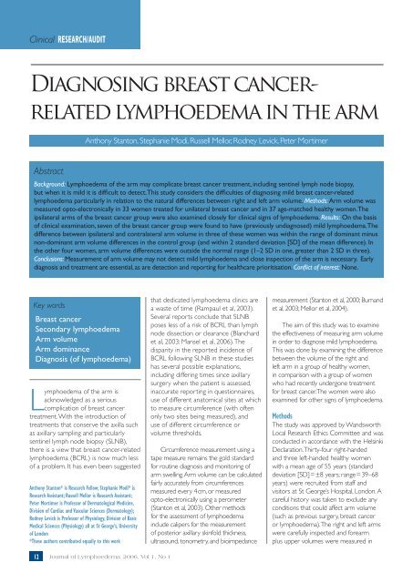
Cl<strong>in</strong>ical RESEARCH/AUDIT<br />
<strong>Diagnos<strong>in</strong>g</strong> <strong>breast</strong> <strong>cancer</strong><strong>related</strong><br />
<strong>lymphoedema</strong> <strong>in</strong> <strong>the</strong> <strong>arm</strong><br />
Anthony Stanton, Stephanie Modi, Russell Mellor, Rodney Levick, Peter Mortimer<br />
Abstract<br />
Background: Lymphoedema of <strong>the</strong> <strong>arm</strong> may complicate <strong>breast</strong> <strong>cancer</strong> treatment, <strong>in</strong>clud<strong>in</strong>g sent<strong>in</strong>el lymph node biopsy,<br />
but when it is mild it is difficult to detect. This study considers <strong>the</strong> difficulties of diagnos<strong>in</strong>g mild <strong>breast</strong> <strong>cancer</strong>-<strong>related</strong><br />
<strong>lymphoedema</strong> particularly <strong>in</strong> relation to <strong>the</strong> natural differences between right and left <strong>arm</strong> volume. Methods: Arm volume was<br />
measured opto-electronically <strong>in</strong> 33 women treated for unilateral <strong>breast</strong> <strong>cancer</strong> and <strong>in</strong> 37 age-matched healthy women. The<br />
ipsilateral <strong>arm</strong>s of <strong>the</strong> <strong>breast</strong> <strong>cancer</strong> group were also exam<strong>in</strong>ed closely for cl<strong>in</strong>ical signs of <strong>lymphoedema</strong>. Results: On <strong>the</strong> basis<br />
of cl<strong>in</strong>ical exam<strong>in</strong>ation, seven of <strong>the</strong> <strong>breast</strong> <strong>cancer</strong> group were found to have (previously undiagnosed) mild <strong>lymphoedema</strong>. The<br />
difference between ipsilateral and contralateral <strong>arm</strong> volume <strong>in</strong> three of <strong>the</strong>se women was with<strong>in</strong> <strong>the</strong> range of dom<strong>in</strong>ant m<strong>in</strong>us<br />
non-dom<strong>in</strong>ant <strong>arm</strong> volume differences <strong>in</strong> <strong>the</strong> control group (and with<strong>in</strong> 2 standard deviation [SD] of <strong>the</strong> mean difference). In<br />
<strong>the</strong> o<strong>the</strong>r four women, <strong>arm</strong> volume differences were outside <strong>the</strong> normal range (1–2 SD <strong>in</strong> one, greater than 2 SD <strong>in</strong> three).<br />
Conclusions: Measurement of <strong>arm</strong> volume may not detect mild <strong>lymphoedema</strong> and close <strong>in</strong>spection of <strong>the</strong> <strong>arm</strong> is necessary. Early<br />
diagnosis and treatment are essential, as are detection and report<strong>in</strong>g for healthcare prioritisation. Conflict of <strong>in</strong>terest: None.<br />
Key words<br />
Breast <strong>cancer</strong><br />
Secondary <strong>lymphoedema</strong><br />
Arm volume<br />
Arm dom<strong>in</strong>ance<br />
Diagnosis (of <strong>lymphoedema</strong>)<br />
Lymphoedema of <strong>the</strong> <strong>arm</strong> is<br />
acknowledged as a serious<br />
complication of <strong>breast</strong> <strong>cancer</strong><br />
treatment. With <strong>the</strong> <strong>in</strong>troduction of<br />
treatments that conserve <strong>the</strong> axilla such<br />
as axillary sampl<strong>in</strong>g and particularly<br />
sent<strong>in</strong>el lymph node biopsy (SLNB),<br />
<strong>the</strong>re is a view that <strong>breast</strong> <strong>cancer</strong>-<strong>related</strong><br />
<strong>lymphoedema</strong> (BCRL) is now much less<br />
of a problem. It has even been suggested<br />
Anthony Stanton* is Research Fellow; Stephanie Modi* is<br />
Research Assistant; Russell Mellor is Research Assistant;<br />
Peter Mortimer is Professor of Dermatological Medic<strong>in</strong>e,<br />
Division of Cardiac and Vascular Sciences (Dermatology);<br />
Rodney Levick is Professor of Physiology, Division of Basic<br />
Medical Sciences (Physiology) all at St George’s, University<br />
of London<br />
*These authors contributed equally to this work<br />
that dedicated <strong>lymphoedema</strong> cl<strong>in</strong>ics are<br />
a waste of time (Rampaul et al, 2003).<br />
Several reports conclude that SLNB<br />
poses less of a risk of BCRL than lymph<br />
node dissection or clearance (Blanchard<br />
et al, 2003; Mansel et al, 2006). The<br />
disparity <strong>in</strong> <strong>the</strong> reported <strong>in</strong>cidence of<br />
BCRL follow<strong>in</strong>g SLNB <strong>in</strong> <strong>the</strong>se studies<br />
has several possible explanations,<br />
<strong>in</strong>clud<strong>in</strong>g differ<strong>in</strong>g times s<strong>in</strong>ce axillary<br />
surgery when <strong>the</strong> patient is assessed,<br />
<strong>in</strong>accurate report<strong>in</strong>g <strong>in</strong> questionnaires,<br />
use of different anatomical sites at which<br />
to measure circumference (with often<br />
only two sites be<strong>in</strong>g measured), and<br />
use of different circumference or<br />
volume thresholds.<br />
Circumference measurement us<strong>in</strong>g a<br />
tape measure rema<strong>in</strong>s <strong>the</strong> gold standard<br />
for rout<strong>in</strong>e diagnosis and monitor<strong>in</strong>g of<br />
<strong>arm</strong> swell<strong>in</strong>g. Arm volume can be calculated<br />
fairly accurately from circumferences<br />
measured every 4 cm, or measured<br />
opto-electronically us<strong>in</strong>g a perometer<br />
(Stanton et al, 2003). O<strong>the</strong>r methods<br />
for <strong>the</strong> assessment of <strong>lymphoedema</strong><br />
<strong>in</strong>clude calipers for <strong>the</strong> measurement<br />
of posterior axillary sk<strong>in</strong>fold thickness,<br />
ultrasound, tonometry, and bioimpedance<br />
measurement (Stanton et al, 2000; Burnand<br />
et al, 2003; Mellor et al, 2004).<br />
The aim of this study was to exam<strong>in</strong>e<br />
<strong>the</strong> effectiveness of measur<strong>in</strong>g <strong>arm</strong> volume<br />
<strong>in</strong> order to diagnose mild <strong>lymphoedema</strong>.<br />
This was done by exam<strong>in</strong><strong>in</strong>g <strong>the</strong> difference<br />
between <strong>the</strong> volume of <strong>the</strong> right and<br />
left <strong>arm</strong> <strong>in</strong> a group of healthy women,<br />
<strong>in</strong> comparison with a group of women<br />
who had recently undergone treatment<br />
for <strong>breast</strong> <strong>cancer</strong>. The women were also<br />
exam<strong>in</strong>ed for o<strong>the</strong>r signs of <strong>lymphoedema</strong>.<br />
Methods<br />
The study was approved by Wandsworth<br />
Local Research Ethics Committee and was<br />
conducted <strong>in</strong> accordance with <strong>the</strong> Hels<strong>in</strong>ki<br />
Declaration. Thirty-four right-handed<br />
and three left-handed healthy women<br />
with a mean age of 55 years (standard<br />
deviation [SD] = ±8 years; range = 39–68<br />
years) were recruited from staff and<br />
visitors at St George’s Hospital, London. A<br />
careful history was taken to exclude any<br />
conditions that could affect <strong>arm</strong> volume<br />
(such as previous surgery, <strong>breast</strong> <strong>cancer</strong><br />
or <strong>lymphoedema</strong>). The right and left <strong>arm</strong>s<br />
were carefully <strong>in</strong>spected and fore<strong>arm</strong><br />
plus upper volumes were measured <strong>in</strong><br />
12 Journal of Lymphoedema, 2006, Vol 1, No 1
Cl<strong>in</strong>ical RESEARCH/AUDIT<br />
<strong>the</strong> horizontal position us<strong>in</strong>g a perometer<br />
model 350 S (Pero-System, Wuppertal,<br />
Germany). We <strong>the</strong>n exam<strong>in</strong>ed <strong>the</strong> <strong>arm</strong>s<br />
of 33 women aged 61 years (SD = ± 9<br />
years; range = 41–75 years), recruited from<br />
<strong>the</strong> <strong>breast</strong> cl<strong>in</strong>ics at St George’s Hospital,<br />
London, and <strong>the</strong> Royal Marsden Hospital,<br />
Surrey, who had recently completed<br />
<strong>breast</strong> <strong>cancer</strong> treatment and had no<br />
record of BCRL. All had been treated by<br />
axillary dissection seven months previously<br />
(SD = ±3 months). We exam<strong>in</strong>ed <strong>the</strong><br />
ipsilateral <strong>arm</strong> for:<br />
8 Decreased visibility of subcutaneous<br />
ve<strong>in</strong>s on <strong>the</strong> ventral fore<strong>arm</strong> and dorsal<br />
hand. This was because sk<strong>in</strong> is thickened<br />
<strong>in</strong> patients with <strong>lymphoedema</strong> and is<br />
<strong>the</strong>refore more opaque<br />
8 Smooth<strong>in</strong>g or fullness of <strong>the</strong> medial<br />
elbow and distal upper-<strong>arm</strong> contours,<br />
where swell<strong>in</strong>g often predom<strong>in</strong>ates<br />
8 Increased sk<strong>in</strong> and subcutis thickness if<br />
<strong>the</strong> tissues are p<strong>in</strong>ched between f<strong>in</strong>ger<br />
and thumb<br />
8 Pitt<strong>in</strong>g oedema upon application of<br />
digital pressure applied for 60 seconds.<br />
Arm volumes were <strong>the</strong>n measured<br />
us<strong>in</strong>g <strong>the</strong> same technique as <strong>the</strong> healthy<br />
group. Comparison of <strong>arm</strong> volumes was<br />
performed us<strong>in</strong>g <strong>the</strong> paired t test.<br />
Results<br />
Healthy group<br />
Differences <strong>in</strong> <strong>the</strong> prom<strong>in</strong>ence of ve<strong>in</strong>s<br />
between <strong>the</strong> right and left fore<strong>arm</strong>s were<br />
sometimes evident on <strong>in</strong>spection of <strong>the</strong><br />
healthy <strong>arm</strong>s, but were not accompanied by<br />
o<strong>the</strong>r signs of <strong>lymphoedema</strong>. Arm volumes<br />
for both groups are shown <strong>in</strong> Table 1. In <strong>the</strong><br />
healthy right-handed group, <strong>the</strong> mean right<br />
<strong>arm</strong> volume was 1.7% (SD = ±3.7%; range=<br />
-4.5–8.5%) greater than left <strong>arm</strong> volume<br />
(n = 34, P = 0.05). Thirteen women (38%)<br />
had a larger non-dom<strong>in</strong>ant <strong>arm</strong>. In <strong>the</strong> three<br />
women who were left-handed, <strong>the</strong> left <strong>arm</strong><br />
was -2.8%, -1.6% and 1.3% (mean = -1%)<br />
larger than <strong>the</strong> right <strong>arm</strong>.<br />
Breast <strong>cancer</strong> group<br />
In <strong>the</strong> group of women who had been<br />
treated for <strong>breast</strong> <strong>cancer</strong>, seven (21%) were<br />
found to have mildly oedematous ipsilateral<br />
<strong>arm</strong>s. Only one woman had noticed any<br />
swell<strong>in</strong>g. She and one o<strong>the</strong>r reported that<br />
<strong>the</strong>ir watch-strap caused <strong>in</strong>dentation at <strong>the</strong><br />
wrist. Cl<strong>in</strong>ical signs of oedema were evident<br />
on exam<strong>in</strong>ation <strong>in</strong> all seven.The ipsilateral<br />
Table 1<br />
Arm volumes (<strong>in</strong> ml) of healthy control subjects and women<br />
treated for <strong>breast</strong> <strong>cancer</strong> (mean; ±SD)<br />
Healthy controls Right <strong>arm</strong> Left <strong>arm</strong> n P*<br />
Right-handed 1,982 (± 545) 1,959 (±527) 34 0.05<br />
Left-handed † 2,317 2,288 3 -<br />
Ipsilateral <strong>arm</strong> Contralateral <strong>arm</strong><br />
All <strong>breast</strong> <strong>cancer</strong> patients<br />
(non-BCRL) 1,875 (± 357) 1,854 (±366) 26 0.3<br />
Breast <strong>cancer</strong> patients<br />
(non-BCRL)<br />
Ipsilateral, dom<strong>in</strong>ant 1,893 (±340) 1,858 (±350) 17 0.04<br />
Ipsilateral, non-dom<strong>in</strong>ant 1,842 (±407) 1,845 (±419) 9 0.8<br />
Patients with <strong>in</strong>cipient<br />
BCRL (all right-handed) 1,623 (± 336) 1,532 (± 328) 7 0.001<br />
*Compar<strong>in</strong>g right and left or ipsilateral and contralateral us<strong>in</strong>g a paired t test<br />
†<br />
Arm volumes (right, left) for <strong>the</strong>se 3 controls were 1,631 ml, 1,652 ml, 1,704 ml,<br />
1,658 ml, 2,610 ml, 2,568 ml<br />
<strong>arm</strong> appeared larger than <strong>the</strong> contralateral<br />
<strong>arm</strong> <strong>in</strong> three women, and <strong>in</strong> one <strong>the</strong>re was<br />
swell<strong>in</strong>g of <strong>the</strong> thumb. There was decreased<br />
visibility of ve<strong>in</strong>s <strong>in</strong> four women; greater<br />
fullness of <strong>the</strong> medial elbow region <strong>in</strong> three;<br />
greater thickness of <strong>the</strong> sk<strong>in</strong> and subcutis <strong>in</strong><br />
two; and pitt<strong>in</strong>g oedema <strong>in</strong> two.<br />
Ipsilateral <strong>arm</strong> volume was 8.1% and<br />
9.4% greater than <strong>the</strong> contralateral <strong>arm</strong><br />
volume for <strong>the</strong> two right-sided ipsilateral<br />
<strong>arm</strong>s, and had a mean percentage of 5.1%<br />
(SD = ±2.8%; range = 1.2–8.8%) greater<br />
for <strong>the</strong> five left-sided ipsilateral <strong>arm</strong>s (n = 7;<br />
P = 0.001). All of <strong>the</strong> women were righthanded.<br />
The difference <strong>in</strong> <strong>arm</strong> volume<br />
for three of <strong>the</strong> seven women with mild<br />
oedema was with<strong>in</strong> <strong>the</strong> normal range of<br />
differences expected due to handedness<br />
based on <strong>the</strong> <strong>arm</strong> volumes of <strong>the</strong> righthanded<br />
control group (one woman was<br />
with<strong>in</strong> -1 SD of <strong>the</strong> mean difference and<br />
two were between ±1 and 2 SD). The<br />
<strong>arm</strong> volume difference for <strong>the</strong> o<strong>the</strong>r four<br />
was beyond <strong>the</strong> normal range (one was<br />
between -1 and -2 SD and three were<br />
greater than ±2 SD). The maximum<br />
difference <strong>in</strong> circumference between <strong>the</strong><br />
ipsilateral and contralateral <strong>arm</strong>s (at any<br />
po<strong>in</strong>t along <strong>the</strong> <strong>arm</strong>) of <strong>the</strong> seven women<br />
with mild swell<strong>in</strong>g, obta<strong>in</strong>ed directly from<br />
<strong>the</strong> perometer images, was 1.4 (SD = ±<br />
0.6 cm; range =0.6–2.2 cm). Only one<br />
woman had a maximum difference <strong>in</strong><br />
circumference of >2 cm. All seven were<br />
advised that <strong>the</strong>y had mild <strong>lymphoedema</strong><br />
and were referred accord<strong>in</strong>gly.<br />
Discussion<br />
The pr<strong>in</strong>cipal f<strong>in</strong>d<strong>in</strong>gs are that mild or<br />
<strong>in</strong>cipient BCRL was present <strong>in</strong> seven out<br />
of 33 women (21%) treated by axillary<br />
dissection for <strong>breast</strong> <strong>cancer</strong> seven months<br />
previously and with no previous record<br />
of BCRL. Most of <strong>the</strong>se women were<br />
unaware that <strong>the</strong>y had <strong>arm</strong> swell<strong>in</strong>g. Cl<strong>in</strong>ical<br />
signs of swell<strong>in</strong>g aris<strong>in</strong>g from thicken<strong>in</strong>g of<br />
<strong>the</strong> sk<strong>in</strong> and subcutis (Mellor et al, 2004)<br />
were identifiable, even though <strong>the</strong> <strong>in</strong>crease<br />
<strong>in</strong> volume of <strong>the</strong> ipsilateral <strong>arm</strong> (compared<br />
with <strong>the</strong> contralateral <strong>arm</strong>) was slight and<br />
with<strong>in</strong> <strong>the</strong> normal range of differences<br />
between dom<strong>in</strong>ant and non-dom<strong>in</strong>ant<br />
<strong>arm</strong>s for age-matched healthy women.<br />
Mild BCRL was only detected by careful<br />
cl<strong>in</strong>ical assessment. Lymphoedema can<br />
<strong>in</strong>volve any tissue with<strong>in</strong> <strong>the</strong> lymphatic bas<strong>in</strong><br />
of <strong>the</strong> damaged nodal group, <strong>in</strong>clud<strong>in</strong>g <strong>the</strong><br />
hand, <strong>breast</strong>, and adjacent trunk, so cl<strong>in</strong>ical<br />
assessment must also <strong>in</strong>clude <strong>the</strong>se regions.<br />
Comparison of ipsilateral and contralateral<br />
<strong>arm</strong> volumes may yield differences of similar<br />
magnitude as found between <strong>the</strong> <strong>arm</strong>s<br />
14 Journal of Lymphoedema, 2006, Vol 1, No 1
Cl<strong>in</strong>ical RESEARCH/AUDIT<br />
of healthy <strong>in</strong>dividuals. The availability of a<br />
pre-surgery measurement of <strong>arm</strong> volume<br />
would <strong>in</strong>crease <strong>the</strong> sensitivity of volume<br />
measurement <strong>in</strong> <strong>the</strong> detection of BCRL. A<br />
fur<strong>the</strong>r limitation when compar<strong>in</strong>g ipsilateral<br />
and contralateral <strong>arm</strong> volumes after<br />
unilateral <strong>breast</strong> surgery is that contralateral<br />
<strong>arm</strong> volume could change, perhaps through<br />
greater use after surgery. In o<strong>the</strong>r words,<br />
<strong>the</strong> relative dom<strong>in</strong>ance of <strong>the</strong> <strong>arm</strong>s may<br />
change. A surpris<strong>in</strong>g f<strong>in</strong>d<strong>in</strong>g was that <strong>the</strong> left<br />
<strong>arm</strong> is bigger than <strong>the</strong> right <strong>in</strong> one-third of<br />
<strong>the</strong> age-matched healthy group of women.<br />
This would make it difficult to take <strong>arm</strong><br />
dom<strong>in</strong>ance <strong>in</strong>to account when assess<strong>in</strong>g <strong>the</strong><br />
severity of BCRL.<br />
Studies purport<strong>in</strong>g to show <strong>the</strong><br />
prevalence of <strong>lymphoedema</strong> after <strong>breast</strong><br />
surgery will underestimate <strong>the</strong> condition<br />
if <strong>the</strong>y are based solely on <strong>the</strong> difference<br />
between ipsilateral and contralateral <strong>arm</strong><br />
size without close cl<strong>in</strong>ical exam<strong>in</strong>ation. The<br />
diagnosis of swell<strong>in</strong>g is often decided on<br />
<strong>the</strong> basis of whe<strong>the</strong>r <strong>the</strong> ipsilateral <strong>arm</strong> is<br />
at least 2 cm greater <strong>in</strong> circumference than<br />
<strong>the</strong> contralateral <strong>arm</strong> at two po<strong>in</strong>ts (one <strong>in</strong><br />
<strong>the</strong> fore<strong>arm</strong> and one <strong>in</strong> <strong>the</strong> upper <strong>arm</strong>). This<br />
threshold for detection is unlikely to detect<br />
mild BCRL. A difference <strong>in</strong> circumference<br />
between <strong>the</strong> ipsilateral and contralateral<br />
<strong>arm</strong>s of more than 2 cm was seen <strong>in</strong><br />
only one of <strong>the</strong> seven cases of BCRL we<br />
detected. Measurement of circumference at<br />
only two sites could result <strong>in</strong> false-negative<br />
report<strong>in</strong>g even of moderate BCRL because<br />
of <strong>the</strong> uneven distribution of swell<strong>in</strong>g<br />
along <strong>the</strong> <strong>arm</strong> (Modi et al, 2005). Although<br />
<strong>the</strong> BCRL seen <strong>in</strong> <strong>the</strong> present study was<br />
<strong>in</strong>cipient and mild, early diagnosis of BCRL<br />
is vital. Treatment and patient education are<br />
likely to stabilise or improve <strong>the</strong> condition,<br />
whereas if ignored, <strong>the</strong> swell<strong>in</strong>g will progress.<br />
Advice on cellulitis and preventive measures<br />
can be given, and measures to stimulate<br />
lymph transport commenced (Mortimer<br />
and Levick, 2004).<br />
Conclusions<br />
BCRL is still common, but probably less<br />
severe than <strong>in</strong> <strong>the</strong> past because of <strong>the</strong><br />
<strong>in</strong>troduction of more conservative surgery,<br />
<strong>in</strong>clud<strong>in</strong>g SLNB, and earlier diagnosis and<br />
treatment. It is important to note that <strong>the</strong><br />
patient may be unaware of <strong>the</strong> swell<strong>in</strong>g.<br />
Careful exam<strong>in</strong>ation of <strong>the</strong> <strong>arm</strong>s of patients<br />
with <strong>breast</strong> <strong>cancer</strong> is vital. Comparison of<br />
<strong>arm</strong> volumes (or circumferences) alone,<br />
will not detect early BCRL and will result<br />
<strong>in</strong> an underestimate of its prevalence <strong>in</strong><br />
studies of <strong>the</strong> complications of axillary<br />
surgery. Accurate detection and report<strong>in</strong>g<br />
are essential to enable prioritisation of<br />
healthcare resources <strong>in</strong> <strong>the</strong> form of<br />
dedicated <strong>lymphoedema</strong> cl<strong>in</strong>ics. JL<br />
AWBS recruited subjects, exam<strong>in</strong>ed and measured<br />
<strong>arm</strong>s, and drafted <strong>the</strong> manuscript. SM recruited subjects,<br />
exam<strong>in</strong>ed and measured <strong>arm</strong>s. RHM exam<strong>in</strong>ed <strong>arm</strong>s.<br />
JRL was co-grant holder for <strong>the</strong> study and advised on<br />
presentation of data. PSM obta<strong>in</strong>ed fund<strong>in</strong>g for <strong>the</strong> study.<br />
All authors read and approved <strong>the</strong> f<strong>in</strong>al manuscript.<br />
The authors would like to thank <strong>the</strong> Wellcome Trust for<br />
f<strong>in</strong>ancial support (grant no. 063025), Mr AK Sh<strong>arm</strong>a (St<br />
George’s Hospital) and Mr G Querci della Rovere (Royal<br />
Marsden Hospital) for access to <strong>the</strong>ir patients, Anita<br />
Wallace (Lymphoedema Support Network) and <strong>the</strong><br />
participants <strong>in</strong> this study.<br />
Armer J, Fu MR, Wa<strong>in</strong>stock JM, Zagar E, Jacobs<br />
LK (2004) Lymphedema follow<strong>in</strong>g <strong>breast</strong><br />
<strong>cancer</strong> treatment, <strong>in</strong>clud<strong>in</strong>g sent<strong>in</strong>el lymph<br />
node biopsy. Lymphology 37: 73–91<br />
Blanchard DK, Donohue JH, Reynolds C, Grant<br />
CS (2003) Relapse and morbidity <strong>in</strong> patients<br />
undergo<strong>in</strong>g sent<strong>in</strong>el lymph node biopsy alone<br />
or with axillary dissection for <strong>breast</strong> <strong>cancer</strong>.<br />
Arch Surg 138: 482–7<br />
Burnand KG, Mortimer PS, Partsch H (2003)<br />
Diagnosis and <strong>in</strong>vestigation of <strong>lymphoedema</strong>.<br />
In: Browse NL, Burnand KG, Mortimer PS, eds.<br />
Diseases of <strong>the</strong> Lymphatics. Arnold, London<br />
110–50<br />
Golshan M, Mart<strong>in</strong> WJ, Dowlatshahi K (2003)<br />
Sent<strong>in</strong>el lymph node biopsy lowers <strong>the</strong> rate of<br />
lymphedema when compared with standard<br />
axillary lymph node dissection. Am Surg 69:<br />
209–11<br />
Langer S, Guen<strong>the</strong>r JM, Haigh PI, Difronzo LA<br />
(2004) Lymphatic mapp<strong>in</strong>g improves stag<strong>in</strong>g<br />
and reduces morbidity <strong>in</strong> women undergo<strong>in</strong>g<br />
total mastectomy for <strong>breast</strong> carc<strong>in</strong>oma. Am Surg<br />
70: 881–5<br />
Leidenius M, Leivonen M, Vironen J, von<br />
Smitten K (2005) The consequences of longtime<br />
<strong>arm</strong> morbidity <strong>in</strong> node-negative <strong>breast</strong><br />
<strong>cancer</strong> patients with sent<strong>in</strong>el node biopsy or<br />
axillary clearance. J Surg Oncol 92: 23–31<br />
Mansel RE, Fallowfield L, Kiss<strong>in</strong> M, et al (2006)<br />
Randomized multicenter trial of sent<strong>in</strong>el node<br />
biopsy versus standard axillary treatment <strong>in</strong><br />
operable <strong>breast</strong> <strong>cancer</strong>: <strong>the</strong> ALMANAC Trial. J<br />
Natl Cancer Inst 98: 599–609<br />
Modi S, Stanton AWB, Mellor RH, Peters<br />
AM, Levick JR, Mortimer PS (2005) Regional<br />
distribution of epifascial swell<strong>in</strong>g and epifascial<br />
lymph dra<strong>in</strong>age rate constants <strong>in</strong> <strong>breast</strong> <strong>cancer</strong><strong>related</strong><br />
lymphedema. Lymphatic Res Biol 3: 3–14<br />
Mellor RH, Bush NL, Stanton AW, Bamber JC,<br />
Levick JR, Mortimer PS (2004) Dual-frequency<br />
ultrasound exam<strong>in</strong>ation of sk<strong>in</strong> and subcutis<br />
Key Po<strong>in</strong>ts<br />
8 Mild <strong>breast</strong> <strong>cancer</strong>-<strong>related</strong><br />
<strong>lymphoedema</strong> of <strong>the</strong> <strong>arm</strong> (BCRL)<br />
can be difficult to detect.<br />
8 Signs to look for when diagnos<strong>in</strong>g<br />
<strong>lymphoedema</strong> <strong>in</strong>clude decreased<br />
visibility of ve<strong>in</strong>s; smooth<strong>in</strong>g of<br />
contours <strong>in</strong> <strong>the</strong> medial elbow<br />
region; <strong>in</strong>creased sk<strong>in</strong> and subcutis<br />
thickness on palpation, and pitt<strong>in</strong>g<br />
oedema. The oedema can be<br />
limited to certa<strong>in</strong> regions, e.g. hand<br />
and wrist only, or upper <strong>arm</strong>.<br />
8 Measurement of <strong>arm</strong> volume is of<br />
little value <strong>in</strong> diagnos<strong>in</strong>g early, mild<br />
BCRL, and reliance on comparison<br />
of <strong>arm</strong> volumes or circumferences<br />
will cause an underestimation of<br />
<strong>the</strong> prevalence of BCRL.<br />
. 8 Accurate detection and report<strong>in</strong>g<br />
of BCRL are essential to enable<br />
prioritisation of healthcare<br />
resources <strong>in</strong> <strong>the</strong> form of dedicated<br />
<strong>lymphoedema</strong> cl<strong>in</strong>ics.<br />
thickness <strong>in</strong> <strong>breast</strong> <strong>cancer</strong>-<strong>related</strong> lymphedema.<br />
Breast J 10: 496–503<br />
Mortimer PS, Levick JR (2004) Chronic<br />
peripheral oedema: <strong>the</strong> critical role of <strong>the</strong><br />
lymphatic system. Cl<strong>in</strong> Med 4: 448–53<br />
Ozaslan C, Kuru B (2004) Lymphedema after<br />
treatment of <strong>breast</strong> <strong>cancer</strong>. Am J Surg 187:<br />
69–72<br />
Rampaul RS, Mull<strong>in</strong>ger K, Macmillan RD, et<br />
al (2003) Incidence of cl<strong>in</strong>ically significant<br />
<strong>lymphoedema</strong> as a complication follow<strong>in</strong>g<br />
surgery for primary operable <strong>breast</strong> <strong>cancer</strong>.<br />
Eur J Cancer 39: 2165–7<br />
Schijven MP, V<strong>in</strong>gerhoets AJ, Rutten HJ, et<br />
al (2003) Comparison of morbidity between<br />
axillary lymph node dissection and sent<strong>in</strong>el<br />
node biopsy. Eur J Surg Oncol 29: 341–50<br />
Stanton AWB, Badger C, Sitzia J (2000) Non<strong>in</strong>vasive<br />
assessment of <strong>the</strong> lymphedematous<br />
limb. Lymphology 33: 122–35<br />
Wilke LG, McCall LM, Pos<strong>the</strong>r KE, et al<br />
(2006) Surgical complications associated<br />
with sent<strong>in</strong>el lymph node biopsy: results<br />
from a prospective <strong>in</strong>ternational cooperative<br />
group trial. Ann Surg Oncol 13: 491–500<br />
Journal of Lymphoedema, 2006, Vol 1, No 1<br />
15