Fluorescent PET (Photoinduced Electron Transfer) sensors ... - Almac
Fluorescent PET (Photoinduced Electron Transfer) sensors ... - Almac
Fluorescent PET (Photoinduced Electron Transfer) sensors ... - Almac
You also want an ePaper? Increase the reach of your titles
YUMPU automatically turns print PDFs into web optimized ePapers that Google loves.
CRITICAL REVIEW<br />
www.rsc.org/analyst | Analyst<br />
<strong>Fluorescent</strong> <strong>PET</strong> (<strong>Photoinduced</strong> <strong>Electron</strong> <strong>Transfer</strong>) <strong>sensors</strong> as potent<br />
analytical tools<br />
A. Prasanna de Silva,* a Thomas S. Moody b and Glenn D. Wright a<br />
First published as an Advance Article on the web 6th October 2009<br />
DOI: 10.1039/b912527m<br />
<strong>Fluorescent</strong> <strong>sensors</strong> are an important part of the analytical scientist’s toolbox. The use of fluorescent<br />
<strong>PET</strong> (<strong>Photoinduced</strong> <strong>Electron</strong> <strong>Transfer</strong>) <strong>sensors</strong> has seen particular growth in recent times. This<br />
Critical Review discusses recent growth areas in fluorescent <strong>PET</strong> <strong>sensors</strong> by emphasizing the modular<br />
features of the ‘fluorophore–spacer–receptor’ design. The occurrence of the dipicolylamine receptor in<br />
<strong>PET</strong> sensor designs is critically examined as a case in point.<br />
Introduction<br />
Analytical science continues to be enriched by the principles of<br />
supramolecular chemistry. 1,2 Hardly a week goes by without<br />
a new fluorescent <strong>PET</strong> (<strong>Photoinduced</strong> <strong>Electron</strong> <strong>Transfer</strong>) sensor<br />
being announced. What are these <strong>sensors</strong> and how do they<br />
operate? How did they evolve? What are the reasons for their<br />
widespread development? This Critical Review briefly addresses<br />
these questions before tracing the recent lineage of a single subfield<br />
of fluorescent <strong>PET</strong> <strong>sensors</strong> and presenting some highlights<br />
in the field from the past year.<br />
a<br />
School of Chemistry and Chemical Engineering, Queen’s University,<br />
Belfast, Northern Ireland BT9 5AG, Northern Ireland. E-mail: a.<br />
desilva@qub.ac.uk; gwright04@qub.ac.uk<br />
b<br />
Biocatalysis Group, <strong>Almac</strong> Sciences, <strong>Almac</strong> House, 20 Seagoe Industrial<br />
Estate, Craigavon, Northern Ireland BT63 5QD. E-mail: tom.moody@<br />
almacgroup.com<br />
Molecular engineering design<br />
A single simple picture (Fig. 1) encapsulates the design of fluorescent<br />
<strong>PET</strong> <strong>sensors</strong>. The ‘fluorophore–spacer–receptor’ format<br />
is a rational combination of three components. The rationale is<br />
contained in Fig. 2a and 2b. In its ‘off’ state, excitation of the<br />
fluorophore component of the sensor produces an electron<br />
transfer from the receptor to the fluorophore as one possibility.<br />
In other words, the excited state energy of the fluorophore needs<br />
to be sufficient to provide both the reduction potential of the<br />
fluorophore and the oxidation potential of the receptor. This is<br />
a thermodynamic condition first derived by Weller. 3 In its ‘on’<br />
state, excitation of the fluorophore results in fluorescence only<br />
because the <strong>PET</strong> process is arrested by the arrival of the analyte<br />
at the receptor site. The arrest can be easily comprehended by<br />
considering H + as the analyte. H + electrostatically attracts<br />
the electron which increases the oxidation potential of the<br />
A. P. de Silva received his early education in chemistry at the University<br />
of Colombo, Sri Lanka, followed by PhD and postdoctoral research in<br />
organic photochemistry at the Queen’s University of Belfast. After<br />
spending a few years lecturing in chemistry at Colombo, he returned to<br />
Belfast where he is a professor and a percussionist. He has been visiting<br />
professor in Louvain-La-Neuve, Cachan, Bordeaux, Strasbourg, Peradeniya,<br />
Kandy, Nara, Bangkok and Shanghai. With his co-workers, he<br />
published the first experimental molecular logic gates in the primary<br />
literature and established the generality of one of the main principles<br />
underlying luminescent <strong>sensors</strong>.<br />
Thomas S. Moody received his 1st Class BSc (Hons) in chemistry and<br />
PhD in physical organic chemistry from the Queen’s University of Belfast<br />
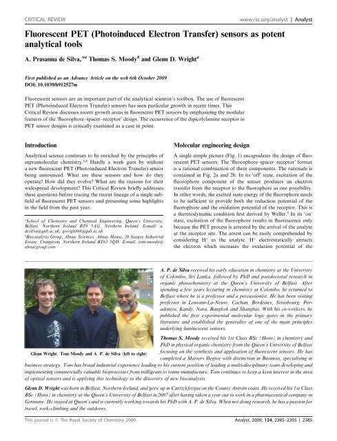
Glenn Wright; Tom Moody and A: P: de Silva ðleft to rightÞ<br />
focusing on the synthesis and application of fluorescent <strong>sensors</strong>. He has<br />
completed a Masters Degree with distinction in Business, specialising in<br />
business strategy. Tom has broad industrial experience leading to his current position of leading a multi-disciplinary team developing and<br />
implementing commercially valuable bioprocesses from milligram to tonne manufacture. Tom continues to keep a keen interest in the area<br />
of optical <strong>sensors</strong> and is applying this technology to the discovery of new biocatalysts.<br />
Glenn D. Wright was born in Belfast, Northern Ireland, and grew up in Carrickfergus on the County Antrim coast. He received his 1st Class<br />
BSc (Hons) in chemistry at the Queen’s University of Belfast in 2007 after having taken a year out to work in a pharmaceutical company in<br />
Germany. He stayed at Queen’s and is currently working towards his PhD with A. P. de Silva. When not doing research, he has a passion for<br />
travel, rock-climbing and the outdoors.<br />
This journal is ª The Royal Society of Chemistry 2009 Analyst, 2009, 134, 2385–2393 | 2385
Fig. 1 The ‘fluorophore–spacer–receptor’ format of fluorescent <strong>PET</strong><br />
<strong>sensors</strong>.<br />
Fig. 2 (a) An electron transfer from the analyte-free receptor to the<br />
photo-excited fluorophore creates the ‘off’ state of the sensor. (b) The<br />
electron transfer from the analyte-bound receptor is blocked resulting in<br />
the ‘on’ state of the sensor.<br />
Fig. 3 Molecular orbital energy diagrams which show the relative<br />
energetic dispositions of the frontier orbitals of the fluorophore and the<br />
receptor in (a) the analyte-free situation and (b) the analyte-bound<br />
situation.<br />
analyte-bound receptor to the point that the thermodynamics for<br />
<strong>PET</strong> are no longer favourable. 4,5<br />
These ideas can also be expressed with the aid of molecular<br />
orbital energy diagrams (Fig. 3a and 3b). We note the bracketing<br />
of the receptor HOMO by the frontier orbitals of the fluorophore<br />
in the ‘off’ state of the sensor and the stabilization of the analytebound<br />
receptor’s HOMO to lie below the fluorophore’s HOMO<br />
in the ‘on’ state. Fig. 3a and 3b allow us to deduce an even<br />
simpler criterion for <strong>PET</strong> sensor design: <strong>PET</strong> occurs if the<br />
oxidation potential of the receptor is smaller in magnitude than<br />
that of the fluorophore. The opposite applies in the ‘on’ state of<br />
the sensor. This rule of thumb is very useful practically, even<br />
though several approximations are involved. More accurate<br />
treatment of <strong>PET</strong> processes are available for the interested<br />
reader. 3,6–8 MO energies and related redox potentials are<br />
increasingly used by <strong>PET</strong> sensor designers. 9–12<br />
The availability of a quantitative design criterion is common in<br />
engineering but rare in chemistry. The case of fluorescent <strong>PET</strong><br />
<strong>sensors</strong> is a rare example of molecular engineering design. Just<br />
like houses and cars, molecular <strong>PET</strong> <strong>sensors</strong> can now be designed<br />
and built for a variety of individual purposes.<br />
Each of the three components within the ‘fluorophore–spacer–<br />
receptor’ format deserves the designer’s attention. The analyte<br />
to be sensed determines the choice of receptor. The reciprocal of<br />
the binding constant for the receptor–analyte interaction determines<br />
the median analyte concentration to be sensed. Consideration<br />
needs to be given at this stage to the selectivity of the<br />
receptor towards the analyte and against anticipated levels of<br />
potential interferents.<br />
Desired colours for excitation and emission help in the choice<br />
of fluorophore. For instance, intracellular studies using glass<br />
microscopy optics will preclude the use of excitation wavelengths<br />
below 340 nm. Tissue experiments will prefer these wavelengths<br />
to be in the red region.<br />
The ease of sensor synthesis dictates the choice of spacer, but<br />
the more fundamental determinant is that the spacer must be<br />
short enough to permit reasonably fast <strong>PET</strong> rates in the ‘off’ state<br />
of the sensor. 13–15 Even virtual spacers can be used provided that<br />
other means, such as sterically-induced orthogonalization, 16<br />
maintains the separation between the fluorophore and the<br />
receptor.<br />
Let us consider an example of simple pH sensing which will<br />
serve as a foundation for a case study (see below). Fig. 4 shows<br />
two of the main options available to the excited <strong>PET</strong> sensor 1. 4 In<br />
order to apply the approximate Weller equation, we note that the<br />
excited state energy of the anthracene fluorophore is 3.0 eV. 17 Its<br />
reduction potential is 2.0 V (vs. sce). The oxidation potential of<br />
the receptor can be estimated from that of triethylamine<br />
(+1.0 V). 17 As the transiting electron falls through these<br />
potentials, the corresponding energies are 2.0 eV and 1.0 eV<br />
respectively (Fig. 5a). So the approximate DG for <strong>PET</strong> is 0.0 eV. 17<br />
This gives a sufficiently fast <strong>PET</strong> rate to overcome fluorescence<br />
(k <strong>PET</strong> [ k Flu ). The same result can be obtained by the rule of<br />
thumb when we note that the oxidation potential of the receptor<br />
is +1.0 V as above and that the oxidation potential of the<br />
anthracene fluorophore is +1.0 V. So DG <strong>PET</strong> is 0.0 eV again.<br />
When we consider the H + -bound amine receptor of 1, its<br />
oxidation potential rises to an immeasurably high value. DG <strong>PET</strong><br />
becomes a large positive number and fluorescence dominates.<br />
2386 | Analyst, 2009, 134, 2385–2393 This journal is ª The Royal Society of Chemistry 2009
Fig. 4 De-excitation pathways open to the photo-excited fluorescent <strong>PET</strong> sensor 1.<br />
Another more recent example shows how these thermodynamic<br />
arguments help even when fluorescent <strong>PET</strong> <strong>sensors</strong> are<br />
constructed without a covalent linkage between the fluorophore<br />
and the receptor. Self-assembly of the trianionic fluorophore 2<br />
within the cavity of the tetracationic receptor 3 produces the ‘off’<br />
state of the sensor system. 18 The reduction potential of the<br />
receptor can be estimated from that of dimethyl viologen<br />
( 0.3 V). 17 The oxidation potential of 2 is 0.8 V and its excited<br />
state energy is 3.0 eV (Fig. 5b). 19 Fast <strong>PET</strong> is made possible by<br />
significantly negative DG <strong>PET</strong> value ( 1.9 eV). Displacement of 2<br />
from its complex with 3 by guanosine triphosphate (GTP) occurs<br />
due to increased synergistic effects, such as electrostatics and<br />
p-stacking inside the cavity. Since the fluorophore 2 is now<br />
distanced from the receptor 3 (besides somewhat less favourable<br />
thermodynamics for <strong>PET</strong>) fluorescence is switched ‘on’. Sensing<br />
of GTP is thus enabled.<br />
The validity of such molecular engineering makes <strong>PET</strong><br />
<strong>sensors</strong> 20 an important segment of research in molecular<br />
devices. 21<br />
Early history<br />
The first case fitting the above description of a fluorescent <strong>PET</strong><br />
sensor was Wang and Morawetz’s dibenzylamine compound<br />
(4; as seen later in Fig. 7b). 22 It contained a small fluorophore<br />
requiring excitation in the deep ultraviolet, spaced with a methylene<br />
group from an aliphatic amine receptor. Naturally, H + was<br />
the chosen target, though it was also engaged with Zn 2+ and also<br />
reacted with acetic anhydride. Several cases 23–28 followed with<br />
Fig. 5 The relative energetic dispositions of the frontier orbitals of the<br />
fluorophore and the receptor in the analyte-free situation for <strong>PET</strong> sensor<br />
systems (a) 1 and (b) 2$3. Redox potentials (V vs. sce) are given in<br />
parentheses as a measure of molecular orbital energies. The energies are<br />
not to scale.<br />
spacers ranging from trimethylene to none at all. The latter<br />
situation occurred due to the sterically-enforced twisting of an<br />
aniline receptor from an anthracene fluorophore. The generality<br />
of the sensing principle was established with a set of related<br />
cases 28–36 carrying different fluorophores (or phosphors), spacers<br />
and receptors. Reviews of this phase are available. 4,5,37,38<br />
Current uptake<br />
As may be expected, a principle that is general, flexible and<br />
extensible tends to be put to use by people seeking solutions to<br />
various analytical problems. A sensing principle would naturally<br />
be applied to target various analytes in various situations. A<br />
commercially successful example which measures blood<br />
components like H + , Na + , K + and Ca 2+ deserves a special<br />
mention. 39 There is a growing body of work where <strong>PET</strong> <strong>sensors</strong><br />
are operating within living cells. 40,41 The current situation is<br />
perhaps best shown graphically. Fig. 6a and 6b show the sources<br />
for fluorescent <strong>PET</strong> <strong>sensors</strong> and switches around the world as<br />
deduced from the literature. Some of these laboratories may have<br />
produced a single publication in this field or several dozen. It is<br />
clear that research in fluorescent <strong>PET</strong> <strong>sensors</strong> is now a delocalized<br />
activity.<br />
A case study: dipicolylamine-based <strong>sensors</strong><br />
It is educational to track how a single avenue of fluorescent <strong>PET</strong><br />
<strong>sensors</strong> has evolved. It illustrates how different people considering<br />
different problems can exploit a single structural motif.<br />
Consider di(2-picolyl)amine {IUPAC name: 2-pyridinemethanamine,<br />
N-(2-pyridinylmethyl)-} which is a popular receptor 42 for<br />
d-block cations among coordination chemists. All of the structures<br />
discussed are contained within Fig. 7a and 7b.<br />
Though 5 (Fig. 7a) was a previously known compound, 43<br />
S. A. de Silva et al. 44 were the first to recognize its ‘fluorophore–<br />
spacer–receptor’ format, its <strong>PET</strong> potential and its di(2-picolyl)-<br />
amine receptor 42 for Zn 2+ . Indeed a strong Zn 2+ -induced<br />
switching ‘on’ of fluorescence (fluorescence enhancement factor,<br />
FE ¼ 77) is seen in acetonitrile. No wavelength shift of the<br />
emission band is seen as befits a <strong>PET</strong> system. The replacement of<br />
the terminal methyl groups of 1 by 2-pyridyl units will make the<br />
DG <strong>PET</strong> only slightly more positive. The thermodynamic conditions<br />
for cation sensing discussed in a previous paragraph are<br />
maintained. More recent crystallographic and computational<br />
studies have also backed up the Zn 2+ -binding of 5. 45<br />
The modularity of the <strong>PET</strong> sensing system 5 can now be<br />
exploited in many ways and we track structural mutations where<br />
the dipicolylamine unit is conserved. The number of laboratories<br />
that have followed this path is remarkable, spurred on by the<br />
This journal is ª The Royal Society of Chemistry 2009 Analyst, 2009, 134, 2385–2393 | 2387
Fig. 6 (a,b) Sources of fluorescent <strong>PET</strong> <strong>sensors</strong>. Only the names of corresponding authors from the literature are given. The corresponding authors will<br />
be happy to receive evidence of errors and omissions so that future versions of the maps can be improved.<br />
need for monitoring the neurophysiology of Zn 2+ and its role in<br />
degenerative disease. 40,41,46<br />
The extension of <strong>PET</strong> systems by adding an extra ‘spacer–<br />
receptor’ component can lead to improvements in FE values. 4,47<br />
This is seen when we go from 5 to 6, 48,49 with the requirement that<br />
each receptor captures Zn 2+ . However, ethanol:water (1:1) was<br />
needed as the solvent.<br />
The hydrophobicity of the anthracene fluorophore would<br />
hamper the use of 5 for monitoring Zn 2+ in the cytosol. Hydrophobic<br />
<strong>PET</strong> <strong>sensors</strong> can localize in intracellular membranes and<br />
lose their ion-sensing ability. 12 Therefore, replacement of<br />
anthracene by hydrophilic heterocyclic fluorophores is logical,<br />
and is discussed in the very next paragraph. On the other hand,<br />
the hydrophobicity of 5 or its cousin 7 50 can be exploited for Zn 2+<br />
measurement in nanospaces adjoining membranes. The fluorophore<br />
is expected to embed in detergent micelles with the more<br />
hydrophilic receptor being accessible to water neighbouring the<br />
membrane 51,52 and any Zn 2+ therein. In the event, a strong Zn 2+ -<br />
induced FE value of 7 is found for 7 in neutral Tween 20<br />
micelles. 50<br />
The mutation of 5 is easiest to see in 8, 53 where the tricyclic<br />
anthracene has become the tricyclic fluorescein. The chloro<br />
substituents serve to reduce 8’s response to H + in physiological<br />
conditions. 54 Sensor 8 gives a Zn 2+ -induced FE value of 2 in<br />
2388 | Analyst, 2009, 134, 2385–2393 This journal is ª The Royal Society of Chemistry 2009
Fig. 7 (a) Structural formulae of the dipicolylamine-based <strong>sensors</strong> discussed in the case study. In all these cases, the R group represents the<br />
di(2-pyridylmethyl)amino moiety. (b) Structural formulae for the compounds highlighted from the past year. Following on from Fig. 2a and 2b, the<br />
following colour scheme is employed in both Fig. 7a and 7b: fluorophores in blue, spacers in red and receptors in green. When atoms in the fluorophore<br />
can also ligate to the analyte, these are shown in green.<br />
This journal is ª The Royal Society of Chemistry 2009 Analyst, 2009, 134, 2385–2393 | 2389
physiological media and permits fluorescence microscopy in<br />
living cells. However, the residual fluorescence response to H + is<br />
still quite large, since scavenging of Zn 2+ does not noticeably<br />
reduce the fluorescence. Nevertheless, 8 was the vanguard of<br />
a strong programme 55 which included cases like 9 and 10. Sensor<br />
9 56 locates the dipicolylamine unit somewhat remote from the<br />
fluorophore. However, the aniline NH group is just one methylene<br />
unit away from the fluorophore so that <strong>PET</strong> would occur at<br />
a reasonably rapid rate. 57 The NH unit joins in Zn 2+ -binding<br />
along with the dipicolylamine receptor. Interestingly, another<br />
strong program begun in 2000 58 has a similar juxtaposition of an<br />
aniline NH and a dipicolylamine, as well as a fluorescein fluorophore,<br />
e.g. sensor 11. 59<br />
Sensor 10 60 contains 8 as the Zn 2+ -responsive component with<br />
green emission of dichlorofluorescein as the output. It also<br />
carries an aminocoumarin fluorophore whose blue emission is<br />
Zn 2+ -independent, which is released by hydrolysis of the ester<br />
bond of 10 by intracellular esterases. Virtually the same aminocoumarin<br />
fluorophore is present in 12 61,62 (though closer to the<br />
receptor) and so the Zn 2+ -independent emission intensity might<br />
be expected. However, the positioning of the dipicolylamine near<br />
the coumarin carbonyl oxygen allows the latter to participate in<br />
Zn 2+ -binding. Since this push-pull fluorophore has an internal<br />
charge transfer (ICT) excited state whose d pole lies at the<br />
carbonyl oxygen, Zn 2+ causes an emission red-shift due to the<br />
electrostatic attraction. Alkoxycoumarin fluorophores when<br />
coupled to amine receptors have suitable <strong>PET</strong> thermodynamics.<br />
63,64 Cases like 13 61,62 and 14 65 show Zn 2+ -induced FE<br />
values of 22 (in methanol) and 8 (in acetonitrile) respectively.<br />
While blue (e.g. coumarin) and green (e.g. fluorescein) emissions<br />
remain the workhorses of fluorescent sensor research, redemitting<br />
fluorophores are sought after for intracellular and tissue<br />
studies due to easy transmission of light. Zn 2+ <strong>sensors</strong> 15 66 and<br />
16 67 address this need. <strong>PET</strong> has been achieved with cyanine<br />
fluorophores when coupled with electron-rich anilinic receptors,<br />
68 but is harder to produce with the dipicolylamine receptor.<br />
Sensor 16 succeeds at this with a Zn 2+ -induced FE value of 7<br />
(in water) whereas 15 only shows wavelength shifts in fluorescence<br />
excitation spectra typical of ICT excited states, e.g. Tsien’s<br />
classical Ca 2+ <strong>sensors</strong>. 69<br />
The blue-green region is also represented by several more Zn 2+<br />
<strong>sensors</strong> 17–21. 70–75 All of them show significant Zn 2+ -induced FE<br />
values, though some of them involve the fluorophore’s participation<br />
in the Zn 2+ -coordination sphere. System 22 76 is clearly of<br />
the ‘fluorophore–spacer–dipicolylamine’ format but <strong>PET</strong><br />
appears to be thermodynamically unfavourable. So, 22 is highly<br />
emissive to begin with and only the quenching metal ion Cu 2+<br />
signals its presence. Zn 2+ has no effect.<br />
Another blue-green-emitting Zn 2+ sensor 23 77 is distinguished<br />
by operating over a large dynamic range. Besides the<br />
dipicolylamine receptor, 23 also contains a 2,2 0 -bipyridine as<br />
a low-affinity binding site which is engaged at high Zn 2+<br />
concentrations. This incurs positive charging and planarization<br />
of the bipyridine rings and so the ICT character of the excited<br />
push-pull fluorophore is enhanced with a significant red-shift.<br />
The switching ‘on’ of the blue emission and that of the green<br />
emission occur at two different Zn 2+ concentration ranges.<br />
The conjugation of an electron-rich aminophenyl substituent<br />
with a furoquinoline unit increases its reduction potential to<br />
weaken <strong>PET</strong> activity of 24. 78 However, this produces a push-pull<br />
fluorophore with considerable ICT character in its excited state.<br />
The quinoline nitrogen atom of the fluorophore clearly participates<br />
in the Zn 2+ -coordination sphere, so that the ICT character<br />
of the excited state increases, resulting in a Zn 2+ -induced red-shift<br />
from green to orange. This allows ratiometric measurement of<br />
Zn 2+ in living cells.<br />
The story doesn’t end here. Some of these Zn 2+ <strong>sensors</strong> have<br />
led to another valuable line of research. It started with the filling<br />
of the unsaturated coordination sphere of dipicolylamine-Zn 2+<br />
(mentioned above) with anionic ligands such as phosphates.<br />
When di-receptor cases such as 6 are studied in neat water,<br />
binding of two Zn 2+ ions cannot be achieved under experimental<br />
conditions unless the mutual repulsion between the two metal<br />
centres is reduced by inserting a bridging anion. Therefore, <strong>PET</strong><br />
suppression and fluorescence switching ‘on’ requires the presence<br />
of Zn 2+ as well as the phosphate anion. Phosphorylation of<br />
tyrosines in peptides can be neatly signalled in this way. Of<br />
course, dephosphorylation can be followed fluorimetrically<br />
too. 79,80 Sensor 6 with Zn 2+ also switches ‘on’ when uridine<br />
5 0 -diphosphate is made available from uridine 5 0 -diphosphate<br />
glycoside during glycosyl transfer to sugar derivatives catalyzed<br />
by glycosyltransferases. 81 The latter enzymes are measurable in<br />
a label-free manner. Sensor 6 with Zn 2+ also lights up when<br />
phosphatidylserine is brought to the outer face of cell membranes<br />
when the cell is ready to die. Thus, apoptosis can be detected in<br />
a simple way. 82<br />
Phosphates can also be detected (though in acetonitrile solution)<br />
by the quenching of the emission of the Zn 2+ complex of 25<br />
by a factor of 70. 83 Sensor 25 itself is poorly emissive and Zn 2+<br />
causes an FE value of 5. A <strong>PET</strong> mechanism is probable. The<br />
coordination sphere of the single dipicolylamine-Zn 2+ is made up<br />
by the aniline nitrogen nearby and, importantly, also by phosphate<br />
which reduces the positive charge density so that the Zn 2+ -<br />
induced <strong>PET</strong> suppression is weakened. The ICT nature of the<br />
push-pull fluorophore is clearly signalled by the Zn 2+ -induced<br />
blue-shift.<br />
Compound 8 53 is re-incarnated in the form of its di-Zn 2+<br />
complex for pyrophosphate sensing. 84 An FE value of 3 is achieved.<br />
It appears that, as seen perhaps more strongly for 6, 79 the<br />
full binding of both dipicolylamine sites and the full <strong>PET</strong><br />
suppression is only achieved when the pyrophosphate bridges the<br />
two Zn 2+ centres. In this case, pyrophosphate is of just the right<br />
length. Pyrophosphate bridging two Zn 2+ -dipicolylamine units is<br />
also found in the ground state dimer of 26 85 (indicated by redshifts<br />
in the absorption spectrum) which leads to a corresponding<br />
red-shifted emission compared to the pyrene monomer.<br />
It is clear that the first <strong>PET</strong> sensing experiment on 5 44 keeps on<br />
putting out new shoots – a testament to the flexibilities arising<br />
from the modular nature of <strong>PET</strong> <strong>sensors</strong>.<br />
Some highlights from the past year<br />
Besides the sustained success of Zn 2+ sensing with <strong>PET</strong> <strong>sensors</strong><br />
carrying bis(picolyl)amine receptors, recent cases also include<br />
macrocycles. An oxa-bridged version of older azacrown etherbased<br />
<strong>PET</strong> <strong>sensors</strong>, 29 27 (Fig. 7b), 86 contains an amine <strong>PET</strong><br />
donor and an anthracene fluorophore. It produces a Zn 2+ -<br />
induced FE of 100 but does not respond to alkali cations.<br />
2390 | Analyst, 2009, 134, 2385–2393 This journal is ª The Royal Society of Chemistry 2009
Interestingly, Na + in sufficient concentration displaces Zn 2+ and<br />
decreases the FE value of 27, as the smaller ion coordinates to the<br />
oxygen atoms rather than the nitrogen.<br />
Cases based on tetraazamacrocycle receptors are also useful<br />
for Zn 2+ sensing. 87 A new example is 28, 88 where the triazole<br />
group also plays a receptor role to cause selective binding of Zn 2+<br />
even over Cd 2+ , with a Zn 2+ -induced FE value of 6. Interestingly,<br />
it detects the intracellular flux of Zn 2+ during cell apoptosis.<br />
In spite of the successes concerning selective Zn 2+ sensing, Cd 2+<br />
was an interferent in many of these cases. The rarity of intracellular<br />
Cd 2+ has been the saviour in this regard. Selective Cd 2+<br />
sensing, which would be useful in studies of Cd 2+ toxicity, has<br />
been a harder nut to crack. 89–91 <strong>PET</strong> sensor 29 with a polyamide<br />
receptor provides a neat solution even within HeLa cells. 92 A<br />
Cd 2+ -induced FE value of 100 is attained. Steric hindrance<br />
between the receptor arms can produce a virtual spacer so that<br />
the usual <strong>PET</strong> behaviour can occur.<br />
Organic chemical reactions rather than ion-coordination can<br />
also lead to <strong>PET</strong>-based fluorescence switching ‘on’, as seen in<br />
30. 93 When solubilized in aqueous Tween 20 micellar solution, its<br />
amine group serves as the <strong>PET</strong> donor so that the fluorophore’s<br />
emission is ‘off’. The dangerous alkylating agent chloromethyl<br />
methyl ether forms a quaternary ammonium product which<br />
prevents <strong>PET</strong> and fluorescence is switched ‘on’. Related cases 94<br />
are known.<br />
As we saw with the 2$3 system, 18 covalent linking of a fluorophore<br />
and a <strong>PET</strong>-active receptor is not necessary for a working<br />
<strong>PET</strong> sensing system. There is no <strong>PET</strong> within 31,<br />
a tris(2,2 0 -bipyridyl)Ru(II) lumophore which is connected to<br />
a mannose-capped dendrimer. 95 Lectins, those exquisite sugarbinding<br />
proteins, rely on polyvalency. Therefore, lectins such as<br />
Concanavalin A associate strongly with 31. However, this does<br />
not produce a luminescence switch unless an extra component is<br />
provided. The extra component 32 contains arylboronic acid<br />
groups for sugar binding and a 4,4 0 -bipyridinium unit as a <strong>PET</strong><br />
acceptor. In the absence of lectin, 31 and 32 associate so that <strong>PET</strong><br />
takes place and the luminescence is ‘off’. The addition of<br />
Concanavalin A displaces 32 from the mannose caps and<br />
a resultant increase in luminescence occurs.<br />
Though perhaps not as grand as lectin sensing, synthetic<br />
macrocycles can also be sensed via <strong>PET</strong> schemes. A pyrene fluorophore<br />
and a pyridinium <strong>PET</strong> acceptor can be discerned<br />
within 33. 96 The phosphonate-bridged resorcinarene 34 can<br />
engulf the pyridinium unit just like simpler resorcinarenes do. 97<br />
Upon complexation, <strong>PET</strong> becomes energetically unfavourable,<br />
causing an increase in fluorescence.<br />
As seen in the previous pages, <strong>PET</strong> <strong>sensors</strong> usually consist of<br />
molecular lumophores. An important extension to this idea has<br />
been reported 98 where the lumophore component represents<br />
a quantum dot, the darling of nanotechnology. This particular<br />
quantum dot is a ZnS–CdSe core–shell structure. Sensor<br />
35 possesses a thiourea receptor which binds to CH 3 CO 2 with<br />
two hydrogen bonds and this increases the reduction potential of<br />
the receptor, thus enhancing <strong>PET</strong> to the lumophore.<br />
We finish off with two cases where ‘fluorophore–spacer–<br />
receptor’ systems are aimed at monitoring intermediates in<br />
catalytic cycles in solution. For instance, a transient Lewis acid<br />
centre could bind a receptor to switch ‘on’ the fluorescence of<br />
a <strong>PET</strong> sensor. This would be a valuable analytical tool. Example<br />
36 99 attacks this problem at the single molecule level since the<br />
required sensitivity of similar <strong>sensors</strong> based on perylenediimide<br />
fluorophores has been demonstrated. 100 Though this ambitious<br />
goal is yet to be achieved, the H + -induced switching ‘on’ of<br />
fluorescence is demonstrated by negating the <strong>PET</strong> donor amine<br />
by protonation. The single molecule studies show this pH<br />
dependence, though the presence of relatively long-lived dark<br />
states at low pH leading to ‘blinking’ is a complication.<br />
The dimethylaminonaphthalenesulfonylamide fluorophore<br />
undergoes <strong>PET</strong> from neighbouring amines as seen in the case of<br />
17. 101 A similar ‘fluorophore–spacer–receptor’ motif can be<br />
found within 37. This motif is tagged to an N-heterocyclic carbene<br />
Pd(II) complex with the intent of monitoring its catalytic<br />
activity in a Suzuki coupling reaction between an aryl bromide<br />
and a boronic acid. As the reaction progresses, the halide ion<br />
product quenches 37’s fluorescence due to the heavy atom effect.<br />
Such monitoring of a product formation is not the main point in<br />
the present context of monitoring of catalytic intermediates.<br />
However, the smaller fluorescence loss observed upon addition<br />
of base to prepare the active Pd(0) catalytic species before the<br />
addition of the aryl halide is potentially more interesting. If the<br />
mopping up of trace Brønsted acids can be ruled out, a ‘steppingstone’<br />
mechanism could be imagined, where an electron is<br />
transferred from Pd(0) to the fluorophore via the amine receptor<br />
to quench fluorescence, but not from Pd(II).<br />
Conclusion<br />
The preceding pages have summarized a few of the current<br />
growth areas in fluorescent <strong>PET</strong> <strong>sensors</strong> where problems in<br />
analytical science are being attacked. We hope the quantitative<br />
design basis of <strong>PET</strong> <strong>sensors</strong> where molecules can be viewed as<br />
engineering objects will appeal to bright analytical minds so that<br />
more of them will join in this venture. With such a combination<br />
of forces, even more analytical solutions will emerge from the<br />
versatile fluorescent <strong>PET</strong> system as the days go on.<br />
Acknowledgements<br />
We thank the Allen McClay trust for support.<br />
References<br />
1 J.-M. Lehn, Supramolecular Chemistry, VCH, Weinheim, 1995.<br />
2 E. V. Anslyn, J. Org. Chem., 2007, 72, 687–699.<br />
3 A. Weller, Pure Appl. Chem., 1968, 16, 115–123.<br />
4 R. A. Bissell, A. P. de Silva, H. Q. N. Gunaratne, P. L. M. Lynch,<br />
G. E. M. Maguire and K. R. A. S. Sandanayake, Chem. Soc. Rev.,<br />
1992, 21, 187–195.<br />
5 R. A. Bissell, A. P. de Silva, H. Q. N. Gunaratne, P. L. M. Lynch,<br />
G. E. M. Maguire, C. P. McCoy and K. R. A. S. Sandanayake,<br />
Top. Curr. Chem., 1993, 168, 223–264.<br />
6 <strong>Photoinduced</strong> electron transfer, ed. M. A. Fox and M. Chanon,<br />
Elsevier, Amsterdam, 1988.<br />
7 G. J. Kavarnos, Fundamentals of photoinduced electron transfer,<br />
VCH, Weinheim, New York, 1993.<br />
8 <strong>Electron</strong> <strong>Transfer</strong>, ed. V. Balzani, Wiley-VCH, Weinheim, 2003.<br />
9 A. Chatterjee, T. M. Suzuki, Y. Takahashi and D. A. P. Tanaka,<br />
Chem.–Eur. J., 2003, 9, 3920–3929.<br />
10 S. Uchiyama, T. Santa and K. Imai, Analyst, 2000, 125, 1839–1845.<br />
11 T. Ueno, Y. Urano, K. Setsukinai, H. Takakusa, H. Kojima,<br />
K. Kikuchi, K. Ohkubo, S. Fukuzumi and T. Nagano,<br />
J. Am. Chem. Soc., 2004, 126, 14079–14085.<br />
This journal is ª The Royal Society of Chemistry 2009 Analyst, 2009, 134, 2385–2393 | 2391
12 C. J. Fahrni, L. C. Yang and D. G. VanDerveer, J. Am. Chem. Soc.,<br />
2003, 125, 3799–3812.<br />
13 G. L. Closs and J. R. Miller, Science, 1988, 240, 440–447.<br />
14 J. C. Beeson, M. A. Huston, D. A. Pollard, T. K. Venkatachalam<br />
and A. W. Czarnik, J. Fluoresc., 1993, 3, 65–69.<br />
15 M. Onoda, S. Uchiyama, T. Santa and K. Imai, Luminescence, 2002,<br />
17, 11–14.<br />
16 S. A. Jonker, F. Ariese and J. W. Verhoeven, Rec. Trav. Chem. Pays<br />
Bas, 1989, 108, 109–115.<br />
17 M. Montalti, A. Credi, L. Prodi and M. T. Gandolfi, Handbook of<br />
photochemistry, CRC Press, Boca Raton, 3rd edn, 2006.<br />
18 P. P. Neelakandan, M. Hariharan and D. Ramaiah, J. Am. Chem.<br />
Soc., 2006, 128, 11334–11335.<br />
19 N. Tarumoto, N. Miyagawa, S. Takahara and T. Yamaoka,<br />
Polym. J., 2005, 37, 545–549.<br />
20 A. P. de Silva, H. Q. N. Gunaratne, T. Gunnlaugsson,<br />
A. J. M. Huxley, C. P. McCoy, J. T. Rademacher and T. E. Rice,<br />
Chem. Rev., 1997, 97, 1515–1566.<br />
21 V. Balzani, A. Credi and M. Venturi, Molecular devices and<br />
machines, VCH, Weinheim, 2nd edn, 2008.<br />
22 Y. C. Wang and H. Morawetz, J. Am. Chem. Soc., 1976, 98,<br />
3611–3615.<br />
23 B. K. Selinger, Aust. J. Chem., 1977, 30, 2087–2094.<br />
24 G. S. Beddard, R. S. Davidson and T. D. Whelan, Chem. Phys. Lett.,<br />
1978, 56, 54–58.<br />
25 H. Shizuka, M. Nakamura and T. Morita, J. Phys. Chem., 1979, 83,<br />
2019–2024.<br />
26 H. Shizuka, T. Ogiwara and E. Kimura, J. Phys. Chem., 1985, 89,<br />
4302–4306.<br />
27 J. P. Konopelski, F. Kotzyba-Hibert, J.-M. Lehn, J.-P. Desvergne,<br />
F. Fages, A. Castellan and H. Bouas-Laurent, J. Chem. Soc.,<br />
Chem. Commun., 1985, 433–436.<br />
28 A. P. de Silva and R. A. D. D. Rupasinghe, J. Chem. Soc., Chem.<br />
Commun., 1985, 1669–1670.<br />
29 A. P. de Silva and S. A. de Silva, J. Chem. Soc., Chem. Commun.,<br />
1986, 1709–1710.<br />
30 M. E. Huston, K. W. Haider and A. W. Czarnik, J. Am. Chem. Soc.,<br />
1988, 110, 4460–4462.<br />
31 A. P. de Silva, S. A. de Silva, A. S. Dissanayake and<br />
K. R. A. S. Sandanayake, J. Chem. Soc., Chem. Commun., 1989,<br />
1054–1056.<br />
32 A. P. de Silva and K. R. A. S. Sandanayake, J. Chem. Soc., Chem.<br />
Commun., 1989, 1183–1185.<br />
33 A. P. de Silva and H. Q. N. Gunaratne, J. Chem. Soc., Chem.<br />
Commun., 1990, 186–187.<br />
34 A. P. de Silva and K. R. A. S. Sandanayake, Angew. Chem., Int. Ed.<br />
Engl., 1990, 29, 1173–1175.<br />
35 E. U. Akkaya, M. E. Huston and A. W. Czarnik, J. Am. Chem. Soc.,<br />
1990, 112, 3590–3593.<br />
36 R. A. Bissell and A. P. de Silva, J. Chem. Soc., Chem. Commun.,<br />
1991, 1148–1150.<br />
37 A. J. Bryan, A. P. de Silva, S. A. de Silva, R. A. D. D. Rupasinghe<br />
and K. R. A. S. Sandanayake, Bio<strong>sensors</strong>, 1989, 4, 169–179.<br />
38 <strong>Fluorescent</strong> Chemo<strong>sensors</strong> of Ion and Molecule Recognition, ACS<br />
Symp. Ser. 538, ed. A. W. Czarnik, American Chemical Society,<br />
Washington DC, 1993.<br />
39 J. K. Tusa and H. He, J. Mater. Chem., 2005, 15, 2640–2647.<br />
40 D. W. Domaille, E. L. Que and C. J. Chang, Nat. Chem. Biol., 2008,<br />
4, 168–175.<br />
41 E. L. Que, D. W. Domaille and C. J. Chang, Chem. Rev., 2008, 108,<br />
1517–1549.<br />
42 R. M. Smith and A. E. Martell, in Critical Stability Constants,<br />
Plenum, New York, 1975, vol. 2, p. 246.<br />
43 S. Bhattacharya and S. S. Mandal, Chem. Commun., 1996,<br />
1515–1516.<br />
44 S. A. de Silva, A. Zavaleta, D. E. Baron, O. Allam, E. V. Isidor,<br />
N. Kashimura and J. M. Percarpio, Tetrahedron Lett., 1997, 38,<br />
2237–2240.<br />
45 S. A. de Silva, M. L. Kasner, M. A. Whitener and S. L. Pathirana,<br />
Int. J. Quantum Chem., 2004, 100, 753–757.<br />
46 C. J. Frederickson, Int. Rev. Neurobiol., 1989, 31, 145–238.<br />
47 A. P. de Silva, T. P. Vance, M. E. S. West and G. D. Wright, Org.<br />
Biomol. Chem., 2008, 6, 2468–2480.<br />
48 K. Kubo and A. Mori, Chem. Lett., 2003, 32, 926–927.<br />
49 K. Kubo and A. Mori, J. Mater. Chem., 2005, 15, 2902–2907.<br />
50 S. Bhattacharya and A. Gulyani, Chem. Commun., 2003, 1158–1159.<br />
51 R. A. Bissell, A. J. Bryan, A. P. de Silva and C. P. McCoy, J. Chem.<br />
Soc., Chem. Commun., 1994, 405–407.<br />
52 S. Uchiyama, K. Iwai and A. P. de Silva, Angew. Chem., Int. Ed.,<br />
2008, 47, 4667–4669.<br />
53 S. C. Burdette, G. K. Walkup, B. Spingler, R. Y. Tsien and<br />
S. J. Lippard, J. Am. Chem. Soc., 2001, 123, 7831–7841.<br />
54 R. Y. Tsien, Am. J. Physiol. Cell Physiol., 1992, 263, C723–C728.<br />
55 E. M. Nolan and S. J. Lippard, Acc. Chem. Res., 2009, 42, 193–203.<br />
56 S. C. Burdette, C. J. Frederickson, W. M. Bu and S. J. Lippard,<br />
J. Am. Chem. Soc., 2003, 125, 1778–1787.<br />
57 R. A. Bissell, A. P. de Silva, W. T. M. L. Fernando,<br />
S. T. Patuwathavithana and T. K. S. D. Samarasinghe,<br />
Tetrahedron Lett., 1991, 32, 425–428.<br />
58 T. Hirano, K. Kikuchi, Y. Urano, T. Higuchi and T. Nagano,<br />
J. Am. Chem. Soc., 2000, 122, 12399–12400.<br />
59 K. Komatsu, K. Kikuchi, H. Kojima, Y. Urano and T. Nagano,<br />
J. Am. Chem. Soc., 2005, 127, 10197–10204.<br />
60 C. C. Woodroofe and S. J. Lippard, J. Am. Chem. Soc., 2003, 125,<br />
11458–11459.<br />
61 N. C. Lim and C. Bruckner, Chem. Commun., 2004, 1094–1096.<br />
62 N. C. Lim, J. V. Schuster, M. C. Porto, M. A. Tanudra, L. Yao,<br />
H. C. Freake and C. Bruckner, Inorg. Chem., 2005, 44, 2018–2030.<br />
63 K. Sasamoto, T. Ushijima, M. Saito and Y. Ohkura, Anal. Sci.,<br />
1996, 12, 189–193.<br />
64 A. P. de Silva, H. Q. N. Gunaratne, P. L. M. Lynch, A. L. Patty and<br />
G. L. Spence, J. Chem. Soc., Perkin Trans. 2, 1993, 1611–1616.<br />
65 C. P. Kulatilleke, S. A. de Silva and Y. Eliav, Polyhedron, 2006, 25,<br />
2593–2596.<br />
66 K. Kiyose, H. Kojima, Y. Urano and T. Nagano, J. Am. Chem. Soc.,<br />
2006, 128, 6548–6549.<br />
67 B. Tang, H. Huang, K. H. Xu, L. L. Tong, G. W. Yang, X. Liu and<br />
L. G. An, Chem. Commun., 2006, 3609–3611.<br />
68 B. Ozmen and E. U. Akkaya, Tetrahedron Lett., 2000, 41,<br />
9185–9188.<br />
69 R. Y. Tsien, Biochemistry, 1980, 19, 2396–2404.<br />
70 T. W. Kim, J. H. Park and J. I. Hong, J. Chem. Soc., Perkin Trans. 2,<br />
2002, 923–927.<br />
71 J. L. Fan, Y. K. Wu and X. J. Peng, Chem. Lett., 2004, 33,<br />
1392–1393.<br />
72 J. L. Fan, X. J. Peng, Y. K. Wu, E. H. Lu, J. Hou, H. B. Zhang,<br />
R. Zhang and X. M. Fu, J. Lumin., 2005, 114, 125–130.<br />
73 S. Huang, R. J. Clark and L. Zhu, Org. Lett., 2007, 9, 4999–5002.<br />
74 L. Xue, H. H. Wang, X. J. Wang and H. Jiang, Inorg. Chem., 2008,<br />
47, 4310–4318.<br />
75 Z. P. Liu, C. L. Zhang, Y. L. Li, Z. Y. Wu, F. Qian, X. L. Yang,<br />
W. J. He, X. Gao and Z. J. Guo, Org. Lett., 2009, 11, 795–798.<br />
76 L. B. Li, S. J. Ji and Y. Liu, Chin. J. Chem., 2008, 26, 979–982.<br />
77 L. Zhang, R. J. Clark and L. Zhu, Chem.–Eur. J., 2008, 14,<br />
2894–2903.<br />
78 L. Xue, C. Liu and H. Jiang, Chem. Commun., 2009, 1061–1063.<br />
79 A. Ojida, Y. Mito-oka, K. Sada and I. Hamachi, J. Am. Chem. Soc.,<br />
2004, 126, 2454–2463.<br />
80 A. Ojida, Y. Mito-oka, M. Inoue and I. Hamachi, J. Am. Chem.<br />
Soc., 2002, 124, 6256–6258.<br />
81 J. Wongkongkatep, Y. Miyahara, A. Ojida and I. Hamachi, Angew.<br />
Chem., Int. Ed., 2006, 45, 665–668.<br />
82 A. V. Koulov, K. A. Stucker, C. Lakshmi, J. P. Robinson and<br />
B. D. Smith, Cell Death Differ., 2003, 10, 1357–1359.<br />
83 A. Coskun, E. Deniz and E. U. Akkaya, Tetrahedron Lett., 2007, 48,<br />
5359–5361.<br />
84 Y. J. Jang, E. J. Jun, Y. J. Lee, Y. S. Kim, J. S. Kim and J. Yoon,<br />
J. Org. Chem., 2005, 70, 9603–9606.<br />
85 H. K. Cho, D. H. Lee and J. I. Hong, Chem. Commun., 2005, 1690–<br />
1692.<br />
86 F. A. Khan, K. Parasuraman and K. K. Sadhu, Chem. Commun.,<br />
2009, 2399–2401.<br />
87 T. Hirano, K. Kikuchi, Y. Urano, T. Higuchi and T. Nagano,<br />
Angew. Chem., Int. Ed., 2000, 39, 1052–1054.<br />
88 E. Tamanini, A. Katewa, L. M. Sedger, M. H. Todd and<br />
M. Watkinson, Inorg. Chem., 2009, 48, 319–324.<br />
89 M. E. Huston, C. Engleman and A. W. Czarnik, J. Am. Chem. Soc.,<br />
1990, 112, 7054–7056.<br />
90 T. Gunnlaugsson, T. C. Lee and R. Parkesh, Tetrahedron, 2004, 60,<br />
11239–11249.<br />
2392 | Analyst, 2009, 134, 2385–2393 This journal is ª The Royal Society of Chemistry 2009
91 X. J. Peng, J. J. Du, J. L. Fan, J. Y. Wang, Y. K. Wu, J. Z. Zhao,<br />
S. G. Sun and T. Xu, J. Am. Chem. Soc., 2007, 129, 1500–1501.<br />
92 T. Y. Cheng, Y. F. Xu, S. Y. Zhang, W. P. Zhu, X. H. Qian and<br />
L. P. Duan, J. Am. Chem. Soc., 2008, 130, 16160–16161.<br />
93 J. J. Lee and B. D. Smith, Chem. Commun., 2009, 1962–1963.<br />
94 S. Tal, H. Salman, Y. Abraham, M. Botoshansky and Y. Eichen,<br />
Chem.–Eur. J., 2006, 12, 4858–4864.<br />
95 R. Kikkeri, I. Garcia-Rubio and P. H. Seeberger, Chem. Commun.,<br />
2009, 235–237.<br />
96 E. Biavardi, G. Battistini, M. Montalti, R. M. Yebeutchou, L. Prodi<br />
and E. Dalcanale, Chem. Commun., 2008, 1638–1640.<br />
97 M. Inouye, K. Hashimoto and K. Isagawa, J. Am. Chem. Soc., 1994,<br />
116, 5517–5518.<br />
98 J. F. Callan, R. C. Mulrooney, S. Kamila and B. McCaughan,<br />
J. Fluoresc., 2008, 18, 527–532.<br />
99 R. Ameloot, M. Roeffaers, M. Baruah, G. De Cremer, B. Sels,<br />
D. De Vos and J. Hofkens, Photochem. Photobiol. Sci., 2009, 8,<br />
453–456.<br />
100 L. Zang, R. C. Liu, M. W. Holman, K. T. Nguyen and<br />
D. M. Adams, J. Am. Chem. Soc., 2002, 124, 10640–10641.<br />
101 V. Sashuk, D. Schoeps and H. Plenio, Chem. Commun., 2009,<br />
770–772.<br />
This journal is ª The Royal Society of Chemistry 2009 Analyst, 2009, 134, 2385–2393 | 2393


