Uncommon Breast Mass - OMJ
Uncommon Breast Mass - OMJ
Uncommon Breast Mass - OMJ

You also want an ePaper? Increase the reach of your titles
YUMPU automatically turns print PDFs into web optimized ePapers that Google loves.
Oman Medical Journal (2011) Vol. 26, No. 5:374-375<br />
DOI 10. 5001/omj.2011.93<br />
Clinical Quiz<br />
<strong>Uncommon</strong> <strong>Breast</strong> <strong>Mass</strong><br />
Pankaj Gupta, 1 Manisha Jana, 2 Smriti Hari 2<br />
Received: 29 May 2011/ Accepted: 18 Jun 2011<br />
© OMSB, 2011<br />
Introduction<br />
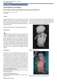
A 40-year old female presented to the <strong>Breast</strong> Clinic with a<br />
painless lump in the right breast for one month with no associated<br />
fever, nipple discharge or skin redness. The patient gave a history<br />
of a hard, slightly painful lump in the left breast for which<br />
she had undergone breast conservation surgery. Review of the<br />
records revealed that the patient had an early stage invasive ductal<br />
carcinoma. On local inspection, the right breast was normal in size<br />
with no nipple retraction and obvious swelling. On palpation; a<br />
5×4 cm soft mobile lump was felt in the upper outer quadrant. It<br />
was non-tender and the skin was freely mobile over it. A separate<br />
2×2 cm firm, non-tender, mobile lump was palpated in the upper<br />
outer quadrant, below the larger lump. There were no palpable<br />
lymph nodes in the axilla. The left breast was asymmetrically<br />
small with a large scar as a result of the previous operation. On<br />
palpation, no lump could be palpated and there was no axillary<br />
lymphadenopathy. The patient was referred to the mammography<br />
unit for bilateral mammograms. Selected mammogram (Figs. 1, 2]<br />
and ultrasound images (Fig. 3) are given as below.<br />
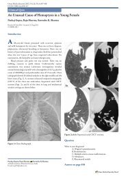
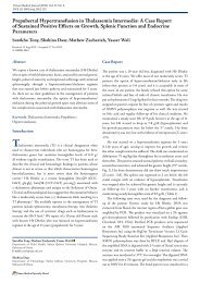
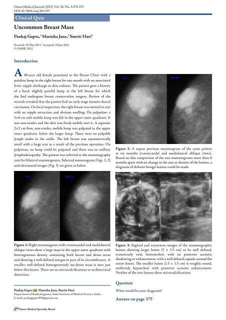
Figure 2: A repeat previous mammogram of the same patient<br />
at six months (craniocaudal and mediolateral oblique views).<br />
Based on this comparison of the two mammograms more than 6<br />
months apart with no change in the size or density of the lesions, a<br />
diagnosis of definite benign lesions could be made.<br />
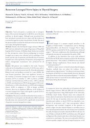
Figure 1: Right mammogram with craniocaudal and mediolateral<br />
oblique views show a large mass in the upper outer quadrant with<br />
heterogeneous density containing both lucent and dense areas<br />
and showing a well-defined margin in part of its circumference. A<br />
smaller, well-defined homogeneously iso-dense mass is seen just<br />
below this lesion. There are no microcalcifications or architectural<br />
distortion.<br />
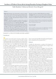
Figure 3: Sagittal and transverse images of the mammographic<br />
lesions showing larger lesion (5 × 3.5 cm) to be well defined,<br />
transversely oval, heteroechoic with no posterior acoustic<br />
shadowing or enhancement, with a well defined capsule around the<br />
entire lesion. The smaller lesion (1.5 × 1.5 cm) is roughly round,<br />
uniformly hypoechoic with posterior acoustic enhancement.<br />
Neither of the two lesions show microcalcifications.<br />
Question<br />
Pankaj Gupta , Manisha Jana, Smriti Hari<br />
Department of Radiodiagnosis, India Institute of Medical Science, India.<br />
E-mail: pankajgupta959@gmail.com<br />
What would be your diagnosis?<br />
Answer on page 375<br />
Oman Medical Specialty Board
Oman Medical Journal (2011) Vol. 26, No. 5:374-375<br />
Answer<br />
Fibroadenoma appears as a well-circumscribed mass<br />
diagnoses are fibroadenoma, lipoma, fat necrosis, and galactocele. 4 with coarse calcifications of varying morphology; however<br />
The mammographic and ultrasound appearance of the lesion<br />
suggests a diagnosis of breast hamartoma.<br />
mammographic appearances can be highly variable. Lipomas are<br />
usually not detected on mammography unless they are large, where<br />
they are seen as well-defined entirely lucent lesion. 5 Fat necrosis<br />
Discussion<br />
has a spectrum of mammographic appearances; however the<br />
pathognomonic appearance is that of a lipid cyst which is seen as<br />
Hamartoma is a rare benign tumor of the breast. It is known<br />
by many synonyms; lipofibroadenoma, fibroadenolipoma or<br />
adenolipoma, which is based on the predominant components<br />
within the mass. 1 The etiology of this disease entity is obscured.<br />
It most commonly presents in middle aged females as a painless<br />
lump. On examination, more than half of these lesions are soft<br />
and non-palpable. Gross pathological examination of a section<br />
gives the appearance of a "slice of salami" or "breast within breast." 1<br />
The tumor is composed of varying admixture of fibrous, glandular<br />
a well-defined, smooth bordered, round or oval lucent mass with a<br />
thin rim which may calcify. 6 Typical mammographic appearance of<br />
a galactocele is a solitary or multiple masses having density similar<br />
to or less than the fibro-glandular parenchyma. The presence of<br />
fat-fluid levels within a well-defined mass is pathognomonic. 7<br />
Malignancy associated with hamartoma is rare but should be kept<br />
in mind. Surgical resection of the mass is the definitive treatment.<br />
Follow up of the lesions is recommended as recurrences have been<br />
reported in several studies. 8<br />
and fatty components. The margin of the tumor is formed by<br />
compressed breast parenchyma known as the "pseudocapsule." Acknowledgements<br />
Histologically; the glandular component forms prominent lobules<br />
in matrix containing fat and fibrous stroma. This organization of<br />
glandular tissue differentiates hamartoma from fibro adenoma.<br />
The authors reported no conflict of interest and no funding was<br />
received for this work.<br />
The mammographic appearance is known as a "piece of cut<br />
sausage." 2 They are well circumscribed masses with both fat and References<br />
soft tissue densities, and are surrounded at least partly by a thin<br />
radio-opaque line which represents the pseudo capsule.<br />
1. Tse GM, Law BK, Ma TK, Chan AB, Pang L-M, Chu WC, et al. Hamartoma<br />
of the breast: a clinicopathological review. J Clin Pathol 2002 Dec;55(12):951-<br />
Based on this classic appearance, a mammographic diagnosis<br />
954.<br />
is possible, as was in our patient. The ultrasound appearance is 2. Paulus DD. Benign diseases of the breast. Radiol Clin North Am 1983<br />
highly variable and it rarely contributes primarily to a differential Mar;21(1):27-50.<br />
3. Gogas J, Markopoulos C, Gogas H, Skandalakis P, Kontzoglou K, Stavridou<br />
diagnosis. However, the typical sonographic appearance is that of a<br />
A. Hamartomas of the breast. Am Surg 1994 Jun;60(6):447-450.<br />
well circumscribed solid mass, which is predominantly hypoechoic 4. Pui MH, Movson IJ. Fatty tissue breast lesions. Clin Imaging 2003 Maybut<br />
has hyper-echogenicity in the form of lines or bands. They have<br />
a variable acoustic shadowing. MRI characteristics of hamartoma<br />
Jun;27(3):150-155.<br />
5. Lanng C, Eriksen BO, Hoffmann J. Lipoma of the breast: a diagnostic<br />
dilemma. <strong>Breast</strong> 2004 Oct;13(5):408-411.<br />
include; fat density within the mass with smooth well defined hypointense<br />
rim and heterogenous contrast enhancement. While fine J Roentgenol 1991 Aug;157(2):271-273.<br />
6. Evers K, Troupin RH. Lipid cyst: classic and atypical appearances. AJR Am<br />
needle aspiration cytology (FNAC) fails to diagnose most of these<br />
lesions, mainly due to the scantly material provided by FNAC,<br />
7. Gómez A, Mata JM, Donoso L, Rams A. Galactocele: three distinctive<br />
radiographic appearances. Radiology 1986 Jan;158(1):43-44.<br />
as well as the lack of any distinctive architectural or cytological<br />
characteristics of hamartoma. 1,3 Even a core biopsy may not prove<br />
8. Daya D, Trus T, D’Souza TJ, Minuk T, Yemen B. Hamartoma of the breast,<br />
an underrecognized breast lesion. A clinicopathologic and radiographic study<br />
of 25 cases. Am J Clin Pathol 1995 Jun;103(6):685-689.<br />
to be useful in the absence of clinical and strong radiological<br />
suspicion. The most important clinical and radiological differential<br />
Oman Medical Specialty Board