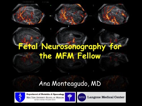
3D Fetal Neurosonography
3D Fetal Neurosonography
3D Fetal Neurosonography
Create successful ePaper yourself
Turn your PDF publications into a flip-book with our unique Google optimized e-Paper software.
<strong>Fetal</strong> <strong>Neurosonography</strong> for<br />
the MFM Fellow<br />
Ana Monteagudo, MD
Outline<br />
• Basic brain scan & fetal neurosonogram<br />
• Advantages of <strong>3D</strong> neurosonography<br />
• Acquisition techniques<br />
• Display modalities I use daily & help me<br />
most in my work are:<br />
– Tomography<br />
– Thick slice mode<br />
– Inversion mode<br />
– <strong>3D</strong> angiography (brain vasculature)<br />
• Examples of pathologies
What is unique about the fetal brain?<br />
• The CNS develops slowly & continuously<br />
• Greatest spurt in cell numbers are in the 1 st<br />
20 wks. After that: mostly increase in size<br />
• CNS anomalies are among the most common<br />
fetal anomalies (second to cardiac anomalies)<br />
Embryonic<br />
<strong>Fetal</strong>
Facts about the Neurosocan<br />
• To do a fetal neuroscan one has to:<br />
–know the CNS embryology &<br />
developmental milestones<br />
–understand the most frequent CNS<br />
anomalies<br />
–use high frequency transducer(s)<br />
–understand & know how to use <strong>3D</strong>
Neurosocan:<br />
The 1 st trimester<br />
NT scan<br />
• Integrity of the<br />
cranium<br />
• Midline falx, choroid<br />
plexus<br />
• Intracranial<br />
translucency<br />
– Lack of visualization<br />
has been associated<br />
with ONTD
11 2/7 weeks<br />
TH<br />
Integrity of the cranium<br />
BS<br />
IT<br />
CP<br />
OB<br />
FCM<br />
Midline falx<br />
Intracranial translucency
9 3/7 wks 12 2/7 wks 12 3/7 wks<br />
Exencephaly-anencephaly sequence<br />
Alobar Holoprosencephaly<br />
Normal<br />
Normal
TH<br />
BS<br />
IT<br />
CP<br />
FCM<br />
OB<br />
TH<br />
Normal<br />
BS<br />
CP<br />
OB<br />
ONTD
Neurosocan: Second Trimester<br />
AIUM guidelines<br />
ISUOG guidelines
Neurosocan: Second Trimester<br />
Anatomical Survey<br />
• Structures that must be included:<br />
– Head & neck<br />
– Cerebellum<br />
– Choroid plexus<br />
– Cisterna magna<br />
– Lateral ventricles<br />
– Midline Falx<br />
– Cavum septi pellucidi<br />
– Biometry: BPD and HC
– Biometry:<br />
• Biparietal diameter<br />
• Head circumference<br />
Basic Brain Scan<br />
Transabdominal sonography: The Axial Plane<br />
• Occipito-frontal diameter<br />
• Atrium of the lateral<br />
ventricle at the level of<br />
the choroid plexus<br />
• Trans-cerebellar diameter<br />
• Depth of the cisterna<br />
magna<br />
• The brain structures:<br />
– Head shape<br />
– Lateral ventricles<br />
– Cavum septi pellucidi<br />
– Thalami<br />
– Cerebellum<br />
– Cisterna magna<br />
– Spine<br />
Sonographic examination of the fetal central nervous system: guidelines for<br />
performing the 'basic examination' and the 'fetal neurosonogram'.<br />
Ultrasound Obstet Gynecol 2007;29:109-16.
Transventricular plane<br />
• Landmarks<br />
– Frontal horns<br />
– Cavum septi<br />
pellucidi<br />
– Posterior horn<br />
• Atrium<br />
• Choroid plexus<br />
– Lateral ventricle<br />
measured at atrium<br />
– Calipers over<br />
glomus<br />
FH<br />
CSP<br />
CP<br />
+<br />
+<br />
PH
Lateral ventricles & Choroid Plexus<br />
20 wks<br />
Up to 3 mm<br />
Transventricular plane:<br />
Normal < 10 mm
Transthalamic plane<br />
• Landmarks<br />
– Frontal horns<br />
– Cavum septi<br />
pellucidi<br />
– Thalami<br />
FH<br />
TH<br />
HG<br />
– Hippocampal gyrus<br />
CSP<br />
– Measure of the<br />
BPD, HC, OFD
Transcerebellar plane<br />
• Landmarks<br />
– Frontal horns<br />
– Cavum septi pellucidi<br />
FH<br />
– Thalami<br />
– Cerebellum<br />
CSP<br />
TH<br />
C<br />
CM<br />
– Cisterna magna<br />
– Measure of the<br />
cerebellar diameter<br />
and cisterna magna<br />
CM: Normal < 10 mm
Summary anatomy:<br />
Axial plane
If an abnormality is detected<br />
during the Basic Scan a<br />
detailed, targeted fetal<br />
neurosonogram is<br />
recommended.
<strong>Fetal</strong> Neurosonogram<br />
=<br />
Multiplanar Imaging<br />
• This scan looks at the brain in greater detail<br />
• Can be performed transabdominally, and/or<br />
transvaginally....<br />
• The coronal and sagittal planes are added to<br />
the scan<br />
• Can be done …..using 2D/ <strong>3D</strong> sonography
2D-TAS vs. 2D-TVS<br />
Axial<br />
• In 2D TAS:<br />
• Axial sections<br />
• Sagittal and coronal may not<br />
be obtained depending on the<br />
fetal lie.<br />
•In 2D TVS:<br />
• Sagittal and coronal sections<br />
• Axial sections are obtained<br />
depending on the fetal lie.<br />
•All sections radiate from a<br />
single point
2DTVS Neuroscan<br />
• It is a transfontanelle<br />
scan<br />
• Can obtain Coronal, &<br />
Sagittal planes<br />
• Remember that all brain<br />
sections “radiate” from<br />
one point (footprint of<br />
transducer tip)<br />
MONTEAGUDO A, REUSS ML, TIMOR-TRITSCH IE. Imaging the fetal brain in the second<br />
and third trimesters using transvaginal sonography. Obstet Gynecol 1991;77:27-32.
<strong>Fetal</strong> Neuroscan Scan<br />
Coronal & Sagittal Planes<br />
• Brain structures imaged<br />
– Anterior horns<br />
– Posterior horns<br />
– 3 rd and 4 th ventricle<br />
– Inter-ventricular foramina<br />
– Cavum septi pellucidi<br />
– Corpus callosum<br />
– Pericallosal artery<br />
– Caudate nuclei<br />
– Thalami<br />
– Cerebellum & vermis<br />
– Cisterna magna<br />
– Interhemispheric<br />
fissure<br />
– Fissures<br />
– Sphenoidal bone<br />
– Ocular orbits<br />
Sonographic examination of the fetal central nervous system: guidelines for<br />
performing the 'basic examination' and the 'fetal neurosonogram'.<br />
Ultrasound Obstet Gynecol 2007;29:109-16.
<strong>Fetal</strong><br />
Neuroscan Scan<br />
Coronal Sections<br />
Sagittal Sections
The Frontal group<br />
Frontal-2 or transfrontal plane<br />
– Interhemispheric<br />
fissure<br />
– Brain parenchyma<br />
anterior lobe<br />
Brain<br />
IHF<br />
Orbits<br />
Sphenoid<br />
Ultrasound Obstet Gynecol 2007;29:109-16
The Midcoronal group<br />
Mid-coronal-1 or transcaudate plane<br />
– Caudate nuclei<br />
– Genu corpus<br />
callosum<br />
– Cavum septi<br />
pellucidi<br />
– Frontal horns<br />
CN<br />
CC<br />
CSP<br />
FH<br />
Ultrasound Obstet Gynecol 2007;29:109-16
The Midcoronal group<br />
Mid-coronal-2 or transthalamic plane<br />
– Thalami<br />
– Interventricular<br />
foramina<br />
– Atrium LV<br />
Atrium<br />
TH<br />
Ultrasound Obstet Gynecol 2007;29:109-16
Mid-Coronal Plane<br />
• Allows visualization<br />
of the leaves of the<br />
septi pellucidi,<br />
which enclose the<br />
space of the cavum<br />
septi pellucidi<br />
• This is the correct<br />
plane to diagnose<br />
agenesis of the<br />
septi pellucidi<br />
Lateral leaves of the<br />
Cavum<br />
septi<br />
pellucidi<br />
Cavum Septi Pellucidi
The Occipital group<br />
Occipital-1& 2 or transcerebellar plane<br />
– Occipital horns<br />
– Interhemispheric<br />
fissure<br />
– Cerebellar<br />
hemispheres<br />
– Vermis<br />
OH<br />
IHF<br />
C<br />
V<br />
Ultrasound Obstet Gynecol 2007;29:109-16
<strong>Fetal</strong><br />
Neuroscan Scan<br />
Coronal Sections<br />
Sagittal Sections
The Median Plane<br />
Median or mid-sagittal plane<br />
– Corpus callosum<br />
– Cavum septi pellucidi<br />
– Brain stem<br />
– Pons<br />
– Posterior fossa<br />
– Vermis<br />
CC<br />
CSP<br />
BS<br />
Pons<br />
V<br />
Ultrasound Obstet Gynecol 2007;29:109-16
Corpus callosum, Cavum Septi<br />
Pellucidi e Vergae<br />
Genu<br />
Corpus<br />
22 w<br />
Splenium<br />
CSP<br />
CV<br />
Rostrum
The Median<br />
Pericallosal Artery
Median plane<br />
16 weeks<br />
22 weeks<br />
34 weeks
The Rt & Lt Oblique<br />
Oblique-1 or Parasagittal plane<br />
– Entire lateral<br />
ventricle<br />
– Choroid plexus<br />
– Periventricular<br />
white matter<br />
FH<br />
TH<br />
CP<br />
PH<br />
– Cortex<br />
IH<br />
Ultrasound Obstet Gynecol 2007;29:109-16
Why <strong>3D</strong> of the brain?<br />
• Better define the anatomy seen by 2D<br />
• To perform sonographic evaluations<br />
unavailable with the conventional 2D<br />
– <strong>3D</strong> reconstructions; serial sections;<br />
inversion & <strong>3D</strong> sono-angiography, etc.<br />
• To use as an additional tool for counseling<br />
patients and communicating with other<br />
specialties (neonatology, neurology,<br />
neurosurgery, radiology, genetics etc.)<br />
• Reassure patients with a NL fetus<br />
• Store digital volumes for future studies
Why <strong>3D</strong> of the brain?<br />
• Obtaining a volume of the fetal brain<br />
(transvaginally or transabdominally) is<br />
relatively fast, easy and diagnostically<br />
valuable.<br />
• If a brain anomaly is seen; <strong>3D</strong> can be<br />
helpful to localize, define, and assess the<br />
extent of lesion<br />
• The results are better than using<br />
conventional 2D US alone; and in<br />
experienced hands <strong>3D</strong> can be comparable<br />
to MRI
Advantages of <strong>3D</strong> <strong>Neurosonography</strong><br />
• Bedside or off-line analysis<br />
• Off-site expert consultation<br />
• Data is easily revisited for additional<br />
evaluation<br />
• Short processing time<br />
• Effective teaching tool (simulating real<br />
time scanning)<br />
• Quick way to image the median plane –<br />
especially the corpus callosum & vermis
<strong>3D</strong> Ultrasound of the Brain<br />
• Volume Acquisition<br />
– Transabdominal<br />
– Transvaginal<br />
• Manipulation<br />
– Zooming<br />
– Chroma mapping<br />
– Marker dot<br />
– Rotation<br />
• Display modalities<br />
– Tomography<br />
– Thick slice mode<br />
– Static volume contrast<br />
imaging (VCI TM )<br />
– Inversion mode<br />
– <strong>3D</strong> angiography (brain<br />
vasculature)<br />
• Anomalies
Basics of Volume Acquisition<br />
• Acquisition of a good<br />
quality volume is the<br />
basis and the secret<br />
of any <strong>3D</strong> analysis<br />
• However, you must<br />
start with a good<br />
quality 2D image
Basics of Volume Acquisition<br />
Acquisition should attempt to overcome<br />
some of the inherent limitations of <strong>3D</strong> US:<br />
1. If there is acoustic shadowing in the acquisition<br />
plane it will be embedded in the volume<br />
2. If fetal movements are present during the<br />
acquisition; the result is motion artifacts<br />
3. If surface rendering is desired a fluid-tissue<br />
interface is needed<br />
4. Other factors such as fetal position and<br />
maternal obesity may challenge <strong>3D</strong> imaging just<br />
as in 2D
Transabdominal Acquisition<br />
• The volume is acquired through<br />
the acoustic window of<br />
– the sphenoid fontanel & the<br />
squamosal suture<br />
– Shorten acquisition time to minimize<br />
motion artifacts (breath-holding)
TAS- Acquisition-Display<br />
A<br />
B<br />
C<br />
Axial – Plane of acquisition<br />
Coronal Plane<br />
Box A: always displays<br />
the plane of acquisition<br />
Box C: displays the<br />
reconstructed plane<br />
Sagittal – reconstructed plane
Transvaginal Acquisiton<br />
• With TVS, the volume is<br />
acquired either in the sagittal<br />
or the coronal plane<br />
• The reconstructed plane is the<br />
axial one.<br />
• (For a detailed fetal brain scan<br />
TVS is the preferred mode of<br />
volume acquisition due to its<br />
excellent resolution).
Transvaginal acquisition<br />
• The volume is acquired through the<br />
acoustic window of<br />
– the anterior fontanelle, metopic<br />
or sagittal suture
Transvaginal Acquisition<br />
• Access fontanelle – align transducer<br />
• By rotating the vaginal probe 90<br />
• Sagittal Coronal<br />
• Hold head steady with<br />
abdominal hand
Transvaginal Acquisition
TVS – Acquisition<br />
A<br />
B<br />
C<br />
Sagittal – Acquisition plane<br />
Axial – Reconstructed plane<br />
Box A: always displays<br />
the plane of acquisition;<br />
regardless of the<br />
position of the fetal<br />
spine; face the fetus to<br />
the left of the screen
<strong>3D</strong>-TAS<br />
vs. <strong>3D</strong>-TVS<br />
• Acquisition: Axial<br />
plane.<br />
•The sagittal plane is a<br />
reconstructed one<br />
•Near field blurred due<br />
to bone attenuation<br />
• Acquisition: Coronal<br />
& sagittal planes<br />
• The axial plane is a<br />
reconstructed one<br />
•Excellent resolution<br />
on median plane<br />
decreasing laterally
<strong>3D</strong> Ultrasound of the Brain<br />
• Volume Acquisition<br />
– Transabdominal<br />
– Transvaginal<br />
• Manipulation<br />
– Zooming<br />
– Chroma mapping<br />
– Marker dot<br />
– Rotation<br />
• Display modalities<br />
– Tomography<br />
– Thick slice mode<br />
– Static volume contrast<br />
imaging (VCI TM )<br />
– Inversion mode<br />
– <strong>3D</strong> angiography (brain<br />
vasculature)<br />
• Anomalies
TAS- Manipulations: Zooming
It may be a “gimmick”, but at times emphasizes structures<br />
TAS- Manipulations:<br />
Chroma Mapping<br />
Black /white<br />
Candle light<br />
Sepia<br />
Blue
TAS- Manipulations: ‘Dot’<br />
Orients Marker you Dot in in all the 3 cavum planes.<br />
Represents septi pellucidi the between same exact the<br />
place thalami on all three planes
TAS- Manipulations: Rotation<br />
Box A<br />
Box B<br />
Box C<br />
The X, Y and Z controls<br />
Goal of rotations is<br />
(knobs) perform<br />
rotations to better around orient their<br />
respective the observer axis in each<br />
box<br />
+ Rotation - Rotation
<strong>3D</strong> Ultrasound of the Brain<br />
• Volume Acquisition<br />
– Transabdominal<br />
– Transvaginal<br />
• Manipulation<br />
– Zooming<br />
– Chroma mapping<br />
– Marker dot<br />
– Rotation<br />
• Display modalities<br />
– Tomography<br />
– Thick slice mode<br />
– Static volume contrast<br />
imaging (VCI TM )<br />
– Inversion mode<br />
– <strong>3D</strong> angiography (brain<br />
vasculature)<br />
• Anomalies
Display Modalities<br />
Inversion<br />
The<br />
volume<br />
Localize<br />
structures<br />
Tomography<br />
Angiography
Orthogonal Planes:<br />
Hydrocephaly & IVH<br />
All 3 scanning planes:<br />
coronal, sagittal and<br />
axial simultaneously<br />
imaged.
Thick Slice:<br />
Enhances edge detection: The software “collapses” a pre-selected number of<br />
successive slices and displays it as a rendered image
Tomography: Serial Coronal Sections
Tomography: Serial Axial Sections
Inversion:<br />
The fluid filled lateral ventricles of the acquired volume can be “inverted”;<br />
therefore, the areas which were initially anechoic become echogenic.
The inversion mode<br />
The fluid filled lateral ventricles of the acquired volume can be “inverted”;<br />
therefore, the areas which were initially anechoic become echogenic.
The inversion mode<br />
Anterior horn<br />
Posterior horn<br />
3 rd ventricle<br />
Inferior horn<br />
Useful in imaging fluid filled<br />
structures such as the<br />
ventricular system<br />
The lateral ventricles as seen in<br />
3 planes
-Colpocephaly, parallel & widely<br />
separated AH, inter-hemispheric<br />
fissure drops into 3 rd V<br />
Orthogonal Planes :AGCC<br />
Direct signs:<br />
-missing corpus callosum & CSP<br />
Indirect signs:
Middle<br />
cerebral<br />
Missing Pericallosal<br />
artery<br />
Circle of<br />
Willis<br />
<strong>3D</strong> color angiogram<br />
of AGCC<br />
Power Doppler + <strong>3D</strong> = <strong>3D</strong> color angiogram.<br />
Enables selective imaging of blood flow<br />
Normal
Tomography: Serial Sagittal Sections
Power Doppler +Tomography:<br />
Serial Sagittal Sections
Absent CSP – r/o SOD<br />
Normal
Absent CSP – r/o SOD
Absent CSP – r/o SOD
Absent CSP – r/o SOD
Posterior Cephalocele
Posterior Cephalocele
Posterior Cephalocele
<strong>3D</strong> Helpful Hints<br />
• Always start with a good quality 2D<br />
image<br />
– TAS acquisition: BPD plane<br />
– TVS acquisition: median or coronal plane<br />
– Angle of acquisition: 45 to 80°<br />
• <strong>Fetal</strong> head must fill the entire box
<strong>3D</strong> Helpful Hints<br />
• After volume is acquired:<br />
– Save<br />
– Zoom the image as large as possible<br />
– Consider using chroma mapping<br />
– Marker Dot – in CSP or between thalami<br />
– Rotate volume as needed-<br />
– Use different display modalities to help<br />
• e.g. Serial tomographic sections, inversion,<br />
thick slice, <strong>3D</strong> angiography
In Conclusion…<br />
• When evaluating the fetal brain –<br />
– First start TAS in the axial plane<br />
• Basic anatomy, Biometry<br />
– Second TVS for the coronal and sagittal<br />
planes<br />
• Coronal- mid-coronal visualization of the leaves of<br />
the septi pellucidi<br />
• Median section for the midbrain (corpus callosum!)<br />
and posterior fossa (vermis!)<br />
• <strong>3D</strong> is a useful complement of the 2D<br />
targeted scan
Thank You!!


