3D Fetal Neurosonography
3D Fetal Neurosonography
3D Fetal Neurosonography
You also want an ePaper? Increase the reach of your titles
YUMPU automatically turns print PDFs into web optimized ePapers that Google loves.
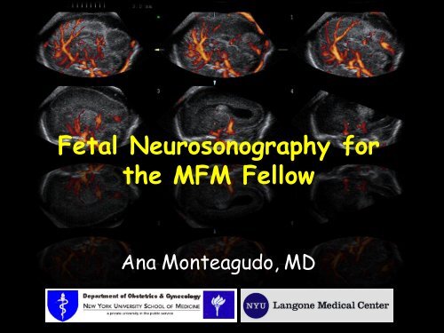
<strong>Fetal</strong> <strong>Neurosonography</strong> for<br />
the MFM Fellow<br />
Ana Monteagudo, MD
Outline<br />
• Basic brain scan & fetal neurosonogram<br />
• Advantages of <strong>3D</strong> neurosonography<br />
• Acquisition techniques<br />
• Display modalities I use daily & help me<br />
most in my work are:<br />
– Tomography<br />
– Thick slice mode<br />
– Inversion mode<br />
– <strong>3D</strong> angiography (brain vasculature)<br />
• Examples of pathologies
What is unique about the fetal brain?<br />
• The CNS develops slowly & continuously<br />
• Greatest spurt in cell numbers are in the 1 st<br />
20 wks. After that: mostly increase in size<br />
• CNS anomalies are among the most common<br />
fetal anomalies (second to cardiac anomalies)<br />
Embryonic<br />
<strong>Fetal</strong>
Facts about the Neurosocan<br />
• To do a fetal neuroscan one has to:<br />
–know the CNS embryology &<br />
developmental milestones<br />
–understand the most frequent CNS<br />
anomalies<br />
–use high frequency transducer(s)<br />
–understand & know how to use <strong>3D</strong>
Neurosocan:<br />
The 1 st trimester<br />
NT scan<br />
• Integrity of the<br />
cranium<br />
• Midline falx, choroid<br />
plexus<br />
• Intracranial<br />
translucency<br />
– Lack of visualization<br />
has been associated<br />
with ONTD
11 2/7 weeks<br />
TH<br />
Integrity of the cranium<br />
BS<br />
IT<br />
CP<br />
OB<br />
FCM<br />
Midline falx<br />
Intracranial translucency
9 3/7 wks 12 2/7 wks 12 3/7 wks<br />
Exencephaly-anencephaly sequence<br />
Alobar Holoprosencephaly<br />
Normal<br />
Normal
TH<br />
BS<br />
IT<br />
CP<br />
FCM<br />
OB<br />
TH<br />
Normal<br />
BS<br />
CP<br />
OB<br />
ONTD
Neurosocan: Second Trimester<br />
AIUM guidelines<br />
ISUOG guidelines
Neurosocan: Second Trimester<br />
Anatomical Survey<br />
• Structures that must be included:<br />
– Head & neck<br />
– Cerebellum<br />
– Choroid plexus<br />
– Cisterna magna<br />
– Lateral ventricles<br />
– Midline Falx<br />
– Cavum septi pellucidi<br />
– Biometry: BPD and HC
– Biometry:<br />
• Biparietal diameter<br />
• Head circumference<br />
Basic Brain Scan<br />
Transabdominal sonography: The Axial Plane<br />
• Occipito-frontal diameter<br />
• Atrium of the lateral<br />
ventricle at the level of<br />
the choroid plexus<br />
• Trans-cerebellar diameter<br />
• Depth of the cisterna<br />
magna<br />
• The brain structures:<br />
– Head shape<br />
– Lateral ventricles<br />
– Cavum septi pellucidi<br />
– Thalami<br />
– Cerebellum<br />
– Cisterna magna<br />
– Spine<br />
Sonographic examination of the fetal central nervous system: guidelines for<br />
performing the 'basic examination' and the 'fetal neurosonogram'.<br />
Ultrasound Obstet Gynecol 2007;29:109-16.
Transventricular plane<br />
• Landmarks<br />
– Frontal horns<br />
– Cavum septi<br />
pellucidi<br />
– Posterior horn<br />
• Atrium<br />
• Choroid plexus<br />
– Lateral ventricle<br />
measured at atrium<br />
– Calipers over<br />
glomus<br />
FH<br />
CSP<br />
CP<br />
+<br />
+<br />
PH
Lateral ventricles & Choroid Plexus<br />
20 wks<br />
Up to 3 mm<br />
Transventricular plane:<br />
Normal < 10 mm
Transthalamic plane<br />
• Landmarks<br />
– Frontal horns<br />
– Cavum septi<br />
pellucidi<br />
– Thalami<br />
FH<br />
TH<br />
HG<br />
– Hippocampal gyrus<br />
CSP<br />
– Measure of the<br />
BPD, HC, OFD
Transcerebellar plane<br />
• Landmarks<br />
– Frontal horns<br />
– Cavum septi pellucidi<br />
FH<br />
– Thalami<br />
– Cerebellum<br />
CSP<br />
TH<br />
C<br />
CM<br />
– Cisterna magna<br />
– Measure of the<br />
cerebellar diameter<br />
and cisterna magna<br />
CM: Normal < 10 mm
Summary anatomy:<br />
Axial plane
If an abnormality is detected<br />
during the Basic Scan a<br />
detailed, targeted fetal<br />
neurosonogram is<br />
recommended.
<strong>Fetal</strong> Neurosonogram<br />
=<br />
Multiplanar Imaging<br />
• This scan looks at the brain in greater detail<br />
• Can be performed transabdominally, and/or<br />
transvaginally....<br />
• The coronal and sagittal planes are added to<br />
the scan<br />
• Can be done …..using 2D/ <strong>3D</strong> sonography
2D-TAS vs. 2D-TVS<br />
Axial<br />
• In 2D TAS:<br />
• Axial sections<br />
• Sagittal and coronal may not<br />
be obtained depending on the<br />
fetal lie.<br />
•In 2D TVS:<br />
• Sagittal and coronal sections<br />
• Axial sections are obtained<br />
depending on the fetal lie.<br />
•All sections radiate from a<br />
single point
2DTVS Neuroscan<br />
• It is a transfontanelle<br />
scan<br />
• Can obtain Coronal, &<br />
Sagittal planes<br />
• Remember that all brain<br />
sections “radiate” from<br />
one point (footprint of<br />
transducer tip)<br />
MONTEAGUDO A, REUSS ML, TIMOR-TRITSCH IE. Imaging the fetal brain in the second<br />
and third trimesters using transvaginal sonography. Obstet Gynecol 1991;77:27-32.
<strong>Fetal</strong> Neuroscan Scan<br />
Coronal & Sagittal Planes<br />
• Brain structures imaged<br />
– Anterior horns<br />
– Posterior horns<br />
– 3 rd and 4 th ventricle<br />
– Inter-ventricular foramina<br />
– Cavum septi pellucidi<br />
– Corpus callosum<br />
– Pericallosal artery<br />
– Caudate nuclei<br />
– Thalami<br />
– Cerebellum & vermis<br />
– Cisterna magna<br />
– Interhemispheric<br />
fissure<br />
– Fissures<br />
– Sphenoidal bone<br />
– Ocular orbits<br />
Sonographic examination of the fetal central nervous system: guidelines for<br />
performing the 'basic examination' and the 'fetal neurosonogram'.<br />
Ultrasound Obstet Gynecol 2007;29:109-16.
<strong>Fetal</strong><br />
Neuroscan Scan<br />
Coronal Sections<br />
Sagittal Sections
The Frontal group<br />
Frontal-2 or transfrontal plane<br />
– Interhemispheric<br />
fissure<br />
– Brain parenchyma<br />
anterior lobe<br />
Brain<br />
IHF<br />
Orbits<br />
Sphenoid<br />
Ultrasound Obstet Gynecol 2007;29:109-16
The Midcoronal group<br />
Mid-coronal-1 or transcaudate plane<br />
– Caudate nuclei<br />
– Genu corpus<br />
callosum<br />
– Cavum septi<br />
pellucidi<br />
– Frontal horns<br />
CN<br />
CC<br />
CSP<br />
FH<br />
Ultrasound Obstet Gynecol 2007;29:109-16
The Midcoronal group<br />
Mid-coronal-2 or transthalamic plane<br />
– Thalami<br />
– Interventricular<br />
foramina<br />
– Atrium LV<br />
Atrium<br />
TH<br />
Ultrasound Obstet Gynecol 2007;29:109-16
Mid-Coronal Plane<br />
• Allows visualization<br />
of the leaves of the<br />
septi pellucidi,<br />
which enclose the<br />
space of the cavum<br />
septi pellucidi<br />
• This is the correct<br />
plane to diagnose<br />
agenesis of the<br />
septi pellucidi<br />
Lateral leaves of the<br />
Cavum<br />
septi<br />
pellucidi<br />
Cavum Septi Pellucidi
The Occipital group<br />
Occipital-1& 2 or transcerebellar plane<br />
– Occipital horns<br />
– Interhemispheric<br />
fissure<br />
– Cerebellar<br />
hemispheres<br />
– Vermis<br />
OH<br />
IHF<br />
C<br />
V<br />
Ultrasound Obstet Gynecol 2007;29:109-16
<strong>Fetal</strong><br />
Neuroscan Scan<br />
Coronal Sections<br />
Sagittal Sections
The Median Plane<br />
Median or mid-sagittal plane<br />
– Corpus callosum<br />
– Cavum septi pellucidi<br />
– Brain stem<br />
– Pons<br />
– Posterior fossa<br />
– Vermis<br />
CC<br />
CSP<br />
BS<br />
Pons<br />
V<br />
Ultrasound Obstet Gynecol 2007;29:109-16
Corpus callosum, Cavum Septi<br />
Pellucidi e Vergae<br />
Genu<br />
Corpus<br />
22 w<br />
Splenium<br />
CSP<br />
CV<br />
Rostrum
The Median<br />
Pericallosal Artery
Median plane<br />
16 weeks<br />
22 weeks<br />
34 weeks
The Rt & Lt Oblique<br />
Oblique-1 or Parasagittal plane<br />
– Entire lateral<br />
ventricle<br />
– Choroid plexus<br />
– Periventricular<br />
white matter<br />
FH<br />
TH<br />
CP<br />
PH<br />
– Cortex<br />
IH<br />
Ultrasound Obstet Gynecol 2007;29:109-16
Why <strong>3D</strong> of the brain?<br />
• Better define the anatomy seen by 2D<br />
• To perform sonographic evaluations<br />
unavailable with the conventional 2D<br />
– <strong>3D</strong> reconstructions; serial sections;<br />
inversion & <strong>3D</strong> sono-angiography, etc.<br />
• To use as an additional tool for counseling<br />
patients and communicating with other<br />
specialties (neonatology, neurology,<br />
neurosurgery, radiology, genetics etc.)<br />
• Reassure patients with a NL fetus<br />
• Store digital volumes for future studies
Why <strong>3D</strong> of the brain?<br />
• Obtaining a volume of the fetal brain<br />
(transvaginally or transabdominally) is<br />
relatively fast, easy and diagnostically<br />
valuable.<br />
• If a brain anomaly is seen; <strong>3D</strong> can be<br />
helpful to localize, define, and assess the<br />
extent of lesion<br />
• The results are better than using<br />
conventional 2D US alone; and in<br />
experienced hands <strong>3D</strong> can be comparable<br />
to MRI
Advantages of <strong>3D</strong> <strong>Neurosonography</strong><br />
• Bedside or off-line analysis<br />
• Off-site expert consultation<br />
• Data is easily revisited for additional<br />
evaluation<br />
• Short processing time<br />
• Effective teaching tool (simulating real<br />
time scanning)<br />
• Quick way to image the median plane –<br />
especially the corpus callosum & vermis
<strong>3D</strong> Ultrasound of the Brain<br />
• Volume Acquisition<br />
– Transabdominal<br />
– Transvaginal<br />
• Manipulation<br />
– Zooming<br />
– Chroma mapping<br />
– Marker dot<br />
– Rotation<br />
• Display modalities<br />
– Tomography<br />
– Thick slice mode<br />
– Static volume contrast<br />
imaging (VCI TM )<br />
– Inversion mode<br />
– <strong>3D</strong> angiography (brain<br />
vasculature)<br />
• Anomalies
Basics of Volume Acquisition<br />
• Acquisition of a good<br />
quality volume is the<br />
basis and the secret<br />
of any <strong>3D</strong> analysis<br />
• However, you must<br />
start with a good<br />
quality 2D image
Basics of Volume Acquisition<br />
Acquisition should attempt to overcome<br />
some of the inherent limitations of <strong>3D</strong> US:<br />
1. If there is acoustic shadowing in the acquisition<br />
plane it will be embedded in the volume<br />
2. If fetal movements are present during the<br />
acquisition; the result is motion artifacts<br />
3. If surface rendering is desired a fluid-tissue<br />
interface is needed<br />
4. Other factors such as fetal position and<br />
maternal obesity may challenge <strong>3D</strong> imaging just<br />
as in 2D
Transabdominal Acquisition<br />
• The volume is acquired through<br />
the acoustic window of<br />
– the sphenoid fontanel & the<br />
squamosal suture<br />
– Shorten acquisition time to minimize<br />
motion artifacts (breath-holding)
TAS- Acquisition-Display<br />
A<br />
B<br />
C<br />
Axial – Plane of acquisition<br />
Coronal Plane<br />
Box A: always displays<br />
the plane of acquisition<br />
Box C: displays the<br />
reconstructed plane<br />
Sagittal – reconstructed plane
Transvaginal Acquisiton<br />
• With TVS, the volume is<br />
acquired either in the sagittal<br />
or the coronal plane<br />
• The reconstructed plane is the<br />
axial one.<br />
• (For a detailed fetal brain scan<br />
TVS is the preferred mode of<br />
volume acquisition due to its<br />
excellent resolution).
Transvaginal acquisition<br />
• The volume is acquired through the<br />
acoustic window of<br />
– the anterior fontanelle, metopic<br />
or sagittal suture
Transvaginal Acquisition<br />
• Access fontanelle – align transducer<br />
• By rotating the vaginal probe 90<br />
• Sagittal Coronal<br />
• Hold head steady with<br />
abdominal hand
Transvaginal Acquisition
TVS – Acquisition<br />
A<br />
B<br />
C<br />
Sagittal – Acquisition plane<br />
Axial – Reconstructed plane<br />
Box A: always displays<br />
the plane of acquisition;<br />
regardless of the<br />
position of the fetal<br />
spine; face the fetus to<br />
the left of the screen
<strong>3D</strong>-TAS<br />
vs. <strong>3D</strong>-TVS<br />
• Acquisition: Axial<br />
plane.<br />
•The sagittal plane is a<br />
reconstructed one<br />
•Near field blurred due<br />
to bone attenuation<br />
• Acquisition: Coronal<br />
& sagittal planes<br />
• The axial plane is a<br />
reconstructed one<br />
•Excellent resolution<br />
on median plane<br />
decreasing laterally
<strong>3D</strong> Ultrasound of the Brain<br />
• Volume Acquisition<br />
– Transabdominal<br />
– Transvaginal<br />
• Manipulation<br />
– Zooming<br />
– Chroma mapping<br />
– Marker dot<br />
– Rotation<br />
• Display modalities<br />
– Tomography<br />
– Thick slice mode<br />
– Static volume contrast<br />
imaging (VCI TM )<br />
– Inversion mode<br />
– <strong>3D</strong> angiography (brain<br />
vasculature)<br />
• Anomalies
TAS- Manipulations: Zooming
It may be a “gimmick”, but at times emphasizes structures<br />
TAS- Manipulations:<br />
Chroma Mapping<br />
Black /white<br />
Candle light<br />
Sepia<br />
Blue
TAS- Manipulations: ‘Dot’<br />
Orients Marker you Dot in in all the 3 cavum planes.<br />
Represents septi pellucidi the between same exact the<br />
place thalami on all three planes
TAS- Manipulations: Rotation<br />
Box A<br />
Box B<br />
Box C<br />
The X, Y and Z controls<br />
Goal of rotations is<br />
(knobs) perform<br />
rotations to better around orient their<br />
respective the observer axis in each<br />
box<br />
+ Rotation - Rotation
<strong>3D</strong> Ultrasound of the Brain<br />
• Volume Acquisition<br />
– Transabdominal<br />
– Transvaginal<br />
• Manipulation<br />
– Zooming<br />
– Chroma mapping<br />
– Marker dot<br />
– Rotation<br />
• Display modalities<br />
– Tomography<br />
– Thick slice mode<br />
– Static volume contrast<br />
imaging (VCI TM )<br />
– Inversion mode<br />
– <strong>3D</strong> angiography (brain<br />
vasculature)<br />
• Anomalies
Display Modalities<br />
Inversion<br />
The<br />
volume<br />
Localize<br />
structures<br />
Tomography<br />
Angiography
Orthogonal Planes:<br />
Hydrocephaly & IVH<br />
All 3 scanning planes:<br />
coronal, sagittal and<br />
axial simultaneously<br />
imaged.
Thick Slice:<br />
Enhances edge detection: The software “collapses” a pre-selected number of<br />
successive slices and displays it as a rendered image
Tomography: Serial Coronal Sections
Tomography: Serial Axial Sections
Inversion:<br />
The fluid filled lateral ventricles of the acquired volume can be “inverted”;<br />
therefore, the areas which were initially anechoic become echogenic.
The inversion mode<br />
The fluid filled lateral ventricles of the acquired volume can be “inverted”;<br />
therefore, the areas which were initially anechoic become echogenic.
The inversion mode<br />
Anterior horn<br />
Posterior horn<br />
3 rd ventricle<br />
Inferior horn<br />
Useful in imaging fluid filled<br />
structures such as the<br />
ventricular system<br />
The lateral ventricles as seen in<br />
3 planes
-Colpocephaly, parallel & widely<br />
separated AH, inter-hemispheric<br />
fissure drops into 3 rd V<br />
Orthogonal Planes :AGCC<br />
Direct signs:<br />
-missing corpus callosum & CSP<br />
Indirect signs:
Middle<br />
cerebral<br />
Missing Pericallosal<br />
artery<br />
Circle of<br />
Willis<br />
<strong>3D</strong> color angiogram<br />
of AGCC<br />
Power Doppler + <strong>3D</strong> = <strong>3D</strong> color angiogram.<br />
Enables selective imaging of blood flow<br />
Normal
Tomography: Serial Sagittal Sections
Power Doppler +Tomography:<br />
Serial Sagittal Sections
Absent CSP – r/o SOD<br />
Normal
Absent CSP – r/o SOD
Absent CSP – r/o SOD
Absent CSP – r/o SOD
Posterior Cephalocele
Posterior Cephalocele
Posterior Cephalocele
<strong>3D</strong> Helpful Hints<br />
• Always start with a good quality 2D<br />
image<br />
– TAS acquisition: BPD plane<br />
– TVS acquisition: median or coronal plane<br />
– Angle of acquisition: 45 to 80°<br />
• <strong>Fetal</strong> head must fill the entire box
<strong>3D</strong> Helpful Hints<br />
• After volume is acquired:<br />
– Save<br />
– Zoom the image as large as possible<br />
– Consider using chroma mapping<br />
– Marker Dot – in CSP or between thalami<br />
– Rotate volume as needed-<br />
– Use different display modalities to help<br />
• e.g. Serial tomographic sections, inversion,<br />
thick slice, <strong>3D</strong> angiography
In Conclusion…<br />
• When evaluating the fetal brain –<br />
– First start TAS in the axial plane<br />
• Basic anatomy, Biometry<br />
– Second TVS for the coronal and sagittal<br />
planes<br />
• Coronal- mid-coronal visualization of the leaves of<br />
the septi pellucidi<br />
• Median section for the midbrain (corpus callosum!)<br />
and posterior fossa (vermis!)<br />
• <strong>3D</strong> is a useful complement of the 2D<br />
targeted scan
Thank You!!


