Extensive Idiopathic Scrotal Calcinosis: A Case Report - JCDR
Extensive Idiopathic Scrotal Calcinosis: A Case Report - JCDR
Extensive Idiopathic Scrotal Calcinosis: A Case Report - JCDR

Create successful ePaper yourself
Turn your PDF publications into a flip-book with our unique Google optimized e-Paper software.
<strong>Case</strong> <strong>Report</strong><br />
<strong>Extensive</strong> <strong>Idiopathic</strong> <strong>Scrotal</strong> <strong>Calcinosis</strong>:<br />
A <strong>Case</strong> <strong>Report</strong><br />
DOI: <strong>JCDR</strong>/2012/3945:2071<br />
Pathology Section<br />
Vijay D. Dombale, S. I. Basarkod, H.B. Kotabagi, Unaiza Farheen<br />
ABSTRACT<br />
A 54-year-old male diabetic presented with multiple swellings in<br />
the scrotal region with a duration of 15 years, which was initially<br />
peanut sized and painless and which gradually progressed<br />
to 1-1.5 cm in diameter and more than 100 in number. His<br />
haematological and biochemical parameters were within normal<br />
limits. Fine needle aspiration of the nodule revealed hypocellular<br />
smears with a pale pink to bluish coloured granular material in<br />
the background. On microscopy, the sections showed calcified<br />
nodules and masses with a peripheral giant cell reaction within<br />
the dermis and the subcutaneous tissue, which were positive<br />
for the Von Kossa stain. It was reported as <strong>Idiopathic</strong> calcinosis<br />
Cutis – Scrotum. Herewith, we are presenting a rare case of<br />
extensive idiopathic scrotal calcinosis.<br />
Key Words: <strong>Scrotal</strong> calcinosis, <strong>Idiopathic</strong> calcinosis cutis, <strong>Calcinosis</strong>, Cutaneous calcinosis<br />
478<br />
Introduction<br />
<strong>Idiopathic</strong> scrotal calcinosis ( also known as “<strong>Idiopathic</strong> calcified<br />
nodules of the scrotum”) is a cutaneous condition which is characterized<br />
by calcification of the skin, resulting from the deposition<br />
of calcium and phosphorus, which occurs on the scrotum [1]. The<br />
main dispute in the pathogenesis of this condition is whether it is a<br />
dystrophic calcification of the pre-existing structures like epidermal<br />
cyst, etc. or whether it is truly idiopathic [2,3].<br />
<strong>Case</strong> <strong>Report</strong><br />
A 54-year-old male diabetic patient presented with a history of<br />
multiple swellings in the scrotal region with a duration of 15 years.<br />
The patient was on oral hypoglycaemic agents. The nodules were<br />
initially peanut-sized and painless, but they gradually progressed<br />
to 1-1.5 cm in diameter. There were approximately 220 nodules.<br />
They were tender, without any discharging sinuses. The skin which<br />
overlay the nodules was fixed.<br />
Investigations<br />
1. Complete blood picture - within normal limits<br />
2. Serum and urinary calcium and phosphorus – normal<br />
3. Blood sugar (Fasting and Post-prandial) – within normal limits<br />
4. Fine needle aspiration- It was difficult to penetrate the nodule.<br />
The needle entered with a gritty sensation and the granular<br />
material was aspirated. The resultant smear was hypocellular<br />
with refractile irregular crystals in groups and they were<br />
dispersed singly in a haemorrhagic background.<br />
“En Bloc excision of the lesion was done and it was sent for a<br />
histopathological examination.” The patient’s sugar levels were well<br />
controlled with insulin at the time of the surgery. There were no<br />
local or post-operative complications.<br />
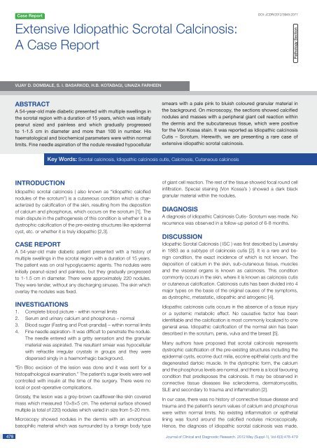
Grossly, the lesion was a grey-brown cauliflower-like skin covered<br />
mass which measured 10×8×5 cm. The external surface showed<br />
multiple (a total of 220) nodules which varied in size from 5–20 mm.<br />
Microscopy showed nodules in the dermis with an amorphous<br />
basophilic material which was surrounded by a foreign body type<br />
of giant cell reaction. The rest of the tissue showed focal round cell<br />
infiltration. Special staining (Von Kossa’s ) showed a dark black<br />
granular material within the nodules.<br />
Diagnosis<br />
A diagnosis of idiopathic <strong>Calcinosis</strong> Cutis- Scrotum was made. No<br />
recurrence was observed in a follow-up period of 6-8 months.<br />
Discussion<br />
<strong>Idiopathic</strong> <strong>Scrotal</strong> <strong>Calcinosis</strong> ( ISC ) was first described by Lewinsky<br />
in 1883 as a subtype of calcinosis cutis [2]. It is a rare and benign<br />
condition, the exact incidence of which is not known. The<br />
deposition of calcium in the skin, sub-cutaneous tissue, muscles<br />
and the visceral organs is known as calcinosis. This condition<br />
commonly occurs in the skin, where it is known as calcinosis cutis<br />
or cutaneous calcification. <strong>Calcinosis</strong> cutis has been divided into 4<br />
major types on the basis of the original causes of the symptoms,<br />
as dystrophic, metastatic, idiopathic and iatrogenic [4].<br />
<strong>Idiopathic</strong> calcinosis cutis occurs in the absence of a tissue injury<br />
or a systemic metabolic effect. No causative factor has been<br />
identifiable and the calcification is most commonly localized to one<br />
general area. <strong>Idiopathic</strong> calcification of the normal skin has been<br />
described in the scrotum, penis, vulva and the breast [5].<br />
Many authors have proposed that scrotal calcinosis represents<br />
dystrophic calcification of the pre-existing structures including the<br />
epidermal cysts, eccrine duct milia, eccrine epithelial cysts and the<br />
degenerated dartoic muscle. In the dystrophic form, the calcium<br />
and the phosphorus levels are normal, and there is a local favouring<br />
condition that predisposes the calcinosis. It may be observed in<br />
connective tissue diseases like scleroderma, dermatomyositis,<br />
SLE and secondary to trauma and inflammation [2].<br />
In our case, there was no history of connective tissue disease and<br />
trauma and the patient’s serum values of calcium and phosphorus<br />
were within normal limits. No existing inflammation or epithelial<br />
lining was found around the calcified nodules microscopically.<br />
Hence, the diagnosis of idiopathic scrotal calcinosis was made.<br />
Journal of Clinical and Diagnostic Research. 2012 May (Suppl-1), Vol-6(3):478-479
www.jcdr.net<br />
Vijay D. Dombale et al., <strong>Extensive</strong> <strong>Idiopathic</strong> <strong>Scrotal</strong> <strong>Calcinosis</strong><br />
[Table/Fig-1]: Multiple scrotal nodules with a<br />
buried penis<br />
[Table/Fig-2]: FNAC showing refractile crystals<br />
[Table/Fig-3]: Gross picture showing<br />
multiple nodules and cut section of single<br />
nodule showing chalky white areas<br />
[Table/Fig-4]: Microscopy showing nodule in<br />
dermis surrounded by foreign body giant cell<br />
reaction<br />
[Table/Fig-5]: Von Kossa stain demonstrating<br />
black granular deposits of calcium within the<br />
dermis<br />
The result of the surgical excision was satisfactory and there was<br />
no recurrence in a follow-up period of 6 months. The uniqueness<br />
of this case lies in the extensive involvement of the scrotum with<br />
220 nodules, the latest case which has been reported having 51<br />
nodules as was reported by Song et al. [6].<br />
References<br />
[1] http:// en.wikipedia.org/wiki/idiopathic_<strong>Scrotal</strong>_calcinosis.<br />
[2] Kelten EC, Akbulut M, Colakoglu N, Bayramoglu H, Duzcan SE.<br />
<strong>Scrotal</strong> <strong>Calcinosis</strong>: is it idiopathic or dystrophic? Aegean Pathology<br />
Journal. 2005; 2:4-7.<br />
[3] Hicheri J, Badri T, Fazaa B, Zermani R, Kourda N, Jilani SB, et al.<br />
<strong>Scrotal</strong> <strong>Calcinosis</strong>: Pathogenesis and case report. Acta Dermatoven<br />
APA. 2005; 4(2):53-56.<br />
[4] http://www.dermnetnz.org/systemic/calcinosis.html.<br />
[5] Kanishwar VS, Waghmare RS, Puranik GV. <strong>Calcinosis</strong> Cutis in the<br />
CREST Syndrome. Bombay Hospital Journal. 2010; 52(1):108-110.<br />
[6] Song DH, Lee KH, Kang WH. <strong>Idiopathic</strong> calcinosis of the scrotum:<br />
histopathologic observations of fifty-one nodules. Journal of the<br />
American Academy of Dermatology. 1990 Jul;23(1):150-51.<br />
AUTHOR(S):<br />
1. Dr. Vijay D. Dombale<br />
2. Dr. S. I. Basarkod<br />
3. Dr. H.B. Kotabagi<br />
4. Unaiza Farheen<br />
PARTICULARS OF CONTRIBUTORS:<br />
1. Department of Pathology, S. Nijalingappa Medical College,<br />
Bagalkot, Karnataka, India.<br />
2. Department of Surgery, S. Nijalingappa Medical College,<br />
Bagalkot, Karnataka, India.<br />
3. Department of Pathology, S. Nijalingappa Medical College,<br />
Bagalkot, Karnataka, India.<br />
4. Department of Pathology, S. Nijalingappa Medical College,<br />
Bagalkot, Karnataka, India.<br />
NAME, ADDRESS, E-MAIL ID OF THE CORRESPONDING<br />
AUTHOR:<br />
Dr. Vijay.D.Dombale<br />
Professor and HOD, Department of Pathology,<br />
S.N.Medical college, Bagalkot-587101, Karnataka, India.<br />
Ph: 09480598017<br />
Mail: drvijay@gmail.com, unaiza85f@rediffmail.com<br />
Financial OR OTHER COMPETING INTERESTS:<br />
None.<br />
Date of Submission: Aug 25, 2011<br />
Date of Peer Review: Nov 03, 2012<br />
Date of Acceptance: Jan 12, 2012<br />
Date of Publishing: May 01, 2012<br />
Journal of Clinical and Diagnostic Research. 2012 May (Suppl-1), Vol-6(3):478-479 479