Fine Needle Aspiration Cytology of Intra-Abdominal Lesions - JCDR
Fine Needle Aspiration Cytology of Intra-Abdominal Lesions - JCDR
Fine Needle Aspiration Cytology of Intra-Abdominal Lesions - JCDR
Create successful ePaper yourself
Turn your PDF publications into a flip-book with our unique Google optimized e-Paper software.
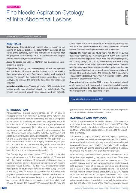
Original Article<strong>Fine</strong> <strong>Needle</strong> <strong>Aspiration</strong> <strong>Cytology</strong><strong>of</strong> <strong>Intra</strong>-<strong>Abdominal</strong> <strong>Lesions</strong>Pathology SectionSIDHALINGREDDY, SAINATH K. ANDOLAAbstractBackground: <strong>Intra</strong>-abdominal masses always remain as anenigma in surgical practice. A documentary evidence <strong>of</strong> thenature <strong>of</strong> the pathology before the institution <strong>of</strong> therapy and forthe prognosis is mandatory. FNAC is a substitute for surgicalprocedures like diagnostic laparotomy.Aims: To assess the utility <strong>of</strong> FNAC in the diagnosis <strong>of</strong> intraabdominallesions.Objectives: To study the cytomorphological features, age andsex distribution <strong>of</strong> intra-abdominal lesions and to categorizethem organwise and as inflammatory, benign and malignantlesions. To classify the malignant lesions according to theircell type. To evaluate the sensitivity, specificity and diagnosticaccuracy.Materials and Methods: The study included 245 intra-abdominallesions which were detected clinically or radiologically. Thelesions were divided clinically into palpable and non-palpablelumps. USG or CT were used for all the non-palpable lesionsand for a few palpable lesions and direct in selected palpablelesions. Giemsa’s and Papanicolaou’s stains were used.Results: The mean age was 45.16 years, with M:F <strong>of</strong> 1:1.3. Thediagnostic yield was 92.1% in USG guided, 100% in CT guidedand 95% in direct aspiration. There were 148 (60.3%) malignant,55 (22.4%) benign, 25 (10.2%) inflammatory and one (0.6%)suspicious lesions and 16 (6.5%) unsatisfactory smears. The liverand the ovary were the most common sites . Adenocarcinomasand hepatocellular carcinomas were the most common malignantlesions. This study showed 94.1% sensitivity, 100% specificity,100% positive predictive value, 92.3% negative predictive valueand 96.5% diagnostic accuracy .Conclusion: <strong>Intra</strong>-abdominal FNA is a simple, economical anda safe procedure with high sensitivity, specificity and diagnosticaccuracy and it can be utilized as a pre-operative procedure forthe management <strong>of</strong> intra-abdominal lesions.Key Words: <strong>Intra</strong>-abdominal, FNA758INtroduction<strong>Intra</strong>-abdominal masses always remain as an enigma insurgical practice. A documentary evidence <strong>of</strong> the nature <strong>of</strong> thepathology before the institution <strong>of</strong> therapy and also for prognosisis mandatory. In a majority <strong>of</strong> cases, the diagnosis which isobtained by FNAC, is the substitute for surgical procedureslike diagnostic laparotomy [1,2]. Most <strong>of</strong> the intra abdominalmasses are non – palpable and even if they are palpable, theidea <strong>of</strong> their size and shape and the extent <strong>of</strong> the lesion is notpossible. Therefore, various imaging modalities like fluoroscopy,CT and USG are used as a guide for fine needle aspiration [2].Most studies have shown it as a highly sensitive, highly specific,accurate and a cost effective diagnostic procedure witha negligible complication rate [2-16]. Uncorrectable severecoagulopathy is an absolute contraindication [17].The FNA cytology was shown to be 100% specific for the diagnosis<strong>of</strong> malignancy [4,8]. The non –availability <strong>of</strong> CT, coupledwith the higher incidence <strong>of</strong> advanced malignancy due topublic awareness and overburdened surgical units with meagreresources require the USG – guided FNAC procedure for cancermanagement in developing countries like India [6,18]. The aim<strong>of</strong> our study was to assess its usefulness as a pre-operativediagnostic procedure in management <strong>of</strong> intra-abdominal lesions.Our objectives were to assess the cytomorphological features,age and sex distribution <strong>of</strong> the patients with intra abdominallesions, to classify the malignant lesions according to their celltype and to evaluate the sensitivity, specificity and the diagnosticaccuracy in different lesions wherever possible.MATERIALS AND METHODSThis study was carried out in the Department <strong>of</strong> Pathology fora period <strong>of</strong> three years (36 months) from June–2005 to May2008. Patients with intra-abdominal lesions which were detectedclinically or under radiological guidance, presented to the Department<strong>of</strong> <strong>Cytology</strong>.<strong>Intra</strong>-abdominal organs including the liver, spleen, pancreas,stomach, gallbladder, the small and large intestines, the omentum,mesentery, the retroperitoneum, kidney, adrenals, lymph nodes, s<strong>of</strong>ttissues and the ovary were included in the study. Parietal swellingsarising from the skin and the abdominal wall, the uterus, the cervix,the prostate and the bone were excluded from the study.After a thorough clinical examination, consent was obtained fromthe patients after explaining the procedure to them. The caseswere divided clinically into palpable and non palpable lumps. Theselected palpable lesions were subjected to direct aspiration andUSG guidance in case <strong>of</strong> on palpable and deep seated lesionsand in case <strong>of</strong> a few selected palpable lesions. The puncturesite was marked. Under aseptic precautions, a 22–23G needlefor superficial lumps and a lumbar puncture needle <strong>of</strong> the samethickness for deep seated lumps, which was fitted with a 10mlsyringe, was introduced immediately under radiological guidanceand the aspiration was done under negative pressure. On anJournal <strong>of</strong> Clinical and Diagnostic Research. 2011 August, Vol-5(4): 758-765
www.jcdr.netSidhalingreddy and Sainath K. Andola, <strong>Fine</strong> needle aspiration cytologyInflammatory Benign Malignant Suspicious UnsatisfactoryAge M F M F M F M F M F Total %1–10 – 1 2 – 2 5 – – 1 – 11 4.811–20 4 1 2 6 5 5 – – – – 23 9.421–30 3 1 – 7 6 4 – – 1 1 23 9.431–40 1 1 0 14 5 14 – – – 2 37 15.141–50 2 5 0 10 19 16 – – 1 1 54 2251–60 1 2 2 8 17 19 – – 3 3 55 22.461–70 – 1 2 1 12 9 – – 1 2 28 11.371–80 1 1 – 1 6 – – – – – 9 3.681–90 – – – – 4 – 1 – – – 5 2Total 12 13 8 47 76 72 1 0 7 9 245% 4.9 5.3 3.3 19.2 31 29.4 0.4 0 2.8 3.7 100Total (%) 25 (10.2%) 55(22.5%) 148(60.4%) 1(0.4%) 16(6.5%) 245 (100%)[Table/Fig-1]: Age and Sex distribution <strong>of</strong> <strong>Intra</strong>-abdominal <strong>Lesions</strong>average, two to three needle passes were made in each case toobtain adequate material. The sample was expelled onto slides,air-dried and stained with Giemsa or it was fixed in 95% ethanoland stained with Papanicolaou’s stain. Special stains were usedwherever required.The cases were analyzed, based on the cytological features. Thefinal diagnosis was arrived at in corroboration with the clinicaland the radiological features. The smears were classified asinflammatory, benign, malignant, suspicious <strong>of</strong> malignancy andunsatisfactory for interpretation.RESULTSDuring the study period, 2624 fine needle aspirations were performed,<strong>of</strong> which 234 cases were intra abdominal lesions, accounting for8.9% <strong>of</strong> the total cases. There were 245 lesions in 234 patients. Therewere 147 palpable and 98 non-palpable lesions. Histopathologicalcorrelation and confirmation was available in 29 cases.Out <strong>of</strong> the 234 cases, there were 101 males and 133 females witha male to female ratio <strong>of</strong> 1:1.3. The youngest patient in the studywas 20 days old and the oldest was 88 years old. A majority <strong>of</strong> thepatients i.e. 146 (59.6%) were in the age group <strong>of</strong> 30-60 years, out<strong>of</strong> 245 lesions. The mean age was 45.16 years (Standard deviation– 18.48). Among 104 lesions in the male patients, a majority weremalignant, accounting for 76 (73.1%) lesions and 12 (11.5%)lesions were inflammatory lesions, eight (7.7%) lesions were benignand one (0.9%) was suspicious for malignancy. In seven (6.8%)cases, the smears were unsatisfactory for evaluation. Among the141 lesions in the female patients, 72 (51.1%) were malignant, 47(33.3%) were benign and 13 (9.2%) were inflammatory. In nine(6.4%) cases, the smears were unsatisfactory for evaluation. Out<strong>of</strong> 245 lesions in the 234 patients, 148 (60.3%) were malignant,55 (22.4%) were benign, 25 (10.2%) were inflammatory and one(0.6%) was a suspicious lesion. There were about 16 (6.5%)unsatisfactory smears. The benign lesions were more common infemales than in males, whereas the malignant lesions had a slightmale preponderance. The incidence <strong>of</strong> the lesions increased inboth the sexes after 30 years [Table/Fig-1].Out <strong>of</strong> 245 lesions, 164 were aspirated under ultrasonographicalguidance and one was aspirated under computed tomographicguidance. Of the 80 cases which were properly selected, thepalpable cases were aspirated directly without any guidance. Thediagnostic yield <strong>of</strong> USG was 92.7% i.e. out <strong>of</strong> 163 USG guidedprocedures and adequate material was obtained in 152 cases. Inthe CT guided procedure, the diagnostic yield was 100%. It was95% in the direct unguided procedure, which was higher than that<strong>of</strong> the USG guided procedure. Overall, the diagnostic yield was93.5% in the 245 lesions [Table/Fig-2].In the present study, most <strong>of</strong> the aspirates were cellular (41.6%) andhaemorrhagic cellular (28.6%). It was a fluid aspirate in 11.8% lesions,followed by a necrotic aspirate in 6.5% lesions, a purulent aspirate in2.9% lesions and a cellular haemorrhagic aspirate in 8.6% lesions.Out <strong>of</strong> the 21 haemorrhagic and acellular haemorrhagic aspirates,most were (16 cases) unsatisfactory for evaluation and five wereinterpreted as hemangiomas. Of the 245 lesions, the aspirate wassatisfactory in 229 (93.5%) lesions. Unsatisfactory aspirates wereobtained in 16 (6.5%) lesions [Table/Fig-3].A majority <strong>of</strong> the lesions were located in the liver and most <strong>of</strong>them were malignant lesions. The most common malignant lesionin the liver was hepatocellular carcinoma (HCC) (34), followedby metastatic carcinoma (25). In seven cases, we could notMethodOrgansNo. <strong>of</strong> CasesInflammatoryBenign SuspiciousMalignantTotal %Liver 11 8 1 67 87 38Gallbladder – – – 6 6 2.6Stomach – – – 2 2 0.9Large bowel 1 – – 2 3 1.3Pancreas – 2 – 5 7 3Spleen – 1 – 2 3 1.3Kidney – – – 12 12 5.2Adrenal – 1 – 2 3 1.3Ovary 2 33 – 13 48 21.1S<strong>of</strong>t tissue – 1 – 8 9 3.9Omentum – – – 4 4 1.8Mesentery – 1 – – 1 0.4Lymphnodes 9 – – 9 18 7.9Unclassified 2 8 – 16 26 11.3Total 25 55 1 148 229 100[Table/Fig-3]: Organ Distribution <strong>of</strong> <strong>Lesions</strong>No. <strong>of</strong> CasesDiagnostic yieldPercentageUSG 164 152 92.7CT 1 1 100Unguided 80 76 95Total 245 229 93.5[Table/Fig-2]: Type <strong>of</strong> Radiological Guidance and Diagnostic YieldJournal <strong>of</strong> Clinical and Diagnostic Research. 2011 August, Vol-5(4): 758-765 759
Sidhalingreddy and Sainath K. Andola, <strong>Fine</strong> needle aspiration cytologywww.jcdr.netMalignant <strong>Lesions</strong>Hepatocellularcarcinoma0–10 11–20 21–40 41–60 61–80 > 80M F M F M F M F M F M F Total %– – – 1 2 1 14 6 8 1 1 – 34 23Adenocarcinoma – – – – 1 6 9 16 4 1 2 – 39 26.3Renal cell carcinoma – – – – 1 2 2 1 1 – – – 7 4.7Adreno cortical– – – – – – 1 – – – – – 1 0.7carcinomaPheochromocytoma – – – – – 1 – – – – – – 1 0.7Serous cystadeno – – – – – 1 – 4 – 2 – – 7 4.7carcinomaMalignant granulosa – – – – – – – 1 – – – – 1 0.7cell tumorMalignant germ cell – – – 1 – – – – – – – – 1 0.7tumorDysgerminoma/– – – 1 1 – – – – – – – 2 1.35SeminomaCholangio carcinoma – – – – – – – 1 – – – – 1 0.7Hodgkin’s lymphoma – – 2 – – – – – – – – – 2 1.35Non–Hodgkin– – – – – 1 – – 1 – – – 2 1.35lymphomaNephroblastoma 1 1 2 – – – – – – – – – 4 2.7Rhabdomyo sarcoma – 1 – – 1 – – – – – – – 2 1.35Malignant round cell – – 2 – 1 – – – – – – – 3 2tumorSquamous cell– – – – – 1 – – – – – – 1 0.7carcinomaSmall cell carcinoma – – – – – – – – 2 1 – – 3 2(metastatic to liver)Anaplastic carcinoma – – – – – 2 – – – – – – 2 1.35(Metastatic)Poorly differentiated – – – 1 3 4 11 5 1 4 – – 29 19.6carcinomaPleomorphic sarcoma – 1 – – – – 1 1 – 1 – – 4 2.7Malignant– 1 – – – – – – – – – – 1 0.7undifferentiated tumorGanglioneuroblastoma l – 1 – – – – – – – – – – 1 0.7Total 1 5 6 4 10 19 38 35 17 10 3 0 148Total 6 10 29 73 27 3 148% 0.7 3.4 4 2.7 6.8 12.8 25.7 23.6 11.5 6.8 2 0% 4.1 6.7 19.6 49.3 18.3 2 100[Table/Fig-4]: Age and Sex Distribution <strong>of</strong> Malignant <strong>Lesions</strong> According to Cell Type760differentiate between primary HCC and the metastatic lesions andthese were labeled as poorly differentiated carcinoma. One casewas diagnosed as cholangiocarcinoma [Table/Fig-2] in a 50 yearsold female. The next most common site was the ovary (48), wheremost <strong>of</strong> the lesions were benign lesions (33). Other common organswhich were involved were the lymph nodes (18), kidney (12), thegallbladder (6) and pancreas (6). There were two cystic lesions inthe pancreas and four adenocarcinomas <strong>of</strong> the pancreas. In thegall bladder, all the lesions were adenocarcinomas.Abscess constituted the most common inflammatory lesion. Out<strong>of</strong> eight abscesses, six were located in liver, one in lymph nodesand one in the appendix. There were five tubercular lymphadenitis[Table/Fig-3] and three reactive lymphadenitis cases. Four cases <strong>of</strong>diffuse parenchymal lesions <strong>of</strong> the liver were seen.Most <strong>of</strong> the benign lesions i.e., 33 lesions were located in the ovaryand most <strong>of</strong> them (11) were diagnosed as cystadenomas. Amongthe remaining benign lesions, nine (16.5%) lesions were diagnosedas cyst contents, six (10.9%) as serous cystadenomas, three (5.4%)as mucinous cystadenomas, one as a simple serous cyst, one asa twisted ovarian cyst, three (5.4%) as benign teratomas, one asan ovarian fibroma and one as a benign, mixed epithelial stromaltumour [Table/Fig-4]. Two cystic lesions were in the pancreasand one was in the liver (calcified cyst content). There were fivehemangiomas, all <strong>of</strong> which were located in the liver, out <strong>of</strong> 55 casesi.e. 9.1% <strong>of</strong> the benign lesions. There was one angiomyolipoma,one adrenal cortical adenoma, one benign trophoblastic lesion andone mucinous cyst <strong>of</strong> the mesentery and the colon each [Table/Fig-5].Adenocarcinomas [Table/Fig-6] were the most common malignantlesions, followed by hepatocellular carcinomas. Adenocarcinomaswere more common in females (23) than in males (16). Hepatocellularcarcinomas were more common in males (25) than in females(nine). Lymphomas [Table/Fig-7], renal cell carcinomas [Table/Fig-8], nephroblastomas [Table/Fig-9] and small cell carcinomaswere more common in males than in females. Pleomorphicsarcomas were more common in females (three) than in the males(one). Serous cystadenocarcinoma (seven) was the most commonmalignant lesion in the ovary, followed by one malignant granulosacell tumour [Table/Fig-10] and one dysgerminoma [Table/Fig-11]Journal <strong>of</strong> Clinical and Diagnostic Research. 2011 August, Vol-5(4): 758-765
www.jcdr.netOrganBiradar et al 141994 ( %)Zawar MPet al 6 2007 (%)Present Study(%)Liver 36 45 38Gallbladder – 2.5 2.6Stomach 10 5 0.9Large Bowel 20 17.5 1.3Pancreas 4 2.5 3Spleen – 5 1.3Kidney 6 20 5.2Adrenal – – 1.3Ovary – – 21.1S<strong>of</strong>t Tissue – – 3.9Omentum – – 1.8Mesentery – – 0.4Lymphnodes 6 2.5 7.9Unclassified 8 – 11.3[Table/Fig-5]: Organ distribution <strong>of</strong> <strong>Intra</strong>-<strong>Abdominal</strong> Malignancy –Comparative Analysis.Type <strong>of</strong>LesionBiradaret al 141994 (%)Aftab A.Khanet al 41995 (%)Shamshadet al 172006 (%)Presentstudy (%)10.2Inflammatory 32 630.5Benign 2 0 22.4Malignant 52 88 57.5 60.3Suspicious 0 0 5.5 0.6Unsatisfactory 14 6 6.5 6.5[Table/Fig-6]: Distribution <strong>of</strong> <strong>Intra</strong>-<strong>Abdominal</strong> <strong>Lesions</strong> – ComparativeAnalysis.StudyNo. <strong>of</strong>FNACsSensitivity%Specificity%DiagnosticAccuracy %Sundaram 204 96.3 100 97et al 75 1982Lees et al 76 454 77 100 83.951985Civardi– 95.6 100 97.6et al 77 1988Govind 500 71.4 55.6 77.5Krishnaet al 13 1993Joao236 87 100 100Nobregaet al 7 1994Aftab A. 50 94 100 94Khan et al4 1995Nautiyal S 72 – – 87.5et al 5 2004Shamshad 200 94.11 100 95.7et al 17 2006Zawar MP 40 – – 90et al 6 2007Present 245 94.1 100 96.5study 2008[Table/Fig-7]: Statistical Results – Comparative Analysisand the remaining were metastatic adenocarcinomas. A majority <strong>of</strong>the adenocarcinomas (25) and the hepatocellular carcinomas (20)were seen in the age group <strong>of</strong> 41-60 years. The youngest patientwho was affected by HCC was an 18 years old female and theoldest one was an 85 years old male. Adenocarcinomas constitutedthe most common metastatic lesions in the liver, followed by smallSidhalingreddy and Sainath K. Andola, <strong>Fine</strong> needle aspiration cytologycell carcinomas (three). All nephroblastomas, one malignant smallround cell tumour and one rhabdomyosarcoma were seen belowthe 20 years age group. One rhabdomyosarcoma was seen in a 40years old male [Table/Fig-5].The HbsAg test was done in nine <strong>of</strong> the 32 hepatocellular carcinomacases, <strong>of</strong> which six were positive and three were negative. 66.7%<strong>of</strong> HbsAg positivity was seen in hepatocellular carcinomas.Histopathological correlation and confirmation was available in29 cases. Out <strong>of</strong> the 13 benign cases, seven were confirmed byhistopathological examination. All mucinous and serous cystadenomaswhich were diagnosed cytologically were confirmedhistopathologically, except one mucinous cystadenoma which wasdiagnosed as papillary cystaden<strong>of</strong>ibroma. One serous cystadenomaturned out to be serous cystadenocarcinoma by histopathologicalexamination. The benign, mixed epithelial stromal tumour turnedout to be a Brenner tumour <strong>of</strong> the ovary by histopathologicalexamination. One twisted ovarian cyst and one ovarian fibromawere confirmed histologically. One cystadenoma with haemorrhageand one spindle cell tumour turned out to be a twisted ovarian cystand a leiomyoma <strong>of</strong> the retroperitoneum respectively.11 cases were confirmed histologically out <strong>of</strong> the 16 malignantcases. All serous cystadenocarcinomas <strong>of</strong> the ovary, one malignantgranulosa cell tumour, one clear cell carcinoma <strong>of</strong> the kidney, twonephroblastomas, one gallbladder adenocarcinoma, one largeintestine adenocarcinoma, two adenocarcinomas <strong>of</strong> the ovaryand one ganglioneuroblastoma [Table/Fig: 8-13] were confirmedhistologically. One case <strong>of</strong> clear cell carcinoma <strong>of</strong> the kidneyturned out to be adrenocortical carcinoma by histopathologicalexamination. One case <strong>of</strong> malignant undifferentiated tumour turnedout to be malignant, mixed epithelial cell tumour with dysgerminomaand embryonal carcinoma and one case <strong>of</strong> carcinoma withtuberculosis turned out to be a metastatic adenocarcinoma <strong>of</strong>the ovary by histopathological examination. One case who presentedwith a suprapubic abdominal mass was diagnosed tohave adenocarcinoma, cytologically. But the histological diagnosis<strong>of</strong> the cervical biopsy was squamous cell carcinoma. Onecase <strong>of</strong> metastatic seminoma <strong>of</strong> the lymphnodes which wasdiagnosed cytologically, turned out to be teratocarcinoma by thehistopathological examination <strong>of</strong> the orchidectomy specimen.DISCUSSIONFNAC is a proven technique for the diagnostic evaluation <strong>of</strong> intraabdominallesions. The diagnostic yield which was obtained byUSG was 92.7%, by CT it was 100% and for direct aspiration itwas 95%. Overall, the diagnostic yield was 93.5%. Nautiyal S.,Mishra RK., and Sharma SP,2 in 2004, found a diagnostic yield<strong>of</strong> 64.81% with direct aspiration <strong>of</strong> the palpable lumps and adiagnostic yield <strong>of</strong> 93.06% with USG guided FNAC which wasdone for both palpable and non-palpable lesions. Nyman et al,[12]in 1995, found a diagnostic yield <strong>of</strong> 64% with USG guided FNAC.In comparison with previous studies, the present study was foundto have more diagnostic yield, irrespective <strong>of</strong> whether it was director guided. In the present study, the diagnostic yield was more withdirect aspiration than with USG guided FNAC. This could be dueto the careful and proper selection <strong>of</strong> the cases for direct aspirationand as most <strong>of</strong> the lesions were superficial and easily palpable. A100% diagnostic yield which was obtained with CT-guided FNAC,was comparable to that which was obtained by Joseph T. et al [8].The age incidence in the present study ranged from 20 days to 88years with a majority <strong>of</strong> the cases being in the age group <strong>of</strong> 30-60Journal <strong>of</strong> Clinical and Diagnostic Research. 2011 August, Vol-5(4): 758-765 761
Sidhalingreddy and Sainath K. Andola, <strong>Fine</strong> needle aspiration cytologywww.jcdr.net[Table/Fig-8]: Hepatocellular carcinoma [Table/Fig-11]: Tubercular granuloma[Table/Fig-9]: Hepatocellular carcinoma with incidental filarial parasite [Table/Fig-12]: Brenner tumor[Table/Fig-10]: Cholangiocarcinoma [Giemsa, x400] [Table/Fig-13]: Mucinous adenocarcinoma762years (59.6%). The incidence <strong>of</strong> malignancy increased after the age<strong>of</strong> 40 years in males and after the age <strong>of</strong> 30 years in females witha peak incidence between the ages <strong>of</strong> 40-60 years, which wascomparable to the results which were obtained by Zawar MP., etal,[3]and Shamshad et al [14].The male to female ratio <strong>of</strong> 1:1.3 was in accordance with theobservations which were made by Shamshad et al [14], and JoaoNobrega et al [4]. But the observations which were made in thestudies by Zawar MP et al [3] , Govind Krishna et al [10], AftabA Khan et al [1], and Ennis and Mac Erlean, [6] showed a malepreponderance. This could be due to the inclusion <strong>of</strong> the ovary inthis study, as done by Shamshad et al [14].The most common organ which was involved in the presentstudy was the liver an observation which was similar tothe one made by Zawar M.P. et al [3], and Biradar et al [11]. Thenext most common site in the present study was the ovary. Butthe ovary was not included in the studies which were done byZawar M.P. et al [3], and Biradar et al [11]. In their studies, theJournal <strong>of</strong> Clinical and Diagnostic Research. 2011 August, Vol-5(4): 758-765
www.jcdr.netSidhalingreddy and Sainath K. Andola, <strong>Fine</strong> needle aspiration cytology[Table/Fig-14]: Hodgkin’s Lymphoma [Table/Fig-17]: Granulosa cell tumor[Table/Fig-15]: Renal cell carcinoma [Table/Fig-18]: Germ cell tumor[Table/Fig-16]: Nephroblastoma[Table/Fig-19]: Ganglioneuroblastomanext most common site was the large intestine. Biradar et al[14], had included the gallbladder, spleen, adrenal, s<strong>of</strong>t tissue,omentum and the mesentery in the unclassified category [Table/Fig-5].In the present study, malignant lesions constituted the mostcommon diagnostic category, which was in accordance with theobservations which were made by Biradar et al [11], Aftab A. Khanet al [1], and Shamshad et al [14] [Table/Fig-14].In the present study, we observed 6.5% unsatisfactory smears,which was similar to the observations which were made byShamshad et al [14], and Aftab A. Khan et al [1], who observed6.5% and 6% unsatisfactory smears. Biradar et al [11], hadobserved more unsatisfactory smears (14%) as compared to thosein our study [Table/Fig-14].Benign lesions showed a high female preponderance in the presentstudy, because cystic lesions <strong>of</strong> the ovary were most commonlyJournal <strong>of</strong> Clinical and Diagnostic Research. 2011 August, Vol-5(4): 758-765 763
Sidhalingreddy and Sainath K. Andola, <strong>Fine</strong> needle aspiration cytologywww.jcdr.netseen as benign lesions. There was no age or sex predilection forinflammatory lesions in the present study.In the present study, adenocarcinomas were the most commonmalignant cell type (26.3%), followed by hepatocellular carcinoma(23%), renal cell carcinoma (4.7%), serous cystadenocarcinoma(4.7%) and nephroblastoma (2.7%). Poorly differentiated carcinomasconstituted 19.6% <strong>of</strong> the lesions in the present study. Thiswas in accordance with the observations which were made byShamshad et al [14], and Aftab A. Khan et al [1], who observed87.1% and 34% poorly differentiated carcinomas respectively.The second most common malignant type in these studies washepatocellular carcinoma. In the liver, the most common malignantlesion was hepatocellular carcinoma (34), followed by metastaticcarcinoma (25). In the western literature, the most common hepaticmalignancy was metastasic carcinoma [4,6,18,19,20]. This couldbe because <strong>of</strong> the high prevalence <strong>of</strong> Hepatitis B infection and theconsumption <strong>of</strong> chutney which was made up <strong>of</strong> groundnuts, whichwas frequently contaminated with aflatoxins, in this geographicalregion. The observations <strong>of</strong> the present study were similar to those<strong>of</strong> Indian studies, where hepatocellular carcinoma constituted themost common hepatic malignancy [3]. Two studies which wereconducted in Kashmir showed observations which were similar tothat <strong>of</strong> the western literature [1,14].The liver constituted the major site for the malignant lesions, asobserved by Aftab A. Khan et al [1], Stewart et al [5], Zawar MPet al [3], Nyman et al [12], Ennis and MacErlean [6], Joao Nobregaet al [4], and Nautiyal et al [2]. But in an observation which wasmade by Shamshad et al [14], and Joseph et al [8], the most commonorgan sites for the malignant lesions were the gallbladder andthe pancreas respectively. Hepatocellular carcinoma was mostcommonly seen in males, in accordance with previous literaturereports [3,20]. Hepatocellular carcinomas and adenocarcinomashad a peak incidence in the age group between 40-60 years, in accordancewith the observations made by Shamshad et al [14], andZawar MP et al [3]. Malignant tumours which were seen before 20years <strong>of</strong> age, were nephroblastomas and other round cell tumours,Hodgkin’s lymphoma, dysgerminoma and ganglioneuroblastoma.This observation was comparable to that <strong>of</strong> the previous literaturereports [18,20].[Table/Fig-20]: Adrenocortical Carcinoma[Table/Fig-21]: Gastrointestinal Stromal TumorAlthough few studies have reported complications like mild localpain, bleeding and tumour seeding <strong>of</strong> the needle tract, a vastamount <strong>of</strong> literature supports the safety <strong>of</strong> FNAC. There was noreport on complications as a result <strong>of</strong> FNAC in the 20 paperswhich amounted to around 20,000 patients, including those <strong>of</strong> thepresent study.764The sensitivity <strong>of</strong> USG guided FNAC ranged from 71.4% to 96.3%.In the present study, it was 94.1%, which was comparable to that<strong>of</strong> most <strong>of</strong> the studies. All the studies observed 100% specificity,as was found in the present study also. The diagnostic accuracyin various studies ranged from 83.9% to 100%. The present studyfound a diagnostic accuracy <strong>of</strong> 96.5%, which was comparable tothat <strong>of</strong> most <strong>of</strong> the studies [Table/Fig-15-22].CONCLUSION<strong>Intra</strong>-abdominal FNA is a relatively simple, economical, quickand safe procedure for the diagnosis <strong>of</strong> intra-abdominal lesions.It not only helps in differentiating between inflammatory, benignand malignant lesions, but also in categorizing different malignantlesions. <strong>Intra</strong>-abdominal FNA is a reliable, sensitive and specificmethod with a high diagnostic accuracy for the diagnosis <strong>of</strong>[Table/Fig-22]: Pheochromocytomamalignant lesions. It can be utilized as a pre-operative procedurefor the management <strong>of</strong> all intra-abdominal lesions.REFERENCES[1] Aftab Khan A., Jan GM., Wani NA. <strong>Fine</strong> <strong>Needle</strong> <strong>Aspiration</strong> <strong>of</strong> <strong>Intra</strong>abdominalmasses for cytodiagnosis. J. Indian Med Assoc 1996;94(5):167-69.[2] Nautiyal S., Mishra RK, Sharma SP., Routine and ultrasound guidedFNAC <strong>of</strong> intra abdominal lumps – A comparative study. Journal <strong>of</strong><strong>Cytology</strong> 2004;21(3):129-132.Journal <strong>of</strong> Clinical and Diagnostic Research. 2011 August, Vol-5(4): 758-765
www.jcdr.net[3] Dr. Zawar M.P., Dr. Bolde S., Dr. Shete S.S. Correlative study <strong>of</strong> fineneedle aspiration cytology and histology in intra-abdominal lumps.SMJ 2007;4.[4] Joao Nobrega and Guimaraes dos Santos. Aspirative cytology withfine-needle in the abdomen, retroperitoneum and pelvic cavity: a sevenyear experience <strong>of</strong> the Portuguese Institute <strong>of</strong> Oncology, Centre <strong>of</strong>Porto. European Journal <strong>of</strong> Surgical Oncology. 1994;20:37-42.[5] Stewart CJR, Coldewey J., Stewart IS. Comparison <strong>of</strong> fine needleaspiration cytology and needle core biopsy in the diagnosis <strong>of</strong> radiologicallydetected abdominal lesions. J. Clin. Pathol. 2002;55:93-97.[6] Mary Ennis G., MacErlean DP. Percutaneous <strong>Aspiration</strong> Biopsy <strong>of</strong>Abdomen and Retroperitoneum. Radiology 1980;31:611-16.[7] Raul Pereiras V., Walter Meiers, et al. Fluoroscopically guided thinneedle aspiration biopsy <strong>of</strong> the abdomen and retroperitoneum.Am J Roentgenol 1978;131:197-202.[8] Joseph T., Ferrucci Jr. MD., Jack Wittenberg MD. CT Biopsy <strong>of</strong><strong>Abdominal</strong> Tumors: Aids for Lesion Localization. Radiology 1978;129:739-744.[9] Vijaya Reddy B., Paolo Gattuso, Kurian Abraham P., Rogelio Moncada,Melanie Castelli J. Computed Tomography-guided fine needle aspirationbiopsy <strong>of</strong> deep-seated lesions – A four-year experience. Acta Cytol.1991;35(6):753-755.[10] Govind Krishna SR., Ananthakrishanan N., Narasimhan R., Veliath AJ.Accuracy <strong>of</strong> <strong>Fine</strong> <strong>Needle</strong> <strong>Aspiration</strong> <strong>Cytology</strong> <strong>of</strong> <strong>Abdominal</strong> Masseswithout Radiological Guidance. Indian J. Pathol. Microbiol. 1993;36(4):442-52.Sidhalingreddy and Sainath K. Andola, <strong>Fine</strong> needle aspiration cytology[11] VB. Biradar et al. A study <strong>of</strong> fine needle aspiration cytology in abdominallump (dissertation). Gulbarga: University <strong>of</strong> Gulbarga, 1994.[12] Nyman RS., Cappelen-Smith J., et al. Yield and complications inultrasound-guided biopsy <strong>of</strong> abdominal lesions. Acta Radiologica1995;36:485-90.[13] David CC., Darshana Jhala, et al. Endoscpoic Ultrasound-Guided <strong>Fine</strong>-<strong>Needle</strong> <strong>Aspiration</strong> Biopsy. Cancer 2002;96(4):232-239.[14] S. Shamshad Ahmed, Kafil Akhtar, S. Shakeel Akhtar et al. Ultrasoundguided fine needle aspiration biopsy <strong>of</strong> abdominal masses. JK Science.2006; 8(4):200-204.[15] Dilip Das K., Chandra Pant S., et al. <strong>Fine</strong> needle aspiration diagnosis <strong>of</strong>intra-thoracic and intra abdominal lesions: Review <strong>of</strong> experience in thepediatric age group. Diagnostic Cytopathology 2006;9(4):383-93.[16] Ahmed SS., Akhtar K., Akhtar SS. Nasir A., Khalid M., Mansoor T.Ultrasound Guided <strong>Fine</strong> <strong>Needle</strong> <strong>Aspiration</strong> Biopsy <strong>of</strong> RetroperitonealMasses. J <strong>of</strong> <strong>Cytology</strong> 2007;24(1):41-45.[17] Scott Gazelle G., John Haaga R. Guided percutaneous biopsy <strong>of</strong>intraabdominal lesions. AJR 1989;153:929-35.[18] Svante R. Orell, Gregory F. Sterrett, Darrel Whitaker. <strong>Fine</strong> needleaspiration cytology. 4th ed. 2005; Churchill Livingstone.[19] Barbara A. Centeno. Pathology <strong>of</strong> liver metastases. Cancer Control.2006;13(1):13-24.[20] Juan Rosai, VJ. Desmet, GO. Nelson. Rosai and Ackerman’s surgicalpathology. 9th ed. 2005; Mosby.AUTHOR(S):1. Dr. Sidhalingreddy2. Dr. Sainath K. AndolaNAME OF DEPARTMENT(S)/INSTITUTION(S) TO WHICHTHE WORK IS ATTRIBUTED:Department <strong>of</strong> Pathology, M.R.Medical college, Gulbarga,KarnatakaNAME, ADDRESS, TELEPHONE, E-MAIL ID OF THECORRESPONDING AUTHOR:Dr. SidhalingreddyAssistant pr<strong>of</strong>essor, Department <strong>of</strong> pathologyS.N.Medical college ,Navanagar, Bagalkot -587102E-mail: drssreddybenoor@gmail.comMobile: 919886689521DECLARATION ON COMPETING INTERESTS:No competing Interests.Date <strong>of</strong> Submission: Jan 04, 2011Date <strong>of</strong> Peer Review: Apr 12, 2011Date <strong>of</strong> Acceptance: Apr 15, 2011Date <strong>of</strong> Publishing: Aug 08, 2011Journal <strong>of</strong> Clinical and Diagnostic Research. 2011 August, Vol-5(4): 758-765 765


