Fine Needle Aspiration Cytology of Intra-Abdominal Lesions - JCDR
Fine Needle Aspiration Cytology of Intra-Abdominal Lesions - JCDR
Fine Needle Aspiration Cytology of Intra-Abdominal Lesions - JCDR

You also want an ePaper? Increase the reach of your titles
YUMPU automatically turns print PDFs into web optimized ePapers that Google loves.
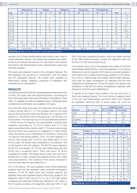
www.jcdr.netSidhalingreddy and Sainath K. Andola, <strong>Fine</strong> needle aspiration cytologyInflammatory Benign Malignant Suspicious UnsatisfactoryAge M F M F M F M F M F Total %1–10 – 1 2 – 2 5 – – 1 – 11 4.811–20 4 1 2 6 5 5 – – – – 23 9.421–30 3 1 – 7 6 4 – – 1 1 23 9.431–40 1 1 0 14 5 14 – – – 2 37 15.141–50 2 5 0 10 19 16 – – 1 1 54 2251–60 1 2 2 8 17 19 – – 3 3 55 22.461–70 – 1 2 1 12 9 – – 1 2 28 11.371–80 1 1 – 1 6 – – – – – 9 3.681–90 – – – – 4 – 1 – – – 5 2Total 12 13 8 47 76 72 1 0 7 9 245% 4.9 5.3 3.3 19.2 31 29.4 0.4 0 2.8 3.7 100Total (%) 25 (10.2%) 55(22.5%) 148(60.4%) 1(0.4%) 16(6.5%) 245 (100%)[Table/Fig-1]: Age and Sex distribution <strong>of</strong> <strong>Intra</strong>-abdominal <strong>Lesions</strong>average, two to three needle passes were made in each case toobtain adequate material. The sample was expelled onto slides,air-dried and stained with Giemsa or it was fixed in 95% ethanoland stained with Papanicolaou’s stain. Special stains were usedwherever required.The cases were analyzed, based on the cytological features. Thefinal diagnosis was arrived at in corroboration with the clinicaland the radiological features. The smears were classified asinflammatory, benign, malignant, suspicious <strong>of</strong> malignancy andunsatisfactory for interpretation.RESULTSDuring the study period, 2624 fine needle aspirations were performed,<strong>of</strong> which 234 cases were intra abdominal lesions, accounting for8.9% <strong>of</strong> the total cases. There were 245 lesions in 234 patients. Therewere 147 palpable and 98 non-palpable lesions. Histopathologicalcorrelation and confirmation was available in 29 cases.Out <strong>of</strong> the 234 cases, there were 101 males and 133 females witha male to female ratio <strong>of</strong> 1:1.3. The youngest patient in the studywas 20 days old and the oldest was 88 years old. A majority <strong>of</strong> thepatients i.e. 146 (59.6%) were in the age group <strong>of</strong> 30-60 years, out<strong>of</strong> 245 lesions. The mean age was 45.16 years (Standard deviation– 18.48). Among 104 lesions in the male patients, a majority weremalignant, accounting for 76 (73.1%) lesions and 12 (11.5%)lesions were inflammatory lesions, eight (7.7%) lesions were benignand one (0.9%) was suspicious for malignancy. In seven (6.8%)cases, the smears were unsatisfactory for evaluation. Among the141 lesions in the female patients, 72 (51.1%) were malignant, 47(33.3%) were benign and 13 (9.2%) were inflammatory. In nine(6.4%) cases, the smears were unsatisfactory for evaluation. Out<strong>of</strong> 245 lesions in the 234 patients, 148 (60.3%) were malignant,55 (22.4%) were benign, 25 (10.2%) were inflammatory and one(0.6%) was a suspicious lesion. There were about 16 (6.5%)unsatisfactory smears. The benign lesions were more common infemales than in males, whereas the malignant lesions had a slightmale preponderance. The incidence <strong>of</strong> the lesions increased inboth the sexes after 30 years [Table/Fig-1].Out <strong>of</strong> 245 lesions, 164 were aspirated under ultrasonographicalguidance and one was aspirated under computed tomographicguidance. Of the 80 cases which were properly selected, thepalpable cases were aspirated directly without any guidance. Thediagnostic yield <strong>of</strong> USG was 92.7% i.e. out <strong>of</strong> 163 USG guidedprocedures and adequate material was obtained in 152 cases. Inthe CT guided procedure, the diagnostic yield was 100%. It was95% in the direct unguided procedure, which was higher than that<strong>of</strong> the USG guided procedure. Overall, the diagnostic yield was93.5% in the 245 lesions [Table/Fig-2].In the present study, most <strong>of</strong> the aspirates were cellular (41.6%) andhaemorrhagic cellular (28.6%). It was a fluid aspirate in 11.8% lesions,followed by a necrotic aspirate in 6.5% lesions, a purulent aspirate in2.9% lesions and a cellular haemorrhagic aspirate in 8.6% lesions.Out <strong>of</strong> the 21 haemorrhagic and acellular haemorrhagic aspirates,most were (16 cases) unsatisfactory for evaluation and five wereinterpreted as hemangiomas. Of the 245 lesions, the aspirate wassatisfactory in 229 (93.5%) lesions. Unsatisfactory aspirates wereobtained in 16 (6.5%) lesions [Table/Fig-3].A majority <strong>of</strong> the lesions were located in the liver and most <strong>of</strong>them were malignant lesions. The most common malignant lesionin the liver was hepatocellular carcinoma (HCC) (34), followedby metastatic carcinoma (25). In seven cases, we could notMethodOrgansNo. <strong>of</strong> CasesInflammatoryBenign SuspiciousMalignantTotal %Liver 11 8 1 67 87 38Gallbladder – – – 6 6 2.6Stomach – – – 2 2 0.9Large bowel 1 – – 2 3 1.3Pancreas – 2 – 5 7 3Spleen – 1 – 2 3 1.3Kidney – – – 12 12 5.2Adrenal – 1 – 2 3 1.3Ovary 2 33 – 13 48 21.1S<strong>of</strong>t tissue – 1 – 8 9 3.9Omentum – – – 4 4 1.8Mesentery – 1 – – 1 0.4Lymphnodes 9 – – 9 18 7.9Unclassified 2 8 – 16 26 11.3Total 25 55 1 148 229 100[Table/Fig-3]: Organ Distribution <strong>of</strong> <strong>Lesions</strong>No. <strong>of</strong> CasesDiagnostic yieldPercentageUSG 164 152 92.7CT 1 1 100Unguided 80 76 95Total 245 229 93.5[Table/Fig-2]: Type <strong>of</strong> Radiological Guidance and Diagnostic YieldJournal <strong>of</strong> Clinical and Diagnostic Research. 2011 August, Vol-5(4): 758-765 759