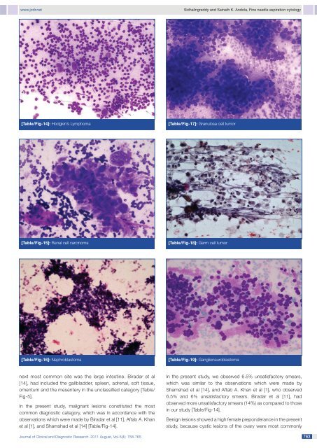
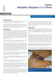
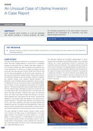
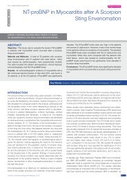
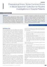
www.jcdr.netSidhalingreddy and Sainath K. Andola, <strong>Fine</strong> needle aspiration cytology[Table/Fig-14]: Hodgkin’s Lymphoma [Table/Fig-17]: Granulosa cell tumor[Table/Fig-15]: Renal cell carcinoma [Table/Fig-18]: Germ cell tumor[Table/Fig-16]: Nephroblastoma[Table/Fig-19]: Ganglioneuroblastomanext most common site was the large intestine. Biradar et al[14], had included the gallbladder, spleen, adrenal, s<strong>of</strong>t tissue,omentum and the mesentery in the unclassified category [Table/Fig-5].In the present study, malignant lesions constituted the mostcommon diagnostic category, which was in accordance with theobservations which were made by Biradar et al [11], Aftab A. Khanet al [1], and Shamshad et al [14] [Table/Fig-14].In the present study, we observed 6.5% unsatisfactory smears,which was similar to the observations which were made byShamshad et al [14], and Aftab A. Khan et al [1], who observed6.5% and 6% unsatisfactory smears. Biradar et al [11], hadobserved more unsatisfactory smears (14%) as compared to thosein our study [Table/Fig-14].Benign lesions showed a high female preponderance in the presentstudy, because cystic lesions <strong>of</strong> the ovary were most commonlyJournal <strong>of</strong> Clinical and Diagnostic Research. 2011 August, Vol-5(4): 758-765 763
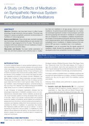
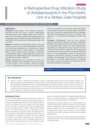
Sidhalingreddy and Sainath K. Andola, <strong>Fine</strong> needle aspiration cytologywww.jcdr.netseen as benign lesions. There was no age or sex predilection forinflammatory lesions in the present study.In the present study, adenocarcinomas were the most commonmalignant cell type (26.3%), followed by hepatocellular carcinoma(23%), renal cell carcinoma (4.7%), serous cystadenocarcinoma(4.7%) and nephroblastoma (2.7%). Poorly differentiated carcinomasconstituted 19.6% <strong>of</strong> the lesions in the present study. Thiswas in accordance with the observations which were made byShamshad et al [14], and Aftab A. Khan et al [1], who observed87.1% and 34% poorly differentiated carcinomas respectively.The second most common malignant type in these studies washepatocellular carcinoma. In the liver, the most common malignantlesion was hepatocellular carcinoma (34), followed by metastaticcarcinoma (25). In the western literature, the most common hepaticmalignancy was metastasic carcinoma [4,6,18,19,20]. This couldbe because <strong>of</strong> the high prevalence <strong>of</strong> Hepatitis B infection and theconsumption <strong>of</strong> chutney which was made up <strong>of</strong> groundnuts, whichwas frequently contaminated with aflatoxins, in this geographicalregion. The observations <strong>of</strong> the present study were similar to those<strong>of</strong> Indian studies, where hepatocellular carcinoma constituted themost common hepatic malignancy [3]. Two studies which wereconducted in Kashmir showed observations which were similar tothat <strong>of</strong> the western literature [1,14].The liver constituted the major site for the malignant lesions, asobserved by Aftab A. Khan et al [1], Stewart et al [5], Zawar MPet al [3], Nyman et al [12], Ennis and MacErlean [6], Joao Nobregaet al [4], and Nautiyal et al [2]. But in an observation which wasmade by Shamshad et al [14], and Joseph et al [8], the most commonorgan sites for the malignant lesions were the gallbladder andthe pancreas respectively. Hepatocellular carcinoma was mostcommonly seen in males, in accordance with previous literaturereports [3,20]. Hepatocellular carcinomas and adenocarcinomashad a peak incidence in the age group between 40-60 years, in accordancewith the observations made by Shamshad et al [14], andZawar MP et al [3]. Malignant tumours which were seen before 20years <strong>of</strong> age, were nephroblastomas and other round cell tumours,Hodgkin’s lymphoma, dysgerminoma and ganglioneuroblastoma.This observation was comparable to that <strong>of</strong> the previous literaturereports [18,20].[Table/Fig-20]: Adrenocortical Carcinoma[Table/Fig-21]: Gastrointestinal Stromal TumorAlthough few studies have reported complications like mild localpain, bleeding and tumour seeding <strong>of</strong> the needle tract, a vastamount <strong>of</strong> literature supports the safety <strong>of</strong> FNAC. There was noreport on complications as a result <strong>of</strong> FNAC in the 20 paperswhich amounted to around 20,000 patients, including those <strong>of</strong> thepresent study.764The sensitivity <strong>of</strong> USG guided FNAC ranged from 71.4% to 96.3%.In the present study, it was 94.1%, which was comparable to that<strong>of</strong> most <strong>of</strong> the studies. All the studies observed 100% specificity,as was found in the present study also. The diagnostic accuracyin various studies ranged from 83.9% to 100%. The present studyfound a diagnostic accuracy <strong>of</strong> 96.5%, which was comparable tothat <strong>of</strong> most <strong>of</strong> the studies [Table/Fig-15-22].CONCLUSION<strong>Intra</strong>-abdominal FNA is a relatively simple, economical, quickand safe procedure for the diagnosis <strong>of</strong> intra-abdominal lesions.It not only helps in differentiating between inflammatory, benignand malignant lesions, but also in categorizing different malignantlesions. <strong>Intra</strong>-abdominal FNA is a reliable, sensitive and specificmethod with a high diagnostic accuracy for the diagnosis <strong>of</strong>[Table/Fig-22]: Pheochromocytomamalignant lesions. It can be utilized as a pre-operative procedurefor the management <strong>of</strong> all intra-abdominal lesions.REFERENCES[1] Aftab Khan A., Jan GM., Wani NA. <strong>Fine</strong> <strong>Needle</strong> <strong>Aspiration</strong> <strong>of</strong> <strong>Intra</strong>abdominalmasses for cytodiagnosis. J. Indian Med Assoc 1996;94(5):167-69.[2] Nautiyal S., Mishra RK, Sharma SP., Routine and ultrasound guidedFNAC <strong>of</strong> intra abdominal lumps – A comparative study. Journal <strong>of</strong><strong>Cytology</strong> 2004;21(3):129-132.Journal <strong>of</strong> Clinical and Diagnostic Research. 2011 August, Vol-5(4): 758-765