Hydrophobic Effect and Hydrogen Bonds Account for the Improved ...
Hydrophobic Effect and Hydrogen Bonds Account for the Improved ...
Hydrophobic Effect and Hydrogen Bonds Account for the Improved ...

Create successful ePaper yourself
Turn your PDF publications into a flip-book with our unique Google optimized e-Paper software.
<strong>Hydrophobic</strong> <strong>Effect</strong> <strong>and</strong> <strong>Hydrogen</strong> <strong>Bonds</strong> <strong>Account</strong> <strong>for</strong> <strong>the</strong> <strong>Improved</strong> Activity of a Complement Inhibitor,<br />
Compstatin<br />
Madan Katragadda, Paola Magotti, Georgia Sfyroera, <strong>and</strong> John D. Lambris<br />
J. Med. Chem.; 2006; 49(15) pp 4616 - 4622; (Article) DOI: 10.1021/jm0603419
4616 J. Med. Chem. 2006, 49, 4616-4622<br />
<strong>Hydrophobic</strong> <strong>Effect</strong> <strong>and</strong> <strong>Hydrogen</strong> <strong>Bonds</strong> <strong>Account</strong> <strong>for</strong> <strong>the</strong> <strong>Improved</strong> Activity of a Complement<br />
Inhibitor, Compstatin<br />
Madan Katragadda, Paola Magotti, Georgia Sfyroera, <strong>and</strong> John D. Lambris*<br />
Protein Chemistry Laboratory, Department of Pathology & Laboratory Medicine, School of Medicine, UniVersity of PennsylVania,<br />
Philadelphia, PennsylVania 19104<br />
ReceiVed March 23, 2006<br />
Tryptophans at positions 4 <strong>and</strong> 7 of compstatin, a peptide complement inhibitor, are crucial <strong>for</strong> its interaction<br />
with C3. However, <strong>the</strong> nature of <strong>the</strong>ir involvement has not been studied to date. Here we investigate <strong>the</strong><br />
molecular <strong>for</strong>ces involved in <strong>the</strong> C3-compstatin interactions, mediated by aromatic residues, by incorporating<br />
in <strong>the</strong>se two positions various tryptophan analogues (5-methyltryptophan, 5-fluorotryptophan, 1-methyltryptophan,<br />
<strong>and</strong> 2-naphthylalanine) <strong>and</strong> assessing <strong>the</strong> resulting peptides <strong>for</strong> activity by enzyme-linked<br />
immunosorbent assay (ELISA) <strong>and</strong> binding by iso<strong>the</strong>rmal titration calorimetry (ITC). Of all <strong>the</strong> compstatin<br />
analogues, peptides containing 1-methyltryptophan at position 4 exhibited <strong>the</strong> highest binding affinity<br />
(K d ) 15 nM) <strong>and</strong> activity (IC 50 ) 0.205 µM), followed by a peptide containing 5-fluorotryptophan at<br />
position 7. Our observations suggest that hydrophobic interactions involving residues at position 4 <strong>and</strong> <strong>the</strong><br />
hydrogen bond initiated by <strong>the</strong> indole nitrogen are primarily responsible <strong>and</strong> crucial <strong>for</strong> <strong>the</strong> increase in<br />
activity. These findings have important implications <strong>for</strong> <strong>the</strong> design of clinically useful complement inhibitors.<br />
* Corresponding author: phone 215-746-5765; fax 215-573-8738; e-mail<br />
lambris@mail.med.upenn.edu.<br />
Introduction<br />
Compstatin is a 13-amino acid cyclic peptide inhibitor that<br />
binds to <strong>the</strong> β-chain of C3, 1 a complement component that is<br />
central to all three pathways of <strong>the</strong> complement system<br />
(classical, alternative, <strong>and</strong> lectin). 2 A number of inhibitors<br />
ranging from small organic compounds to large-size proteins<br />
have been designed to act at several points in <strong>the</strong> complement<br />
cascade, but compstatin, a 13-amino acid cyclic peptide (ICV-<br />
VQDWGHHRCT-NH 2 ), is unique in acting at <strong>the</strong> level of C3. 3<br />
Compstatin has been tested successfully in several in vivo <strong>and</strong><br />
ex vivo animal models <strong>and</strong> has shown enormous potential <strong>for</strong><br />
development as a complement <strong>the</strong>rapeutic. 4-7 The threedimensional<br />
structure of <strong>the</strong> peptide, as determined by NMR,<br />
shows a coil with a type II β-turn involving Q5-D6-W7-G8. 8<br />
With this structure as a template, several combinatorial,<br />
computational, <strong>and</strong> rational design approaches have been carried<br />
out to enhance <strong>the</strong> activity of compstatin. 10,11 These enhancements<br />
have yielded a peptide, having 2-naphthylalanine in<br />
position 4, with 99-fold higher activity than that of <strong>the</strong> parent<br />
molecule. Several of <strong>the</strong> most active analogues have contained<br />
tryptophan or o<strong>the</strong>r aromatic ring structures at positions 4 or-<br />
(<strong>and</strong>) 7, underscoring <strong>the</strong> importance of an aromatic ring<br />
structure at <strong>the</strong>se positions <strong>for</strong> complement inhibitor activity. 12<br />
Despite this increase in activity, very little is known about<br />
<strong>the</strong> underlying molecular <strong>for</strong>ces that impart recognition <strong>and</strong><br />
specificity to compstatin <strong>and</strong> <strong>the</strong>reby dictate its inhibitory<br />
activity. Incorporation of aromatic residues such as tyrosine at<br />
position 4 results in a lower activity than that seen when<br />
tryptophan is present. 10,11 The fact that tryptophan <strong>and</strong> tyrosine<br />
differ in <strong>the</strong> electrostatic potentials of <strong>the</strong>ir ring suggested a<br />
possible correlation between <strong>the</strong> ring current <strong>and</strong> <strong>the</strong> inhibitory<br />
activity; indeed, an increase in inhibitory activity was observed<br />
with an increase in <strong>the</strong> negative electrostatic potential. Such a<br />
relationship is indicative of a cation-π interaction, as has been<br />
described <strong>for</strong> several protein-protein interactions. 13-18 In addition,<br />
<strong>the</strong> higher hydrophobicity of tryptophan when compared<br />
to tyrosine may also be responsible <strong>for</strong> <strong>the</strong> higher activity<br />
observed.<br />
Compstatin contains a tryptophan residue at position 7 that<br />
participates in <strong>the</strong> <strong>for</strong>mation of <strong>the</strong> β turn. 8 Substitution of this<br />
tryptophan residue with alanine or phenylalanine results in loss<br />
of activity, 9 thus indicating that tryptophan is crucial at this<br />
position <strong>for</strong> <strong>the</strong> structural stability <strong>and</strong>/or activity of compstatin.<br />
Loss of activity upon substitution by phenylalanine indicates<br />
that Trp7, in addition to Trp4, may mediate a cation-π<br />
interaction or <strong>the</strong> <strong>for</strong>mation of a hydrogen bond between <strong>the</strong><br />
N-H <strong>and</strong> a hydrogen-bond acceptor on C3. However, <strong>the</strong> role<br />
of Trp7 in <strong>the</strong>se interactions has not been delineated at <strong>the</strong><br />
atomic level.<br />
In this study we have used peptide syn<strong>the</strong>sis <strong>and</strong> iso<strong>the</strong>rmal<br />
titration calorimetry (ITC) a analysis to examine <strong>the</strong> potential<br />
noncovalent interactions involved in this interaction between<br />
C3 <strong>and</strong> compstatin <strong>and</strong> to analyze <strong>the</strong> role of selective hydrogen<br />
bonding at positions 4 <strong>and</strong> 7. Analyses in which we introduced<br />
<strong>the</strong> tryptophan analogues 5-fluorotryptophan, 5-methyltryptophan,<br />
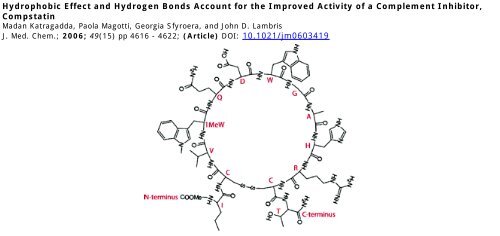
1-methyltryptophan, <strong>and</strong> 2-naphthylalanine (Figure 1)<br />
at positions 4 <strong>and</strong>/or 7 have revealed that <strong>the</strong> incorporation of<br />
1-methyltryptophan at position 4 yields a peptide with a 264-<br />
fold increase in inhibitory activity. Our results suggest that<br />
reducing <strong>the</strong> hydrophobic character of Trp4 results in a dramatic<br />
loss in inhibitory activity. In contrast, at position 7, hydrophobic<br />
residues are not tolerated, <strong>and</strong> <strong>the</strong>y drastically lower <strong>the</strong><br />
inhibitory activity. Instead, an interaction of a polar nature, such<br />
as hydrogen bonding, is preferred, <strong>and</strong> increasing <strong>the</strong> number<br />
of hydrogen bonds increases <strong>the</strong> inhibitory activity of compstatin<br />
analogues.<br />
a Abbreviations: Ac, acetyl group; ELISA, enzyme-linked immunosorbent<br />
assay; 5fW, 5-fluoro-L-tryptophan, 1MeW, 1-methyl-L-tryptophan;<br />
5MeW, 5-methyl-L-tryptophan; MALDI, matrix-assisted laser desorption<br />
ionization; TOF, time-of-flight; HPLC, high-per<strong>for</strong>mance liquid chromatography;<br />
ITC, iso<strong>the</strong>rmal titration calorimetry; 2-Nal, 2-naphthylalanine;<br />
NA, not active; log P, logarithm of partition coefficient.<br />
10.1021/jm0603419 CCC: $33.50 © 2006 American Chemical Society<br />
Published on Web 07/04/2006
C3-Compstatin Interaction Journal of Medicinal Chemistry, 2006, Vol. 49, No. 15 4617<br />
Figure 1. Side-chain structures of analogues used in <strong>the</strong> study: (A) tryptophan (W), (B) 5-fluorotryptophan (5fW), (C) 5-methyltryptophan (5MeW),<br />
(D) 1-methyltryptophan (1MeW), <strong>and</strong> (E) 2-naphthylalanine (2Nal).<br />
Results<br />
Syn<strong>the</strong>sis of Compstatin Analogues. The analogues of<br />
compstatin were syn<strong>the</strong>sized by solid-phase Fmoc-based peptide<br />
chemistry. Because of <strong>the</strong> unavailability of Fmoc-derived<br />
L-derivatives of <strong>the</strong> tryptophan analogues, <strong>the</strong> Fmoc-derived<br />
DL-derivatives were used. The reactions yielded a diastereomeric<br />
mixture of peptides bearing a D-analogue or an L-analogue.<br />
There<strong>for</strong>e, each of <strong>the</strong> peptide mixtures was fur<strong>the</strong>r subjected<br />
to reversed-phase HPLC to separate <strong>the</strong> peptide diastereomers,<br />
as described under Materials <strong>and</strong> Methods. Each peptide with<br />
a single substitution yielded two well-separated peaks, while<br />
<strong>the</strong> peptide with substitutions at both positions 4 <strong>and</strong> 7 yielded<br />
four well-separated peaks. Digesting <strong>the</strong> peptides corresponding<br />
to <strong>the</strong>se peaks with V8 protease <strong>and</strong> subsequent MALDI analysis<br />
made it possible to identify <strong>the</strong> diastereomeric nature of <strong>the</strong><br />
analogues containing tryptophan analogues at position 7. The<br />
rationale <strong>for</strong> using V8 protease was that this enzyme cleaves at<br />
<strong>the</strong> C-terminal side of aspartic acid only when followed by an<br />
L-amino acid. Identification of cleavage products in <strong>the</strong> mass<br />
spectra indicated that <strong>the</strong> L-diastereomeric peptide eluted first,<br />
followed by <strong>the</strong> D-<strong>for</strong>m(s), <strong>for</strong> which no cleavage fragments<br />
were detected (data not shown). On <strong>the</strong> basis of <strong>the</strong>se observations,<br />
we concluded that <strong>the</strong> peptide eluted in <strong>the</strong> first peak is<br />
of <strong>the</strong> L-<strong>for</strong>m <strong>and</strong> in <strong>the</strong> second peak is of <strong>the</strong> D-<strong>for</strong>m. This<br />
conclusion was fur<strong>the</strong>r supported by assessment of <strong>the</strong> complement-inhibiting<br />
activity of <strong>the</strong> peptides eluted in <strong>the</strong> two peaks.<br />
In agreement with previous findings, where any D-amino acid<br />
substitution renders <strong>the</strong> molecule inactive, 9 <strong>the</strong> second peak<br />
showed no activity while <strong>the</strong> first peak inhibited complement<br />
activation (data not shown).<br />
Incorporation of Tryptophan Analogues at Position 4. It<br />
has previously been shown that substitution of valine with<br />
tryptophan at position 4 increases <strong>the</strong> activity of compstatin by<br />
45-fold. 11 In <strong>the</strong> present study we replaced tryptophan with<br />
tryptophan analogues <strong>and</strong> 2-naphthylalanine to study <strong>the</strong> nature<br />
of <strong>the</strong> interaction mediated by <strong>the</strong> residue at position 4 during<br />
<strong>the</strong> course of <strong>the</strong> binding of compstatin analogues to C3.<br />
Previously described ELISA-based assays were used to test <strong>the</strong><br />
activity of all <strong>the</strong> compstatin analogues bearing tryptophan<br />
analogues at position 4. While substitution with 1-methyltryptophan<br />
(1MeW), 5-methyltryptophan (5MeW), <strong>and</strong> 2-naphthylalanine<br />
(2Nal) increased <strong>the</strong> activity of compstatin 264-,<br />
67-, <strong>and</strong> 99-fold, respectively, substitution with 5-fluorotryptophan<br />
(5fW) resulted in a lower activity: 31-fold increase<br />
(Figure 2, Table 1). Figure 4A shows inhibitory constants (IC 50 )<br />
plotted against log P values <strong>for</strong> <strong>the</strong> tryptophan analogues <strong>and</strong><br />
2-naphthylalanine. The plot indicates that <strong>the</strong> activity of<br />
compstatin increases with an increase in <strong>the</strong> hydrophobicity of<br />
<strong>the</strong> amino acid replaced at position 4. However, <strong>the</strong> IC 50<br />
representing <strong>the</strong> activity of compstatin analogue with 1-methyltryptophan<br />
[4(1MeW)7W] did not fall on <strong>the</strong> linear line,<br />
showing an exception to this correlation.<br />
To investigate whe<strong>the</strong>r a correlation exists between <strong>the</strong><br />
activity <strong>and</strong> binding affinity, <strong>the</strong> interaction of <strong>the</strong> compstatin<br />
analogues listed in Table 1 was measured by iso<strong>the</strong>rmal titration<br />
calorimetry. The calorimetric data obtained <strong>for</strong> <strong>the</strong> interaction<br />
of all <strong>the</strong> peptides with C3 fit to “one set of sites” model, with<br />
a stoichiometry close to 1 (Figure 3). These results suggest that<br />
<strong>the</strong> binding of <strong>the</strong>se peptides to C3 occurs in a 1:1 ratio. The<br />
<strong>the</strong>rmodynamic parameters resulting from <strong>the</strong>se fits are shown<br />
in Table 2. As evident from <strong>the</strong> K d values, peptide 4(1MeW)-<br />
7W exhibited higher binding affinity (K d ) 0.015 µM) than<br />
any of <strong>the</strong> o<strong>the</strong>r peptides listed in category I. Plotting <strong>the</strong>se<br />
values against <strong>the</strong> log P values of analogues indicated that a<br />
correlation exists between <strong>the</strong> binding affinity <strong>and</strong> <strong>the</strong> hydrophobic<br />
nature of <strong>the</strong> tryptophan analogues (except <strong>the</strong> 1-methyltryptophan<br />
analogue) <strong>and</strong> 2-naphthylalanine (Figure 4B): <strong>the</strong><br />
binding affinity increased with an increase in <strong>the</strong> hydrophobicity<br />
of <strong>the</strong> analogue incorporated at position 4. This observation is<br />
consistent with <strong>the</strong> correlation observed between log P <strong>and</strong> <strong>the</strong><br />
inhibitory constants, indicating that <strong>the</strong> activity follows binding<br />
affinity. For example, increase in <strong>the</strong> activity observed <strong>for</strong><br />
4(1MeW)7W in comparison to 4W7W is mainly due to its<br />
increased binding affinity to C3. Consistent with <strong>the</strong> correlation<br />
observed between log P <strong>and</strong> IC 50 , <strong>the</strong> exception exhibited by<br />
4(1MeW)7W is reiterated in <strong>the</strong> log P-K d correlation. This<br />
indicates that <strong>the</strong> anomalous behavior of 4(1MeW)7W is not<br />
due to <strong>the</strong> nature of inhibition assays or binding constant<br />
determinations.<br />
All <strong>the</strong> peptides bound to C3 with a negative enthalpy <strong>and</strong><br />
positive entropy, suggesting that <strong>the</strong> binding is enthalpy-driven.<br />
Such binding was previously shown to be characteristic <strong>for</strong> <strong>the</strong><br />
interaction of compstatin with C3. 19 However, binding of all<br />
<strong>the</strong> compstatin analogues in category I was characterized by an<br />
enthalpy change that was lower than that observed <strong>for</strong> 4W7W<br />
<strong>and</strong> by an entropy change shifted toward <strong>the</strong> favorable end.<br />
Figure 4C shows a plot of log P versus -T∆S, which indicates<br />
that, with an increase in <strong>the</strong> hydrophobicity of <strong>the</strong> analogues<br />
incorporated at position 4, entropy is more favored; this situation<br />
has a positive impact on <strong>the</strong> free energy change (Table 2), which<br />
is consistent with an increase in binding affinity.<br />
Incorporation of Tryptophan Analogues at Position 7. It<br />
has been proposed in previous studies that tryptophan at position<br />
7 may <strong>for</strong>m a hydrogen bond with a H-bond acceptor on C3.<br />
In <strong>the</strong> present study we <strong>the</strong>re<strong>for</strong>e replaced tryptophan at position<br />
7 (4W7W) with tryptophan analogues such as those used <strong>for</strong>
4618 Journal of Medicinal Chemistry, 2006, Vol. 49, No. 15 Katragadda et al.<br />
Table 1. Sequences of Peptides Used in This Study<br />
peptide<br />
sequence<br />
IC 50<br />
(µM)<br />
RA a<br />
4W7W Ac-ICVWQDWGAHRCT-NH 2 1.2 45<br />
Substitution at Position 4 (Category I)<br />
4(5fW)7W Ac-ICV(5fW)QDWGAHRCT-NH 2 1.74 31<br />
4(5MeW)7W Ac-ICV(5MeW)QDWGAHRCT-NH 2 0.87 67<br />
4(1MeW)7W Ac-ICV(1MeW)QDWGAHRCT-NH 2 0.205 264<br />
4(2Nal)7W Ac-ICV(2Nal)QDWGAHRCT-NH 2 0.545 99<br />
Substitution at Position 7 (Category II)<br />
4W7(5fW) Ac-ICVWQD(5fW)GAHRCT-NH 2 0.446 121<br />
4W7(5MeW) Ac-ICVWQD(5MeW)GAHRCT-NH 2 NA NA<br />
4W7(1MeW) Ac-ICVWQD(1MeW)GAHRCT-NH 2 NA NA<br />
Double Substitution at Positions 4 <strong>and</strong> 7 (Category III)<br />
4(1MeW)7(5fW) Ac-ICV(1MeW)QD(5fW)GAHRCT-NH 2 0.205 264<br />
a Activity relative to <strong>the</strong> peptide H-I(CVVQDWGHHRC)T-NH 2.<br />
Thus, unlike <strong>the</strong> activity exhibited by <strong>the</strong> peptides listed in<br />
category I, no correlation was observed between <strong>the</strong> activity<br />
<strong>and</strong> hydrophobicity of <strong>the</strong> tryptophan analogues at position 7.<br />
Calorimetric titration of <strong>the</strong> peptides listed in category II<br />
yielded <strong>the</strong> <strong>the</strong>rmodynamic parameters listed in Table 2; since<br />
no binding was detected <strong>for</strong> peptides bearing 5-methyltryptophan<br />
<strong>and</strong> 1-methyltryptophan, binding parameters do not exist <strong>for</strong><br />
<strong>the</strong>se analogues. Only 4W7(5fW) bound to C3, with an affinity<br />
of 0.035 µM; this value is greater than that observed <strong>for</strong> any of<br />
<strong>the</strong> peptides in category I except 4(1MeW)7W. In contrast to<br />
<strong>the</strong> peptides in category I, 4W7(5fW) bound to C3 with a<br />
favorable binding enthalpy change (∆H )-21.83 kcal/mol,<br />
∆∆H ) -3.69 kcal/mol) <strong>and</strong> unfavorable entropy change<br />
(-T∆S ) 11.56 kcal/mol, -T∆∆S ) 2.77 kcal/mol), suggesting<br />
<strong>the</strong> involvement of additional favorable noncovalent interactions<br />
of polar nature.<br />
Incorporation of Tryptophan Analogues at Both Positions<br />
4 <strong>and</strong> 7. Since <strong>the</strong> substitution of tryptophan with 1-methyltryptophan<br />
at position 4 <strong>and</strong> with 5-fluorotryptophan at position<br />
7 yielded compstatin analogues that showed a marked increase<br />
in inhibitory activity, we also created an analogue bearing<br />
simultaneously both <strong>the</strong>se substitutions. The resulting peptide,<br />
4(1MeW)7(5fW), generated an inhibition curve similar to that<br />
<strong>for</strong> <strong>the</strong> single substitution with 1-methyltryptophan [4(1MeW)-<br />
7W] (Figure 2, Table 1). The binding affinity (K d ) 0.017)<br />
observed <strong>for</strong> <strong>the</strong> double analogue was also similar to that <strong>for</strong><br />
4(1MeW)7W. Binding of 4(1MeW)7(5fW) to C3 yielded an<br />
enthalpy change of -17.33 kcal/mol, an entropy change of<br />
-6.73 kcal/mol, <strong>and</strong> a free energy change of -10.6 kcal/mol.<br />
These values are similar to those observed <strong>for</strong> <strong>the</strong> binding of<br />
4(1MeW)7W suggesting that 5-fluorotryptophan has no effect<br />
at position 7 in <strong>the</strong> presence of 1-methyltryptophan at position<br />
4.<br />
Figure 2. Activity of syn<strong>the</strong>tic compstatin analogues: Plots of percent<br />
complement inhibition vs peptide concentration <strong>for</strong> 4W7W compstatin<br />
(2), 4(5fW)7W (1), 4(5MeW)7W (b), 4(1MeW)7W ([), 4(2Nal)7W<br />
(9), 4W7(5fW) (`), <strong>and</strong> 4(1MeW)7(5fW) (triangles pointing left).<br />
position 4. With <strong>the</strong> exception of <strong>the</strong> substitution with 5-fluorotryptophan,<br />
which yielded a 121-fold active peptide, all <strong>the</strong> o<strong>the</strong>r<br />
substitutions rendered compstatin inactive (Figure 2, Table 1).<br />
Discussion<br />
<strong>Hydrogen</strong> <strong>Bonds</strong> at Position 7 Increase <strong>the</strong> Activity of<br />
Compstatin. Tryptophan at position 7 is one of <strong>the</strong> four residues<br />
that make up <strong>the</strong> β turn, which has been shown to be critical<br />
<strong>for</strong> <strong>the</strong> activity of compstatin. 9 It has also been suggested that<br />
Trp7 might stabilize <strong>the</strong> interaction of compstatin with C3<br />
through a hydrogen bond. However, no direct experimental<br />
evidence has been obtained to support this hypo<strong>the</strong>sis. In <strong>the</strong><br />
present study, incorporation of 5-fluorotryptophan at this<br />
position resulted in an increase in <strong>the</strong> inhibitory activity, whereas<br />
incorporation of ei<strong>the</strong>r 5-methyltryptophan or 1-methyltryptophan<br />
rendered compstatin inactive. This loss of activity upon<br />
incorporation of 1-methyltryptophan supports <strong>the</strong> contention that
C3-Compstatin Interaction Journal of Medicinal Chemistry, 2006, Vol. 49, No. 15 4619<br />
Figure 3. Thermodynamic characterization of <strong>the</strong> interaction of compstatin analogues with C3: ITC data representing <strong>the</strong> binding of 4W7W,<br />
4(5fW)7W, 4(5MeW)7W, 4(1MeW)7W, 4(2Nal)7W <strong>and</strong> 4W7(5fW) to C3. The plots were obtained by fitting <strong>the</strong> corrected raw data to “one set<br />
of sites” model in Origin 7.0. All <strong>the</strong> errors reported in Table 2 are results of nonlinear least-squares fit.<br />
Table 2. Thermodynamic Parameters <strong>for</strong> <strong>the</strong> Interaction of Syn<strong>the</strong>tic Compstatin Analogues<br />
peptide<br />
K a<br />
(M -1 )<br />
K d<br />
(µM)<br />
∆H<br />
(kcal/mol)<br />
∆∆H<br />
(kcal/mol)<br />
-T∆S<br />
(kcal/mol)<br />
-T∆∆S<br />
(kcal/mol)<br />
∆G<br />
(kcal/mol)<br />
∆∆G<br />
(kcal/mol)<br />
4W7W 7.036 × 10 6 ( 4.8 × 10 5 0.14 -18.14 ( 0.19 0 8.79 0 -9.4 0<br />
4(5fW)7W 6.65 × 10 6 ( 5.1 × 10 5 0.15 -16.69 ( 0.23 1.45 7.39 -1.4 -9.4 0<br />
4(5MeW)7W 9.59 × 10 6 ( 7.3 × 10 5 0.12 -17.75 ( 0.22 0.34 8.2 -0.54 -9.55 -0.15<br />
4(1MeW)7W 6.56 × 10 7 ( 6.6 × 10 6 0.015 -17.59 ( 0.14 0.81 6.94 -1.85 -10.65 -1.1<br />
4(2Nal)7W 9.21 × 10 6 ( 9.7 × 10 5 0.11 -14.27 ( 0.19 3.87 4.8 -3.99 -9.5 -0.1<br />
4W7(5fW) 2.83 × 10 7 ( 3.7 × 10 6 0.035 -21.83 ( 0.10 -3.69 11.56 2.77 -10.25 -0.8<br />
4(1MeW)7(5fW) 5.91 × 10 7 ( 4.7 × 10 6 0.017 -17.33 ( 0.093 0.81 6.73 -2.06 -10.6 -1.2<br />
<strong>the</strong> hydrogen bond mediated by N-H of Trp 7 is crucial <strong>for</strong><br />
<strong>the</strong> interaction of compstatin with C3. Since we were unable to<br />
observe any binding of <strong>the</strong> peptide containing 1-methyltryptophan<br />
with C3 in <strong>the</strong> calorimeter, <strong>the</strong> enthalpy change<br />
associated with <strong>the</strong> hydrogen bond <strong>for</strong>mation could not be<br />
determined. In addition, <strong>the</strong> complete loss of activity of<br />
compstatin upon incorporation of 5-methyltryptophan strongly<br />
suggests that a hydrophobic amino acid is not tolerated at<br />
position 7, probably because of <strong>the</strong> polar nature of <strong>the</strong> interaction<br />
mediated at position 7.<br />
Incorporation of 5-fluorotryptophan at position 7 increased<br />
<strong>the</strong> enthalpy by -3.69 kcal/mol when compared to 4W7W<br />
(Table 2). This finding suggests that enthalpically favored<br />
interactions are mediated by <strong>the</strong> fluorine atom. Replacing one<br />
of <strong>the</strong> indole hydrogens with a fluorine atom might streng<strong>the</strong>n<br />
<strong>the</strong> hydrogen-bonding character of <strong>the</strong> indole NH as a result of<br />
<strong>the</strong> drop in pK a . However, <strong>the</strong> magnitude of <strong>the</strong> enthalpy<br />
increase was ra<strong>the</strong>r high to be attributable to an increase in <strong>the</strong><br />
hydrogen-bonding strength of <strong>the</strong> indole NH. Ano<strong>the</strong>r possibility<br />
is that <strong>the</strong> fluorine <strong>for</strong>ms a hydrogen bond as a result of its
4620 Journal of Medicinal Chemistry, 2006, Vol. 49, No. 15 Katragadda et al.<br />
Figure 4. Plots showing <strong>the</strong> relationship between hydrophobicity of<br />
<strong>the</strong> analogues at position 4, denoted by log P, <strong>and</strong> <strong>the</strong> inhibitory constant<br />
(A), <strong>the</strong> binding constant (B), <strong>and</strong> entropy, denoted by -T∆S (C).<br />
electron-donating nature, as has been demonstrated <strong>for</strong> <strong>the</strong><br />
structure of <strong>the</strong> tetradeca(3-fluorotyrosyl)glutathione transferase.<br />
20 This possibility would more likely fit our observations,<br />
as <strong>the</strong> energy of <strong>the</strong> hydrogen bond is about 2 kcal/mol.<br />
However, <strong>the</strong> magnitude is twice <strong>the</strong> hydrogen-bond energy.<br />
One possible explanation is that a water molecule is bridging<br />
<strong>the</strong> interaction between <strong>the</strong> fluorine atom <strong>and</strong> a hydrogen<br />
acceptor on C3, in which case two hydrogen bonds (equivalent<br />
to about 4 kcal/mol energy) need to be <strong>for</strong>med. Our studies on<br />
<strong>the</strong> <strong>the</strong>rmodynamics of <strong>the</strong> interaction of C3 with compstatin<br />
suggest that water molecules are involved at <strong>the</strong> binding<br />
interface. 19 Thus, <strong>the</strong> hypo<strong>the</strong>sis that water mediates <strong>the</strong><br />
interaction between <strong>the</strong> fluorine atom <strong>and</strong> a residue on C3 fits<br />
our observations. Fur<strong>the</strong>r support comes from <strong>the</strong> decrease in<br />
entropy observed <strong>for</strong> <strong>the</strong> interaction of <strong>the</strong> position 7-substituted<br />
4W7(5fW) analogue relative to <strong>the</strong> 4W7W analogue (Table 2),<br />
a decrease that could be produced by <strong>the</strong> binding of an additional<br />
water molecule at <strong>the</strong> interface. Such water-mediated interactions<br />
between fluorine atoms <strong>and</strong> o<strong>the</strong>r hydrogen bond acceptors have<br />
been observed in o<strong>the</strong>r systems. 21<br />
A <strong>Hydrophobic</strong> <strong>Effect</strong> at Position 4 Increases <strong>the</strong> Activity<br />
of Compstatin. It has been shown in computational studies <strong>and</strong><br />
ELISA-based activity assays that substitution of valine with<br />
tyrosine at position 4 increased <strong>the</strong> activity of compstatin 14-<br />
fold. 10 Fur<strong>the</strong>r substitution of valine with tryptophan increases<br />
<strong>the</strong> activity of compstatin 45-fold. 11 The basis <strong>for</strong> <strong>the</strong> difference<br />
in activity observed between <strong>the</strong> tyrosine- <strong>and</strong> tryptophancontaining<br />
peptides has not been studied. This difference,<br />
however, suggests that incorporation of an aromatic residue at<br />
position 4 makes compstatin more active. There are four<br />
possibilities to account <strong>for</strong> an increase in <strong>the</strong> activity exhibited<br />
by a tryptophan-containing peptide when compared to a tyrosinecontaining<br />
peptide: (1) A hydrophobic effect at position 4 is<br />
required <strong>for</strong> <strong>the</strong> interaction, as tryptophan is more hydrophobic<br />
than tyrosine. (2) A cation-π interaction could be initiated at<br />
position 4, as tryptophan possesses a greater π electron density<br />
than tyrosine. (3) A hydrogen bond could be <strong>for</strong>med between<br />
tyrosine or tryptophan <strong>and</strong> a residue on C3, <strong>and</strong> toge<strong>the</strong>r with<br />
<strong>the</strong> above-mentioned interactions, this reaction could better<br />
stabilize <strong>the</strong> interaction. (4) Incorporation of analogues changes<br />
<strong>the</strong> structure of compstatin to <strong>the</strong> extent that <strong>the</strong> interaction is<br />
effected.<br />
To investigate <strong>the</strong> possible involvement of <strong>the</strong>se interactions<br />
or changes at position 4, we incorporated 5-methyltryptophan,<br />
5-fluorotryptophan, 1-methyltryptophan, <strong>and</strong> 2-naphthylalanine<br />
at this position. Incorporation of 1-methyltryptophan produced<br />
<strong>the</strong> greatest increase in activity <strong>and</strong> binding affinity. This finding<br />
is in marked contrast to <strong>the</strong> result we obtained when we<br />
incorporated 1-methyltryptophan at position 7. This observation<br />
strongly suggests that an indole N-mediated hydrogen bond is<br />
not necessary at position 4 <strong>for</strong> <strong>the</strong> binding <strong>and</strong> activity of<br />
compstatin. In fact, <strong>the</strong> absence of this hydrogen bond, or a<br />
reduction of <strong>the</strong> polar character by replacing hydrogen with<br />
methyl on <strong>the</strong> indole nitrogen at position 4, seems to be<br />
beneficial <strong>for</strong> <strong>the</strong> binding <strong>and</strong> activity of compstatin. These<br />
findings provide strong evidence that a hydrophobic interaction<br />
or effect at position 4 streng<strong>the</strong>ns <strong>the</strong> interaction of compstatin<br />
with C3. Fur<strong>the</strong>r support <strong>for</strong> this conclusion comes from <strong>the</strong><br />
log P-IC 50 <strong>and</strong> log P-K d correlations observed upon incorporation<br />
of 5-methyltryptophan, 5-fluorotryptophan, <strong>and</strong> 2-naphthylalanine,<br />
as well as <strong>the</strong> entropy differences observed upon<br />
incorporation of <strong>the</strong> analogues. In <strong>the</strong> classical hydrophobic<br />
effect, an increase in entropy is expected; in our case, such a<br />
result is clearly indicated by <strong>the</strong> plot showing <strong>the</strong> relationship<br />
between <strong>the</strong> hydrophobic nature of <strong>the</strong> incorporated analogues<br />
<strong>and</strong> <strong>the</strong> entropy (Figure 4). We suggest that this increase in<br />
entropy could be an effect of <strong>the</strong> bulk solvent.<br />
Tryptophan has been shown to participate in cation-π <strong>and</strong><br />
π-π interactions through <strong>the</strong> π electron density on <strong>the</strong> indole<br />
ring (Figure 5; tryptophan), <strong>and</strong> <strong>the</strong>se interactions are believed
C3-Compstatin Interaction Journal of Medicinal Chemistry, 2006, Vol. 49, No. 15 4621<br />
Figure 5. Representation of <strong>the</strong> electrostatic potential of <strong>the</strong> side chains<br />
of tryptophan, 5-fluorotryptophan, 5-methyltryptophan, 1-methyltryptophan,<br />
<strong>and</strong> 2-naphthylalanine. Red indicates negative potential, <strong>and</strong><br />
blue indicates positive potential. These maps were created with<br />
Chemsketch.<br />
to impart specificity to several intermolecular <strong>and</strong> intramolecular<br />
interactions involving proteins 13,14,18,22 <strong>and</strong> peptides 23,24 <strong>and</strong> also<br />
to catalysis. 16,17,22,25 Thus, reducing <strong>the</strong> π character of <strong>the</strong> indole<br />
by introducing strong electronegative groups such as fluorine<br />
(Figure 5; 5-fluoro-tryptophan) onto <strong>the</strong> indole ring, as in our<br />
case, could lead to a weakening of <strong>the</strong> cation-π <strong>and</strong> π-π<br />
interactions. Such changes in <strong>the</strong> strength of a cation-π<br />
interaction as a function of <strong>the</strong> degree of fluorination of <strong>the</strong><br />
indole ring have previously been demonstrated in <strong>the</strong> interactions<br />
between <strong>the</strong> acetylcholine receptor <strong>and</strong> nicotine <strong>and</strong> between<br />
<strong>the</strong> serotonergic receptor <strong>and</strong> serotonin. 15 In contrast, placement<br />
of electron-rich substituents such as methyl groups on <strong>the</strong> indole<br />
should leave <strong>the</strong> π electron density unperturbed (Figure 5;<br />
5-methyl-tryptophan <strong>and</strong> 1-methyl-tryptophan) <strong>and</strong> would be<br />
expected to leave <strong>the</strong> π electron-mediated interactions unchanged.<br />
Similarly, <strong>the</strong> addition of a benzene ring, as in <strong>the</strong><br />
case of <strong>the</strong> 2-naphthylalanine analogue, should increase <strong>the</strong> π<br />
electron density (Figure 5; 2-naphthylalanine) <strong>and</strong> streng<strong>the</strong>n<br />
<strong>the</strong> cation-π interaction. Contrary to all <strong>the</strong>se expectations,<br />
however, we did not observe a clear correlation between <strong>the</strong> π<br />
electron density <strong>and</strong> <strong>the</strong> activity or binding. In addition, <strong>the</strong><br />
binding enthalpy remained unchanged when such analogues<br />
were incorporated. These observations strongly suggest that a<br />
cation-π or π-π interaction is not mediated by tryptophan at<br />
position 4 <strong>and</strong> is not required <strong>for</strong> <strong>the</strong> activity of compstatin.<br />
It is also possible that incorporation of tryptophan analogues<br />
might change <strong>the</strong> structural features of compstatin <strong>and</strong> affect<br />
binding. However, we have previously reported that <strong>the</strong><br />
substitution of valine with tryptophan <strong>and</strong> 2-naphthylalanine at<br />
position 4 on <strong>the</strong> structure of compstatin has negligible effect<br />
on its structure as examined by NMR. 11 In light of <strong>the</strong>se<br />
observations, we concluded that it is not likely that <strong>the</strong> solution<br />
structure of compstatin changes upon substitution of tryptophan<br />
analogues or 2-naphthylalanine. However, <strong>the</strong>se conclusions do<br />
not exclude <strong>the</strong> possibility of structural changes of bound <strong>for</strong>m<br />
of compsatin, which we were attempting to obtain though<br />
cocrystallization experiments; <strong>the</strong> crystal structure of C3 was<br />
recently published. 26 Con<strong>for</strong>mational changes upon compstatin<br />
binding to C3 have been previously suggested by use of surface<br />
plasmon resonance analysis. 27 In addition, <strong>the</strong> tryptophan side<br />
chain is believed to be facing outward as no interactions between<br />
tryptophan side chain <strong>and</strong> <strong>the</strong> rest of <strong>the</strong> molecule, which could<br />
stabilize <strong>the</strong> fold, were observed in <strong>the</strong> solution structure. This<br />
suggested that placing a fluorine atom or methyl group at<br />
position 5 of <strong>the</strong> indole ring might not affect compstatin’s<br />
structure. Thus, we conclude that <strong>the</strong> log P versus IC 50 <strong>and</strong> log<br />
P versus K d correlations observed are due to <strong>the</strong> modulation of<br />
hydrophobic character of tryptophan, which in effect modulates<br />
<strong>the</strong> interaction of compstatin with C3. However, <strong>the</strong> same may<br />
not be true in case of compstatin analogue bearing 1-methyltryptophan<br />
due to its anomalous behavior (please refer to plots<br />
in Figure 4A,B). 1-Methyltryptophan has a methyl group<br />
attached to <strong>the</strong> indole nitrogen, unlike o<strong>the</strong>r analogues. This<br />
might place <strong>the</strong> methyl group in close proximity to <strong>the</strong> main<br />
chain of compstatin <strong>and</strong> affect <strong>the</strong> structure of compstatin in a<br />
way that it would bind to C3 with high affinity.<br />
In summary, we propose that tryptophan at position 7 imparts<br />
<strong>the</strong> interaction through a hydrogen bond with <strong>the</strong> possibility of<br />
streng<strong>the</strong>ning <strong>the</strong> interaction through incorporation of additional<br />
hydrogen-bond acceptors such as fluorine. In contrast, <strong>the</strong><br />
tryptophan at position 4 prefers to interact through its hydrophobic<br />
nature, <strong>and</strong> <strong>the</strong> increase of hydrophobicity led to an<br />
increase in <strong>the</strong> activity. We suggest, on <strong>the</strong> basis of <strong>the</strong> results<br />
from <strong>the</strong> present study, that fur<strong>the</strong>r optimization of <strong>the</strong> activity<br />
of compstatin can be made possible through introducing<br />
analogues that favor increase in <strong>the</strong> enthalpy change at position<br />
7 <strong>and</strong> <strong>the</strong> analogues that favor entropy change at position 4.<br />
Experimental Procedures<br />
Reagents. Fmoc-5-fluoro-DL-tryptophan, Fmoc-5-methyl-DLtryptophan,<br />
2-naphthylalanine, <strong>and</strong> Fmoc-1-methyl-DL-tryptophan<br />
were obtained from Anaspec Inc. (San Jose, CA).<br />
Peptide Syn<strong>the</strong>sis <strong>and</strong> Purification. Analogues of compstatin<br />
containing 5-fluoro-L-tryptophan, 5-methyl-L-tryptophan, 1-methyl-<br />
L-tryptophan, L-tryptophan, or 2-naphthylalanine at positions 4 <strong>and</strong>/<br />
or 7 were syn<strong>the</strong>sized chemically by solid-phase peptide chemistry<br />
according to st<strong>and</strong>ard protocols. 11 The N- <strong>and</strong> C-termini of <strong>the</strong><br />
peptides were protected with acetyl <strong>and</strong> amide groups. All peptides<br />
were fur<strong>the</strong>r purified on a C18 reversed-phase HPLC column to<br />
95% purity, lyophilized, <strong>and</strong> characterized by MALDI mass<br />
spectrometry. For <strong>the</strong> syn<strong>the</strong>sis of <strong>the</strong> analogues containing<br />
1-methyltryptophan, 5-methyltryptophan, <strong>and</strong> 5-fluorotryptophan<br />
(Table 1), we used <strong>the</strong> Fmoc-DL-derivatives. Fur<strong>the</strong>r separation of<br />
<strong>the</strong> diastereomeric peptides was per<strong>for</strong>med as described by Meyers<br />
et al. 28 In brief, <strong>the</strong> DL mixture of each peptide was separated into<br />
D <strong>and</strong> L isomeric peptides on a C18 reversed-phase HPLC column<br />
with 10% acetonitrile in 0.01 M ammonium acetate, pH 4.1. The<br />
isomeric identity of <strong>the</strong> eluted peptides with incorporations at<br />
position 7 was determined by treating <strong>the</strong> peptides with V8 protease,<br />
followed by analysis by MALDI-TOF mass spectrometry (Micro-<br />
Mass TOFspec2E).<br />
Purification of C3. C3 was purified from fresh human plasma<br />
obtained from <strong>the</strong> blood bank of <strong>the</strong> Hospital of <strong>the</strong> University of<br />
Pennsylvania as described previously. 29 Briefly, <strong>the</strong> plasma was<br />
fractionated with 15% (w/v) PEG 3350, <strong>and</strong> <strong>the</strong> pellet was<br />
resuspended in 20 mM phosphate buffer, pH 7.8, <strong>and</strong> <strong>the</strong>n subjected<br />
to anion-exchange chromatography on a DEAE-HR 40, 50 × 5<br />
cm column (Millipore Inc., Billerica, MA) with <strong>the</strong> same buffer.<br />
The proteins were eluted with 6Lofalinear gradient (15-70%)<br />
with20 mM phosphate buffer, pH 7.8, containing 500 mM NaCl.<br />
The C3 was fur<strong>the</strong>r purified on a size-exclusion column Superdex<br />
200 26/60 (Amersham Biosciences) <strong>and</strong> a Mono S column<br />
(Amersham Biosciences) to separate C3 from C3(H 2 O). 27<br />
Inhibition of Complement Activation. Assays to assess <strong>the</strong><br />
complement inhibitory activity of compstatin <strong>and</strong> its analogues were<br />
per<strong>for</strong>med as described elsewhere. 11 In brief, <strong>the</strong>y involved an
4622 Journal of Medicinal Chemistry, 2006, Vol. 49, No. 15 Katragadda et al.<br />
antigen-antibody complex-mediated complement activation of<br />
human serum, as detected by <strong>the</strong> deposition of C3b/iC3b to<br />
complex, in <strong>the</strong> presence of <strong>the</strong> compstatin analogue. The absorbance<br />
data obtained at 405 nm were translated into percent<br />
inhibition based on <strong>the</strong> absorbance corresponding to 100% complement<br />
activation. The percent inhibition was plotted against <strong>the</strong><br />
peptide concentration, <strong>and</strong> <strong>the</strong> resulting data set was fit to <strong>the</strong><br />
logistic dose-response function by use of Origin 7.0 software. IC 50<br />
values were obtained from <strong>the</strong> fitted parameters that achieved <strong>the</strong><br />
lowest χ 2 value. Plots of IC 50 versus log P were drawn by use of<br />
Origin 7.0 software. Log P values <strong>for</strong> tryptophan, 5-fluorotryptophan,<br />
5-methyltryptophan, 1-methyltryptophan, <strong>and</strong> 2-naphthylalanine<br />
were calculated in JME molecular editor (www.molinspiration.com)<br />
from <strong>the</strong> structures shown in Figure 1.<br />
Iso<strong>the</strong>rmal Titration Calorimetric Analysis of <strong>the</strong> Interaction<br />
of C3 with Compstatin <strong>and</strong> Its Analogues. Iso<strong>the</strong>rmal titration<br />
calorimetry experiments were per<strong>for</strong>med with <strong>the</strong> Microcal VP-<br />
ITC calorimeter (Microcal Inc., Northampton, MA). Protein<br />
concentrations of 3.5-5 µM <strong>and</strong> peptide concentrations of 80-<br />
200 µM were used <strong>for</strong> <strong>the</strong>se experiments. All titrations were<br />
per<strong>for</strong>med in PBS (10 mM phosphate buffer with 150 mM NaCl,<br />
pH 7.4). In each experiment, <strong>the</strong> target protein, C3, was loaded<br />
into <strong>the</strong> cell, <strong>and</strong> peptide was loaded into <strong>the</strong> syringe. All<br />
experiments were per<strong>for</strong>med at 25 °C, <strong>and</strong> <strong>for</strong> each experiment,<br />
2-µL peptide injections were made into <strong>the</strong> cell containing <strong>the</strong><br />
protein. In each experiment, <strong>the</strong> raw iso<strong>the</strong>rms were corrected <strong>for</strong><br />
<strong>the</strong> heats of dilution by subtracting <strong>the</strong> iso<strong>the</strong>rms representing<br />
peptide injections into <strong>the</strong> buffer. The resulting iso<strong>the</strong>rms were fit<br />
to various models by use of Origin 7.0 software, <strong>and</strong> <strong>the</strong> model<br />
that achieved <strong>the</strong> lowest χ 2 value was deemed to be appropriate<br />
<strong>for</strong> <strong>the</strong> respective data set. The binding affinity <strong>and</strong> entropy values<br />
were plotted against log P values by use of Origin 7.0 software.<br />
Acknowledgment. We thank Charissa Wiley, Stacy Feinsot,<br />
<strong>and</strong> Lynn Spruce <strong>for</strong> <strong>the</strong>ir help with peptide syn<strong>the</strong>sis <strong>and</strong> HPLC<br />
purification <strong>and</strong> Lang Fang <strong>for</strong> her help with C3 purification.<br />
We also thank Dr. Piet Gros <strong>and</strong> Bert J. C. Janssen (Utrecht)<br />
<strong>and</strong> Dr. Michael Schuster (Penn) <strong>for</strong> critically reading <strong>the</strong><br />
manuscript <strong>and</strong> Deborah McClellan <strong>for</strong> editorial assistance. This<br />
work was supported by NIH Grants GM 62134 <strong>and</strong> GM 069736.<br />
References<br />
(1) Soulika, A. M.; Holl<strong>and</strong>, M. C. H.; Sfyroera, G.; Sahu, A.; Lambris,<br />
J. D. Compstatin inhibits complement activation by binding to <strong>the</strong><br />
[β]-chain of complement factor 3. Mol. Immunol. 2006, 43, 2023-<br />
2029.<br />
(2) Mastellos, D.; Lambris, J. D. Complement: more than a ′guard′<br />
against invading pathogens? Trends Immunol. 2002, 23, 485-491.<br />
(3) Sahu, A.; Kay, B. K.; Lambris, J. D. Inhibition of human complement<br />
by a C3-binding peptide isolated from a phage-displayed r<strong>and</strong>om<br />
peptide library. J. Immunol. 1996, 157, 884-891.<br />
(4) Nilsson, B.; Larsson, R.; Hong, J.; Elgue, G.; Ekdahl, K. N.; et al.<br />
Compstatin inhibits complement <strong>and</strong> cellular activation in whole<br />
blood in two models of extracorporeal circulation. Blood 1998, 92,<br />
1661-1667.<br />
(5) Fiane, A. E.; Mollnes, T. E.; Videm, V.; Hovig, T.; Hogasen, K.; et<br />
al. Compstatin, a peptide inhibitor of C3, prolongs survival of ex<br />
vivo perfused pig xenografts. Xenotransplantation 1999, 6, 52-65.<br />
(6) Soulika, A. M.; Khan, M. M.; Hattori, T.; Bowen, F. W.; Richardson,<br />
B. A.; et al. Inhibition of heparin/protamine complex-induced<br />
complement activation by Compstatin in baboons. Clin. Immunol.<br />
2000, 96, 212-221.<br />
(7) Mollnes, T. E.; Brekke, O. L.; Fung, M.; Fure, H.; Christiansen, D.;<br />
et al. Essential role of <strong>the</strong> C5a receptor in E. coli-induced oxidative<br />
burst <strong>and</strong> phagocytosis revealed by a novel lepirudin-based human<br />
whole blood model of inflammation. Blood 2002, 100, 1869-<br />
1877.<br />
(8) Morikis, D.; Assa-Munt, N.; Sahu, A.; Lambris, J. D. Solution<br />
structure of Compstatin, a potent complement inhibitor. Protein Sci.<br />
1998, 7, 619-627.<br />
(9) Morikis, D.; Roy, M.; Sahu, A.; Troganis, A.; Jennings, P. A.; et al.<br />
The structural basis of compstatin activity examined by structurefunction-based<br />
design of peptide analogues <strong>and</strong> NMR. J. Biol. Chem.<br />
2002, 277, 14942-14953.<br />
(10) Klepeis, J. L.; Floudas, C. A.; Morikis, D.; Tsokos, C. G.; Argyropoulos,<br />
E. et al. Integrated computational <strong>and</strong> experimental approach<br />
<strong>for</strong> lead optimization <strong>and</strong> design of compstatin variants with improved<br />
activity. J Am Chem Soc 2003, 125, 8422-8423.<br />
(11) Mallik, B.; Katragadda, M.; Spruce, L. A.; Carafides, C.; Tsokos,<br />
C. G. et al. Design <strong>and</strong> NMR characterization of active analogues of<br />
compstatin containing nonnatural amino acids. J Med Chem 2005,<br />
48, 274-286.<br />
(12) Katragadda, M.; Lambris, J. D. Expression of compstatin in Escherichia<br />
coli: Incorporation of unnatural amino acids enhances its<br />
activity. Protein Expression Purif. 2006, 47, 289-295.<br />
(13) Umezawa, Y.; Nishio, M. CH/[π] interactions in <strong>the</strong> crystal structure<br />
of class I MHC antigens <strong>and</strong> <strong>the</strong>ir complexes with peptides. Bioorg.<br />
Med. Chem. 1998, 6, 2507-2515.<br />
(14) Matsushima, A.; Fujita, T.; Nose, T.; Shimohigashi, Y. Edge-to-face<br />
CH/ π interaction between lig<strong>and</strong> Phe-phenyl <strong>and</strong> receptor aromatic<br />
group in <strong>the</strong> thrombin receptor activation. J. Biochem. (Tokyo) 2000,<br />
128, 225-232.<br />
(15) Beene, D. L.; Br<strong>and</strong>t, G. S.; Zhong, W.; Zacharias, N. M.; Lester,<br />
H. A.; et al. Cation-π interactions in lig<strong>and</strong> recognition by<br />
serotonergic (5-HT3A) <strong>and</strong> nicotinic acetylcholine receptors: <strong>the</strong><br />
anomalous binding properties of nicotine. Biochemistry 2002, 41,<br />
10262-10269.<br />
(16) Zacharias, N.; Dougherty, D. A. Cation-π interactions in lig<strong>and</strong><br />
recognition <strong>and</strong> catalysis. Trends Pharmacol. Sci. 2002, 23, 281-<br />
287.<br />
(17) Choi, H. S.; Suh, S. B.; Cho, S. J.; Kim, K. S. Ionophores <strong>and</strong><br />
receptors using cation-π interactions: collarenes. Proc. Natl. Acad.<br />
Sci. U.S.A. 1998, 95, 12094-12099.<br />
(18) Schiefner, A.; Breed, J.; Bosser, L.; Kneip, S.; Gade, J.; et al. Cation-<br />
{π} Interactions as Determinants <strong>for</strong> Binding of <strong>the</strong> Compatible<br />
Solutes Glycine Betaine <strong>and</strong> Proline Betaine by <strong>the</strong> Periplasmic<br />
Lig<strong>and</strong>-binding Protein ProX from Escherichia coli. J. Biol. Chem.<br />
2004, 279, 5588-5596.<br />
(19) Katragadda, M.; Morikis, D.; Lambris, J. D. Thermodynamic Studies<br />
on <strong>the</strong> Interaction of <strong>the</strong> Third Complement Component <strong>and</strong> Its<br />
Inhibitor, Compstatin. J. Biol. Chem. 2004, 279, 54987-54995.<br />
(20) Xiao, G.; Parsons, J. F.; Tesh, K.; Armstrong, R. N.; Gillil<strong>and</strong>, G.<br />
L. Con<strong>for</strong>mational changes in <strong>the</strong> crystal structure of rat glutathione<br />
transferase M1-1 with global substitution of 3-fluorotyrosine <strong>for</strong><br />
tyrosine. J. Mol. Biol. 1998, 281, 323-339.<br />
(21) Abbate, F.; Casini, A.; Scozzafava, A.; Supuran, C. T. Carbonic<br />
anhydrase inhibitors: X-ray crystallographic structure of <strong>the</strong> adduct<br />
of human isozyme II with <strong>the</strong> perfluorobenzoyl analogue of methazolamide.<br />
Implications <strong>for</strong> <strong>the</strong> drug design of fluorinated inhibitors.<br />
J. Enzyme Inhib. Med. Chem. 2003, 18, 303-308.<br />
(22) Lee, S.; Lin, X.; McMurray, J.; Sun, G. Contribution of an active<br />
site cation-π interaction to <strong>the</strong> spectroscopic properties <strong>and</strong> catalytic<br />
function of protein tyrosine kinase Csk. Biochemistry 2002, 41,<br />
12107-12114.<br />
(23) Umezawa, Y.; Nishio, M. CH/[π] Interactions as demonstrated in<br />
<strong>the</strong> crystal structure of guanine-nucleotide binding proteins, Src<br />
homology-2 domains <strong>and</strong> human growth hormone in complex with<br />
<strong>the</strong>ir specific lig<strong>and</strong>s. Bioorg. Med. Chem. 1998, 6, 493-504.<br />
(24) Umezawa, Y.; Tsuboyama, S.; Takahashi, H.; Uzawa, J.; Nishio, M.<br />
CH/[π] interaction in <strong>the</strong> con<strong>for</strong>mation of peptides. A database<br />
study+. Bioorg. Med. Chem. 1999, 7, 2021-2026.<br />
(25) Basran, J.; Mewies, M.; Ma<strong>the</strong>ws, F. S.; Scrutton, N. S. Selective<br />
modification of alkylammonium ion specificity in trimethylamine<br />
dehydrogenase by <strong>the</strong> rational engineering of cation-π bonding.<br />
Biochemistry 1997, 36, 1989-1998.<br />
(26) Janssen, B. J. C.; Huizinga, E. G.; Raaijmakers, H. C. A.; Roos, A.;<br />
Daha, M. R.; et al. Structures of complement component C3 provide<br />
insights into <strong>the</strong> function <strong>and</strong> evolution of immunity. Nature 2005,<br />
437, 505-511.<br />
(27) Sahu, A.; Soulika, A. M.; Morikis, D.; Spruce, L.; Moore, W. T.; et<br />
al. Binding kinetics, structure-activity relationship, <strong>and</strong> biotrans<strong>for</strong>mation<br />
of <strong>the</strong> complement inhibitor compstatin. J. Immunol. 2000,<br />
165, 2491-2499.<br />
(28) Meyers, C. A.; Coy, D. H.; Huang, W. Y.; Schally, A. V.; Redding,<br />
T. W. Highly active position eight analogues of somatostatin <strong>and</strong><br />
separation of peptide diastereomers by partition chromatography.<br />
Biochemistry 1978, 17, 2326-2331.<br />
(29) Hammer, C. H.; Wirtz, G. H.; Renfer, L.; Gresham, H. D.; Tack, B.<br />
F. Large scale isolation of functionally active components of <strong>the</strong><br />
human complement system. J. Biol. Chem. 1981, 256, 3995-4006.<br />
JM0603419