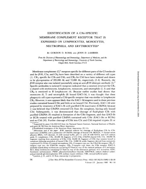
IDENTIFICATION OF A C3bi-SPECIFIC ... - John D. Lambris
IDENTIFICATION OF A C3bi-SPECIFIC ... - John D. Lambris
IDENTIFICATION OF A C3bi-SPECIFIC ... - John D. Lambris

Create successful ePaper yourself
Turn your PDF publications into a flip-book with our unique Google optimized e-Paper software.
<strong>IDENTIFICATION</strong> <strong>OF</strong> A <strong>C3bi</strong>-<strong>SPECIFIC</strong><br />
MEMBRANE COMPLEMENT RECEPTOR THAT IS<br />
EXPRESSED ON LYMPHOCYTES, MONOCYTES,<br />
NEUTROPHILS, AND ERYTHROCYTES*<br />
BY GORDON D. ROSS:I: ANO JOHN D. LAMBRIS<br />
From the Division of Rheumatology and Immunology, Department of Medicine, and the<br />
Department of Bacteriology and Immunology, University of North Caroh'na,<br />
Chapel Hill, North Carolina 27514<br />
Membrane complement (C)1 receptors specific for different parts of the C3 molecule<br />
and for/~IH, C5a, and Clq have been described on a variety of different cell types<br />
(1). CR1, specific for C3b and C4b, and CRz for C3d have been isolated and shown<br />
to be glycoproteins of 205,000 Mr and 72,000 Mr, respectively (2-4). Recently, the<br />
fllH receptor also was isolated successfully using an anti-/~lH idiotypic antibody (5). 2<br />
Specific antibodies to isolated C receptors indicated that a common structure for CR1<br />
is shared with erythrocytes, lymphocytes, monocytes, and neutrophils (2, 3) and that<br />
CR~ is restricted to B lymphocytes (4). Because earlier studies had shown that<br />
monocytes (6, 7) and neutrophils (8) bound EAC1-3d, it was thought that these<br />
phagocytic cell types expressed a C3d-specific receptor that was similar to lymphocyte<br />
CR2. However, it now appears likely that the EAC 1-3d reagents used in these previous<br />
studies contained bound <strong>C3bi</strong> and little or no bound C3d. Previously, EAC 1-3d were<br />
prepared by treatment of EAC 1-3b with purified C3b inactivator (C3bINA) because<br />
it was believed that C3bINA removed C3c from the complexes, leaving only bound<br />
C3d. Subsequently, it was demonstrated that cleavage of fluid-phase C3b with<br />
purified C3bINA (9) resulted in formation of the <strong>C3bi</strong> fragment, and that EAC1-3b<br />
or EC3b treated with purified C3bINA contained only <strong>C3bi</strong> (EAC1-3bi or E<strong>C3bi</strong>)<br />
and no C3d (10). Further cleavage of <strong>C3bi</strong> into C3c and C3d required trypsin (9) or<br />
* Supported by grant CA-25613-03 from the National Cancer Institute, National Institutes of Health,<br />
and grant 80 766 from the American Heart Association.<br />
:~ Established investigator of the American Heart Association (78 155).<br />
1Abbreviations used in this paper." ,81H, essential cofactor for cleavage of fluid-phase C3b by <strong>C3bi</strong>nactivator<br />
(C3bINA) and a potentiator of C3bINA cleavage of bound C3b; BDVEA, I¢~ bovine serum<br />
albumin (BSA), 3.2% dextrose, 35 mM veronal buffer, pH 7.2, with 20 mM EDTA, and 0.2% sodium<br />
azide; C; complement; C3b, 181,000 Mr fragment of C3; <strong>C3bi</strong>, C3bINA-cleaved C3b; C3c, 140,000 Mr<br />
fragment resulting from proteolysis of <strong>C3bi</strong>; C3d, 30,000 Mr fragment of <strong>C3bi</strong> that remains bound to<br />
complexes following proteolysis of bound <strong>C3bi</strong>; C3e, 10,000 Mr acidic fragment derived from extensively<br />
trypsinized C3; C3-ms, C3-coated microspheres; CRI, C-receptor type one, the C3b-C4b receptor; CR2, C-<br />
receptor type two, the C3d-<strong>C3bi</strong> receptor; CRa, C-receptor type three, the <strong>C3bi</strong> receptor; EAC1-3,<br />
antibody-coated sheep erythrocytes containing C3 fixed by way of the classical pathway of C activation;<br />
EC3, sheep erythrocytes containing C3 fixed by way of the alternative pathway of C activation; FITC,<br />
fluorescein isothiocyanate; PMSF, phenylmethylsulfonyl fluoride; SDS-PAGE, sodium dodecyl sulfatepol~acrylamide<br />
gel electrophoresis; STI, soybean trypsin inhibitor; VBS, veronal-buffered saline.<br />
<strong>Lambris</strong>, J. D., and G. D. Ross, manuscript in preparation.<br />
96 J. Exv, MED. © The Rockefeller University Press • 0022-1007/82/01/0096/15 $1.00<br />
Volume 155 ,January 1982 96-110<br />
Downloaded from www.jem.org on November 8, 2004
GORDON D. ROSS AND JOHN D. LAMBRIS 97<br />
plasmin (11), suggesting that if these enzymes were present in serum, EAC prepared<br />
with serum might contain bound C3d as well as <strong>C3bi</strong> or only bound C3d. (Terms are<br />
defined in abbreviations list.)<br />
In the present study, various cell types were examined for binding of complexes<br />
that contained only <strong>C3bi</strong> or only C3d. Neutrophils, monocytes, and erythrocytes were<br />
found to bind <strong>C3bi</strong> but not C3d and to express a receptor for <strong>C3bi</strong> (CR3) that was<br />
distinct from CR2 and specific for a site contained in the <strong>C3bi</strong> molecule that was<br />
outside of the d region.<br />
Materials and Methods<br />
Leukocytes and E~ythrocytes. Heparinized blood was obtained from normal volunteers or<br />
patients with leukemia. Tonsils were obtained from patients undergoing routine tonsillectomy.<br />
Normal blood mononuclear cells and neutrophils were isolated on a two-step Ficoll-Hypaque<br />
density gradient (1.08 g/ml and 1.105 g/ml) (3, 8), and monoeytes were either depleted from<br />
mononuclear cell fractions with Sephadex G- 10 (Pharmacia Fine Chemicals, Div. of Pharmacia<br />
Inc., Piscataway, N. J.) (12) or purified on Percoll gradients (13). Immature myeloid cells were<br />
isolated from leukemic blood on a six-step Ficoll-Hypaque gradient (8). After two washes with<br />
phosphate-buffered saline (PBS), erythrocytes and each leukocyte type were resuspended at 4<br />
× 10 cells/ml in 35 mM veronal buffer, pH 7.2, containing 1% bovine serum albumin (BSA),<br />
20 mM EDTA, 3.2% dextrose, and 0.2% sodium azide (BDVEA; 6 mS at 22°C). Raji and<br />
Daudi leukemic lymphoblastoid cells and the BF lymphoblastoid line derived from normal<br />
lymphocytes were maintained in RPMI 1640 supplemented with 10% fetal bovine serum and<br />
antibiotics.<br />
Purification of C Components and Preparation of C3 Fragments. C3, factor B, fl 1 H, nephritic factor,<br />
and C3bINA were purified as previously described (14, 15). Factor D was purified as described<br />
by LaSavre et al. (16). C3b-Sepharose was generated by mixing together 600 mg of C3 in 10<br />
mM EDTA-PBS with a 0.24% weight ratio of trypsin in the presence of 75 ml of activated-thiol<br />
Sepharose (Pharmacia Fine Chemicals) in a total volume of 150 ml (17). After 15 rain at 37°C,<br />
trypsin was inhibited by addition of a threefold molar excess of soybean trypsin inhibitor (STI)<br />
and then the disulfide-linked C3b-sepharose was washed three times by centrifugation with icecold<br />
10 mM EDTA-PBS. Elution of the C3b-Sepharose with l-cysteine demonstrated 6 mg of<br />
C3b per ml of gel. <strong>C3bi</strong>-Sepharose was formed by treatment of the C3b-Sepharose in 20 mM<br />
EDTA-DGVB (3.7 mS at 22°C), with a weight ratio of 50% ~IH and 4% C3blNA for 6 h at<br />
37~C, followed by four washes with 1.0 M NaC1 in PBS. C3d-Sepharose was formed by<br />
treatment of C3b-Sepharose with trypsin (17) or elastase. With elastase, 8.5 ml of C3b-Sepharose<br />
was treated with an 8% weight ratio of purified porcine elastase (18) in 20 mM Tris/HC1, pH<br />
8.7, for 3 h at 37°C, followed by a second addition of 8% elastase and another 3 h at 37°C. The<br />
C3d-Sepharoses formed with each enzyme were then washed three times with ice-cold veronalbuffered<br />
saline (VBS). The wash supernatants were then concentrated to 10 ml with a UM-2<br />
membrane (Amicon Corp., Scientific Sys. Div. Lexington, Mass.) and chromatographed on a<br />
5 × 90-cm column of Sephadex G-75 (Pharmacia Fine Chemicals) in VBS. Four protein (A280)<br />
peaks were detected, separately pooled, and concentrated with a UM-2 membrane. Analysis by<br />
sodium dodecyl sulfate-polyacrylamide gel electrophoresis (SDS-PAGE) (19) demonstrated that<br />
the first peak from the G-75 column contained C3c (20) and the second peak contained trypsin<br />
or elastase, whereas no Coomasie Blue-stained protein bands were detectable in the third and<br />
fourth peaks with 15% polyacrylamide. These last two small C3 fragment pools contained no<br />
detectable intact C3c or C3d by SDS-PAGE. A small C3 fragments preparation was also<br />
generated by treatment of water-lysed E<strong>C3bi</strong> stroma with plasmin-Sepharose. Purified plasminogen<br />
(21) was coupled to 5 ml of Sepharose CL-4B (22) at a ratio of 10 mg enzyme per ml<br />
of gel and then after activation of a 50% gel suspension with urokinase (14), was mixed on a<br />
o<br />
I0<br />
tube rotator for 60 rain at 37 C with 4 × 10 E<strong>C3bi</strong> stroma that contained 1.2 X 10 ~ <strong>C3bi</strong><br />
molecules per cell. The supernatant was then collected by centrifugation, concentrated with a<br />
UM-2 membrane, and chromatographed on Sephadex G-75 in a similar manner as were the<br />
trypsin and elastase C3 fragments. The C3b-, <strong>C3bi</strong>-, and C3d-Sepharoses were each eluted with<br />
Downloaded from www.jem.org on November 8, 2004
98 MEMBRANE RECEPTORS FOR <strong>C3bi</strong><br />
20 mM cysteine and the liberated C3 fragments dialyzed against BDVEA (prepared without<br />
BSA). Analysis of each C3 fragment by SDS-PAGE under reducing conditions with 12%<br />
polyacrylamide demonstrated the known Mr chain structures (23): 3 C3b, 115,000 a' and 75,000<br />
fl; <strong>C3bi</strong>, 80% iC3b3 (41,000 a'4) and 20% iC3b2 (75,000 fl, 68,000 Or'a, and 43,000 a'2); C3c<br />
(from elastase digest only), 75,000 /~, 43,000 a'2, and 29,000 a'3; and C3d, 30,000 a'5.<br />
Furthermore, no C3b was detected in the <strong>C3bi</strong> nor in the C3c, and no C3d was detected in the<br />
<strong>C3bi</strong>.<br />
Preparation of C3-coated Sheep Erythrocytes (EC3) and Fluorescent Microspheres (C3-ms). EC3b,<br />
E<strong>C3bi</strong>, and EC3d were prepared as previously described (14, 24) and contained 1.5 to 2.5 ×<br />
104 bound C3 molecules per cell, as determined by the uptake of 125I-monoclonal anti-C3<br />
(Bethesda Research Laoraories, Rockville, Md.). Coumarin (green) and rhodamine (red) stained<br />
fluorscent 0.9-#m Diam microspheres (Covalent Technology Corp., Redwood City, Calif.) were<br />
coated with isolated C3b, <strong>C3bi</strong>, and C3d fragments forming C3b-ms, <strong>C3bi</strong>-ms, and C3d-ms<br />
(25). 300/.tl of a 1.4% suspension of microspheres in PBS were mixed with 100/zl of C3b, <strong>C3bi</strong>,<br />
or C3d (400/zg/ml) and incubated at 25°C for 1 h on a tube rotator. The C3-ms were then<br />
washed three times with 1% BSA/PBS by centrifugation for 10 min in a Beckman Microfuge<br />
(Beckman Instruments, Inc., Palo Alto, Calif.), resuspended in 3.0 ml of BDVEA containing<br />
1.0 mM phenylmethylsulfonyl fluoride (PMSF), and sonicated briefly until a single particle<br />
suspension was obtained.<br />
Preparation of Antibodies Specific for CR1, CR2, C3c, and C3d. Rabbits were immunized with<br />
purified CRx (26), CR2 (4), trypsin-generated C3c and C3d (14), and the F(ab')2, Fab', or Fab<br />
fragments of the isolated IgG antibodies were prepared by pepsin or papain cleavage (3, 4, 14).<br />
Assay of C Receptors. C receptors were assayed by rosette formation with EC3 (1) or C3-ms in<br />
BDVEA. For C3-ms rosette assay, 100 ~1 of cells at 4 X 10 s cells/ml were mixed with 100/d of<br />
C3-ms in a 10 × 75-mm plastic tube and placed on a tube rotator with horizontal axis for 15<br />
min at 37°C. Alternatively, the 200-/zl mixture of cells and C3-ms were pelleted together at<br />
1,000 g for 5 rain and incubated as a pellet for 5 rain at 370C. Next, the unbound C3-ms were<br />
removed from the cell suspension (or resuspended cell pellet) by layering the 200/~1 of cells onto<br />
4 ml of 6% BSA in PBS in another 10 × 75-mm plastic tube and centrifuging at 200 g for 10<br />
min at room temperature. After aspiration of the supernatant, the cell pellet was resuspended<br />
in residual wash fluid by shaking the tube gently, and the cells were examined for bound<br />
fluorescent beads by standard fluorescence microscopy techniques. With leukocytes, cells<br />
binding five or more C3-ms were considered positive, whereas with erythrocytes, a positive<br />
cutoff of three or more bound C3-ms was used.<br />
For assay of the morphology of immature myeloid cell EC3 rosettes, Wright-Giemsa stained<br />
smears of rosette suspensions were prepared and analyzed as previously described (8).<br />
Assay for C Receptor Specificity. A pellet of 4 × 10 ~ C-receptor cells in a 10 × 75-mm plastic<br />
tube was resuspended in 100/~1 of either BDVEA or F(ab')2 anti-CR1 (1 mg/ml), F(ab')z-anti-<br />
CR2 (3 mg/ml), C3b (1.0 mg/ml), <strong>C3bi</strong> (0.7 mg/ml), elastase-generated C3c (2.5 mg/ml), or<br />
trypsin-generated C3d (0.5 mg/ml) diluted in BDVEA, incubated at 37°C for 10 min, and<br />
assayed for C-receptors by addition of 100 #1 of EC3 or C3-ms in BDVEA. Alternatively, pellets<br />
of 2 × l0 7 EC3 were treated with 100/d of BDVEA or Fab anti-C3c (100 #g/ml) or Fab anti-<br />
C3d (3 mg/ml) in BDVEA, incubated for 20 rain at 37°C, and then tested for rosette formation<br />
with 100/LI of C-receptor cells.<br />
Assay for Enhancement of E<strong>C3bi</strong> Rosette Formation by Protease Inhibitors and Anti-Elastase. Cell<br />
pellets of 4 × 10⁵ leukocytes in 10 × 75-mm plastic tubes were resuspended in 100/zl of various<br />
concentrations of either protease inhibitors or rabbit IgG anti-human neutrophil elastase (kindly<br />
provided by Dr. <strong>John</strong> Spitznagel, Emory University, Atlanta, Ga.), previously absorbed six<br />
times with a 10% packed volume of sheep erythrocytes. Next, each inhibitor or anti-elastase<br />
treated cell suspension was assayed for E<strong>C3bi</strong> rosette formation in the usual manner.<br />
Double-Label Assay of Lymphocytes for CR~ and Surface Immunoglobulin (I~), or Leu-1 and 3A 1 T Cell<br />
Antigens, or OKM-1 Monocyte-Null Cell Determinant. A pellet of 1 × 10 lymphocytes was treated<br />
simultaneously for 20 rain at room temperature with 25/zl of F(ab')2-anti-CR2 (3 mg/ml) and<br />
Downloaded from www.jem.org on November 8, 2004<br />
3 Ross, G. D., and J. D. <strong>Lambris</strong>. Identification of three forms of iC3b that have distinct structures and<br />
binding site properties. Proceedings of IX International Complement Workshop.J. Imrnunol. In press.
GORDON D. ROSS AND JOHN D. LAMBRIS 99<br />
a fluorescein isothiocyanate (FITC)-linked stain specific for either surface Ig, Leu-1 or 3A1 T<br />
cell antigens, or OKM-1 monocyte-null cell determinant, and then examined for rosette<br />
formation with rhodamine-stained <strong>C3bi</strong>-ms. For Ig staining, cells were treated with 25/zl of<br />
F(ab')2-anti-IgM, IgD, IgA, IgG-fluorescein (N. L. Cappel Laboratories, Cochranville, Pa.). For<br />
T cell staining, 'cells were treated with a 45-p.1 mixture containing 2.0/~g of protein-A-FITC<br />
(Pharmacia Fine Chemicals) and 1.0 p.g of either mouse IgG-anti-Leu-1 (B-D FACS Systems,<br />
Beeton, Dickinson & Co., Sunnyvale, Calif.), mouse IgG-anti-3A1 (27) (kindly donated by Dr.<br />
George Eisenbarth, Duke University, Durham, N. C.), or mouse IgG-anti-OKM-I (Ortho<br />
Pharmaceutical, Raritan, N. J.). Next, to enhance the monoclonal antibody FITC staining, the<br />
washed cells were stained in addition with FITC-F(ab')2-anti-mouse IgG (N. L. Cappel<br />
Laboratories). Finally, the fluorescein-stained cells were resuspended in 100/zl of BDVEA and<br />
assayed for CRs by addition of 100 ~1 of rhodamine-<strong>C3bi</strong>-ms. Cells were examined sequentially<br />
for fluorescein and/or rhodamine staining. In each case the anti-CR2-treated cells were also<br />
tested for complete absence of the ability to rosette with C3d-ms.<br />
Results<br />
Binding of <strong>C3bi</strong> Complexes to Various Leukocyte Types and Erythrocytes. Both E<strong>C3bi</strong> and<br />
<strong>C3bi</strong>-ms bound to a proportion of lymphocytes, erythrocytes, neutrophils, and monocytes<br />
(Table I). In all cases, <strong>C3bi</strong>-ms bound to a greater percentage of cells than did<br />
E<strong>C3bi</strong>. The increased binding of <strong>C3bi</strong>-ms was particularly apparent with erythrocytes,<br />
92% of which bound <strong>C3bi</strong>-ms, and 10% or less bound E<strong>C3bi</strong>. Only lymphoid cells<br />
bound C3d complexes. Immature monocytes and myeloid cells isolated from patients<br />
with either acute monocytic leukemia or chronic myelogenous leukemia were also<br />
negative for binding of C3d complexes.<br />
Specificity of <strong>C3bi</strong>-dependent Rosette Formation. The specificity of <strong>C3bi</strong> complex binding<br />
was examined by assays for inhibition of rosette formation, either by treatment of<br />
the C receptor cells with Fab' anti-C-receptor antibodies or fluid-phase C3 fragments<br />
(Table II) or by treatment of the C3 complexes with Fab anti-C3c or Fab anti-C3d<br />
antibodies (Table III). With all cell types, C3b-ms rosettes were inhibited by anti-<br />
CR1, fluid-phase C3b and fluid-phase C3c but not by anti-CR2, fluid-phase <strong>C3bi</strong>, or<br />
fluid-phase C3d. By contrast, <strong>C3bi</strong>-ms rosettes were not inhibited by anti-CR1, fluidphase<br />
C3b, or fluid-phase C3c. Thus, neither <strong>C3bi</strong>-ms nor fluid-phase <strong>C3bi</strong> bound to<br />
CR1 on any cell type. With erythrocytes, neutrophils, and monocytes, <strong>C3bi</strong>-ms rosettes<br />
were inhibited by fluid-phase <strong>C3bi</strong> but not by anti-CR1, anti-CR2, fluid-phase C3b,<br />
C3c, or C3d. Therefore, with these nonlymphoid cell types, <strong>C3bi</strong>-ms were bound to<br />
Downloaded from www.jem.org on November 8, 2004<br />
TABLE I<br />
EC3 and C3-ms Rosette Formation with Lymphocytes, E(ythrocytes,<br />
Neutrophils, and Monocytes<br />
Blood lymphocytes<br />
Lymphoblastoid lines<br />
Raji 0 0<br />
Daudi 0 0<br />
BF 98 L00<br />
Erythrocytes 75 95<br />
Neutrophils 95 100<br />
Monocytes 85 95<br />
* Assayed in the presence of 1.0 mg/ml of STI.<br />
EC3b C3b-ms E<strong>C3bi</strong> <strong>C3bi</strong>-ms EC3d C3d-ms<br />
% % % % % %<br />
16 17 [0 12 7 9<br />
99 100 98 100<br />
84 96 86 95<br />
98 99 97 98<br />
10 92 0 0<br />
75* 89* 0 0<br />
84 91 0 0
100 MEMBRANE RECEPTORS FOR <strong>C3bi</strong><br />
TAnLE II<br />
Inhibition of C3-ms Rosettes by Anti-C3 Receptor Antibodies and<br />
Fluid-Phase C3 Fragments<br />
Inhibition of rosette formation by<br />
C3<br />
C receptor cell type complex Anti- Anti- Fluid-phase*<br />
CRI CRz C3b <strong>C3bi</strong> C3c C3d<br />
% % % % % %<br />
Erythrocytes C3b-ms 100 0 100 0 48 0<br />
<strong>C3bi</strong>-ms 0 0 0 85 0 0<br />
Neutrophils C3b-ms 100 0 62 0 32 0<br />
<strong>C3bi</strong>-ms 0 0 0 80 0 0<br />
Monocytes C3b-ms 100 0 60 0 27 0<br />
<strong>C3bi</strong>-ms 0 0 0 50 0 0<br />
Raji lymphoblasts <strong>C3bi</strong>-ms 0 78 0 I00 0 71<br />
C3d-ms 0 100 0 100 0 100<br />
* C3b, 500 #g/ml; <strong>C3bi</strong>, 350 #g/ml; C3c (elastase), 1.25 mg/ml; C3d (trypsin),<br />
250 #g/rnl.<br />
TABLE III<br />
Inhibition of EC3 Rosettes with Fab-Anti-C3c and Fab-Anti-C3d<br />
Cell type<br />
EC3 type<br />
Inhibition of rosettes by<br />
Anti-C3c Anti-C3d<br />
% %<br />
Erythrocyte EC3b 100 0<br />
E<strong>C3bi</strong> 100 I00<br />
Neutrophils and EC3b 100 0<br />
Monocytes E<strong>C3bi</strong> 100 100<br />
Raji or Daudi E<strong>C3bi</strong> 100 100<br />
Lymphoblasts EC3d 0 100<br />
Downloaded from www.jem.org on November 8, 2004<br />
a receptor that was distinct from CR1 and CR2, herein designated CR3. Lymphocytes<br />
differed from other cell types in that <strong>C3bi</strong>-ms rosettes were partially inhibited by<br />
anti-CR2 and fluid-phase C3d. Also, lymphocyte-C3d-ms rosettes were completely<br />
inhibited by fluid-phase <strong>C3bi</strong> as well as by anti-CR2 or fluid-phase C3d. Thus, with<br />
lymphocytes that expressed CR2, <strong>C3bi</strong> complexes were bound to CR2 by way of the<br />
d region of the intact <strong>C3bi</strong> molecule. However, with concentrations of up to 5 mg/ml<br />
of Fab' anti-CR2 or 1.0 mg/ml of fluid-phase C3d, lymphocyte <strong>C3bi</strong> complex rosettes<br />
were never inhibited completely. This indicated that lymphocytes expressed CR3 in<br />
addition to CR2 and that both C receptor types were responsible for binding <strong>C3bi</strong><br />
complexes to lymphocytes.<br />
Fab anti-C3c and Fab anti-C3d both inhibited E<strong>C3bi</strong> rosette formation with all<br />
cell types (Table III). Fab anti-C3d did not inhibit EC3b rosette formation, despite<br />
the finding that Fab anti-C3d inhibited the agglutination of EC3b by IgG anti-C3d<br />
and thus bound to the d region of intact C3b.<br />
Because CR3-dependent rosettes were not inhibited by fluid-phase C3c or C3d,
GORDON D. ROSS AND JOHN D. LAMBRIS 101<br />
other smaller C3 fragments generated by proteolysis of C3b or <strong>C3bi</strong> were examined<br />
for inhibition of <strong>C3bi</strong>-ms rosette formation. Inhibition of neutrophil-E<strong>C3bi</strong> rosettes<br />
and human E-<strong>C3bi</strong>-ms rosettes was observed with the fluid-phase small C3 fragments<br />
pool generated with plasmin or trypsin but not with elastase.<br />
Inhibition of <strong>C3bi</strong>-dependent Neutrophil Rosette Formation by Secreted Neutrophil<br />
Elastase. Because of the known proteolytic sensitivity of <strong>C3bi</strong> (9) and because the<br />
bound product of <strong>C3bi</strong> digestion, C3d, was unreactive with neutrophils and monocytes,<br />
protease inhibitors were added to rosette assays to protect the <strong>C3bi</strong>-complexes<br />
from proteolysis into CR3-unreactive C3d complexes (Table IV). Both STI and PMSF<br />
enhanced neutrophil-E<strong>C3bi</strong> rosette formation from 5% up to 76-89%, whereas benzamidine<br />
and epsilon amino caproic acid caused no rosette enhancement. These same<br />
protease inhibitors had no effect on E<strong>C3bi</strong> rosette formation with blood monocytes or<br />
lymphocytes. Because neutrophils were known to secrete elastase in response to<br />
opsonized bacteria (28) and because human neutrophil elastase was known to cleave<br />
<strong>C3bi</strong> into C3c and C3d (20), an antibody to human neutrophil elastase was examined<br />
for its ability to enhance neutrophil-E<strong>C3bi</strong> rosette formation (Table IV). Anti-elastase<br />
produced the same rosette enhancement as did STI and PMSF. In the absence of<br />
protease inhibitors or anti-elastase, E<strong>C3bi</strong> that had been incubated with neutrophils<br />
for 60 min at 37°C were no longer agglutinated by anti-C3c, whereas agglutination<br />
by anti-C3d and Raji rosette formation were undiminished.<br />
Acquisition of Elastase-secreting Ability with Neutrophil Maturation. Neutrophils from<br />
the blood of a patient with chronic myelogenous leukemia and blood count of 2 × 10⁵<br />
neutrophils per/~1 were fractionated into immature (band-form nucleus) and mature<br />
polymorphonuclear cells and examined for E<strong>C3bi</strong> rosette formation with increasing<br />
concentrations of STI (Table V). In the absence of STI, 49% of band-form neutrophils<br />
formed rosettes with E<strong>C3bi</strong>, whereas high-density polymorphs did not form E<strong>C3bi</strong><br />
TABLE IV<br />
Enhancement of Neutrophil-E<strong>C3bi</strong> Rosette Formation with Protease Inhibitors<br />
and Anti-Elastase<br />
Protease inhibitor<br />
Neutrophil-E<strong>C3bi</strong><br />
rosette formation<br />
%<br />
Buffer control 5<br />
STI<br />
1.0 mg/ml 88<br />
0.5 mg/ml 89<br />
0.25 mg/ml 70<br />
0.10 mg/ml 35<br />
0.05 mg/ml 25<br />
PMSF<br />
2.0 mM 36<br />
[.0 mM 76<br />
0.5 mM 40<br />
Anti-elastase<br />
1/5 87<br />
1/10 87<br />
1/20 79<br />
1/40 38<br />
Downloaded from www.jem.org on November 8, 2004
102 MEMBRANE RECEPTORS FOR <strong>C3bi</strong><br />
TABLE V<br />
Neutrophil Cell Density and Maturation-linked Requirement for Increased<br />
Amounts of STI to Allow E<strong>C3bi</strong> Rosette Formation<br />
Neutrophil density in g/ml<br />
(predominant morphology)<br />
1.07 1.09 1.105 1.12<br />
(bands) (PMN) (PMN) (PMN)<br />
%R* %R %R %R<br />
Buffer control 49 40 10 0<br />
ST1<br />
25 ~g/ml 51 54 20 10<br />
50 p.g/ml 50 70 25 19<br />
200/Lg/ml 50 69 49 39<br />
400/zg/m| 51 68 92 68<br />
800 ~g/ml 49 71 95 97<br />
1 mg/ml 51 69 94 98<br />
* Percent E<strong>C3bi</strong> rosette formation.<br />
TABLE VI<br />
Expression of CR3 on Lymphocytes Detected by <strong>C3bi</strong>-ms Rosette Formation<br />
with Anti-CR2-treated Cells<br />
Cell type<br />
Rosette formation with<br />
C3d-ms <strong>C3bi</strong>-ms<br />
% %<br />
Blood lymphocytes (8) 9 12<br />
+ anti-CR2 0 3.5<br />
Tonsil lymphocytes (3) 54 59<br />
+ anti-CR2 0 27<br />
Raji lymphoblasts 99 99<br />
+ anti-CR2 0 40<br />
Daudi lymphoblasts 95 96<br />
+ anti-CR2 0 32<br />
BF lymphoblasts 59 56<br />
+ anti-CR2 0 9<br />
Downloaded from www.jem.org on November 8, 2004<br />
rosettes. With the band-form neutrophils, STI did not increase the proportion of<br />
E<strong>C3bi</strong> rosettes. However, with neutrophils isolated at a 1.09 g/ml density, 50 Ilg/ml<br />
STI was required for maximum enhancement of E<strong>C3bi</strong> rosette formation, whereas<br />
with 1.105 g/ml neutrophils and 1,12 g/ml neutrophils, 400/~g/ml and 800/~g/ml of<br />
STI were required, respectively (Table V).<br />
Expression of CR~ on Different Lymphocyte Subsets. Lymphocytes from blood and<br />
tonsils and various B type lymphoblastoid lines were treated with sufficient anti-CR2<br />
to inhibit C3d-ms binding completely and then assayed for binding of <strong>C3bi</strong>-ms (Table<br />
VI). The majority of <strong>C3bi</strong>-ms-binding cells expressed only CRz and did not express<br />
CRa, as anti-CR2 treatment of ceils produced 54-84% inhibition of <strong>C3bi</strong>-ms rosette<br />
formation. Among normal blood lymphocytes, only 1.5-4.5% (average 3.5%) of cells<br />
expressed CRa, and these were apparently distinct from the CR2-bearing cells that<br />
represented 9.0% of peripheral lymphocytes. Double-label assays with blood lympho-
GORDON D. ROSS AND JOHN D. LAMBRIS 103<br />
cytes from six normal individuals demonstrated that 86% of cells bearing CRz or CR3<br />
also expressed membrane Ig detectable with F(ab')2-anti-Ig. No CR2-positive cells<br />
were detected that expressed either Leu-1 or 3AI T cell determinants. Among the<br />
CRa-positive blood lymphocytes, 5% of cells stained with either anti-Leu-1 or anti-<br />
3A1, and 16% of cells stained with anti-OKM-1. Tonsils contained a considerably<br />
higher proportion of CR3-bearing cells than did peripheral blood. However, unlike<br />
blood lymphocytes, the majority of tonsil CRa-positive cells expressed CR2 because<br />
the percentage of cells binding either <strong>C3bi</strong>-ms or C3d-ms was nearly equal. All three<br />
B type lymphoblastoid cell lines examined expressed CR3 on a proportion of the cells.<br />
Discussion<br />
The major finding in the present study is that lymphocytes, monocytes, neutrophils,<br />
and erythrocytes express a <strong>C3bi</strong>-specific membrane C receptor (CRa) that is distinct<br />
from CR1 and CR2. CRa is specific for <strong>C3bi</strong> and unreactive with C3b, C3c, and C3d.<br />
Neutrophils and monocytes lack detectable CR2 at all stages of maturation and bind<br />
<strong>C3bi</strong> complexes exclusively to CRa. Neutrophils begin to express CRa at the myelocyte<br />
stage and the receptor is fully expressed on polymorphs. Peripheral blood lymphocytes<br />
bind <strong>C3bi</strong> complexes primarily to CRy, and the cells that express CRa are a separate<br />
B cell subset from the CR2-bearing B cells.<br />
C receptors were assayed with either sheep erythrocytes or fluorescent microspheres<br />
coated with specific C3 fragments (EC3 or C3-ms). C3-ms had distinct advantages<br />
over EC3. First, probably because of their smaller size, C3-ms were more sensitive to<br />
cells known to have a low number of C-receptors per cell. C3b-ms and C3d-ms bound<br />
to nearly all human erythrocytes and Daudi cells respectively, whereas EC3b and<br />
EC3d bound to fewer of these two cell types. Also, C3-ms could be prepared with very<br />
small amounts of pure C3 fragments that had been previously characterized fully by<br />
SDS-PAGE.<br />
The receptor specificity of C3 complex binding to different cell types was investigated<br />
by rosette inhibition studies in which either the C receptor cells were treated<br />
with anti-CRa, anti-CR2, fluid-phase C3b, <strong>C3bi</strong>, C3c, or C3d, or alternatively the C3<br />
complexes were treated with anti-C3c or anti-C3d. In experiments that examined the<br />
binding specificity of C3b complexes or fluid-phase C3b, it was essential to use<br />
inhibitors of proteolysis of C3b in the rosette assay buffer. Lymphocytes (14, 29),<br />
monocytes (30, 31), and neutrophils (32) secrete endogenous fllH and C3bINA that<br />
may convert bound or fluid-phase C3b into <strong>C3bi</strong>. Neutrophils also secrete elastase<br />
that may cleave C3b into C3d (20). EDTA and sodium azide were used in the<br />
rosetting buffer (BDVEA) because they had been shown previously to inhibit the<br />
release of B cell C3bINA and fllH (14) and also seemed to inhibit monocyte and<br />
neutrophil release of these components. In addition, STI was added to the BDVEA<br />
buffer to inhibit neutrophil elastase activity. <strong>C3bi</strong> complexes did not bind to CR1<br />
because <strong>C3bi</strong>-ms rosettes were not inhibited by anti-CR1, fluid-phase C3b, or fluidphase<br />
C3c, whereas these same materials did inhibit C3b-ms binding to CR1. In<br />
addition, isolated fluid-phase a25I-labeled CRx does not bind to E<strong>C3bi</strong>, whereas fluidphase<br />
CR1 does bind to EC3b (3). The <strong>C3bi</strong> complex binding activity of erythroeytes,<br />
monocytes, and neutrophils was also distinct from CR2 activity because these cell<br />
types did not bind C3d complexes, nor were <strong>C3bi</strong>-ms rosettes inhibited by anti-CR~<br />
or fluid-phase C3d. Lymphocytes differed from these other cell types in that <strong>C3bi</strong><br />
Downloaded from www.jem.org on November 8, 2004
104 MEMBRANE RECEPTORS FOR <strong>C3bi</strong><br />
complexes were bound primarily to CR2. Lymphocyte binding of <strong>C3bi</strong>-ms was<br />
inhibited by anti-CR2 and fluid-phase C3d as well as by fluid <strong>C3bi</strong>. Furthermore,<br />
lymphocyte-C3d-ms rosette formation was inhibited completely by fluid-phase <strong>C3bi</strong><br />
as well as by fluid C3d and anti-CR2. A portion of lymphocytes did bind <strong>C3bi</strong><br />
complexes independently of CRz because treatment of lymphocytes with amounts of<br />
F(ab')2 anti-CR2 or fluid-phase C3d that were twofold to fourfold greater than that<br />
required for complete inhibition of C3d-ms binding only produced 50-84% inhibition<br />
of <strong>C3bi</strong>-ms binding. Thus, lymphocytes bound <strong>C3bi</strong> complexes either to CRz by way<br />
of the d region of the intact <strong>C3bi</strong> molecule or by way of a distinct <strong>C3bi</strong>-specific<br />
receptor that was distinct from CR2. The <strong>C3bi</strong>-specific binding activity that was<br />
distinct from CR2 was designated CR3 with all cell types.<br />
Because C3b does not bind to CR3, the CR3 binding site in the <strong>C3bi</strong> molecule must<br />
be exposed by cleavage of C3b with the C3bINA. Thus, CR3 has a similar binding<br />
specificity as bovine conglutinin (33). Unlike conglutinin (34) however, CRz activity<br />
was not inhibited by EDTA or n-acetyl-o-glucosamine (G. D. Ross, unpublished<br />
observation). Because neither fluid-phase C3b, C3c, nor C3d inhibited the CR3-<br />
binding activity of <strong>C3bi</strong>-ms, the CR3 binding site in the <strong>C3bi</strong> molecule must be either<br />
destroyed, liberated, or covered by proteolysis of <strong>C3bi</strong>. To determine whether a smalI<br />
CR3-specific fragment could be generated that was distinct from intact C3c and C3d,<br />
bound C3b or <strong>C3bi</strong> was digested into C3c and C3d with elastase, trypsin, or plasmin,<br />
and then after removal of the intact C3c and C3d fragments, the remaining small<br />
fragment pools were examined for inhibition of <strong>C3bi</strong> complex binding to neutrophil<br />
or erythrocyte CR3. Inhibition of CR3 was demonstrated by the small C3 fragments<br />
pool generated with plasmin and trypsin but not with elastase. Elastase is known to<br />
have a more limited number of digestion sites in the C3 molecule than has either<br />
plasmin or trypsin (11, 20, 23). In particular, elastase digests only the a chain of C3<br />
or C3b (20), whereas trypsin cleaves both the 0t and the/3 chains of C3 or C3b, and<br />
plasmin cleaves both the a and fl chains of <strong>C3bi</strong> (11). Because none of the three<br />
proteases digest C3d, the data suggests the possibility that the CR3 binding site may<br />
be folded within the intact C3c fragment and not exposed until cleaved from the<br />
molecule with trypsin or plasmin. Likewise, the CR1 binding site is apparently folded<br />
within the <strong>C3bi</strong> molecule and then re-exposed in the C3c fragment that is excised by<br />
proteolysis of <strong>C3bi</strong>. This is because fluid C3c but not fluid <strong>C3bi</strong> inhibited CR1-C3bms<br />
rosette formation. Available data (35, 36) suggest the possibility that the C3e<br />
fragment may contain the CR3 binding site. C3e was shown to be removed from C3c<br />
by extensive trypsin digestion, and 1251-C3e was shown to bind to neutrophils (36).<br />
Previously, it had been shown (8) that neutrophils acquired the ability to bind<br />
EAC 1-3bi at approximately the myelocyte stage of maturation, and then as the cells<br />
matured into polymorphonuclear cells this ability was lost. In these former studies, an<br />
EAC l-3d reagent was used that is now recognized to have actually been an EAC 1-3bi<br />
reagent because it was prepared with purified C3bINA without the additional<br />
proteolysis required to cleave the bound <strong>C3bi</strong> fully to C3d. The present study<br />
demonstrated that the loss of ability of mature neutrophils to rosette with E<strong>C3bi</strong> (or<br />
EAC1-3bi) was not due to a loss of CR3 but rather to the maturation-linked acquisition<br />
of the ability to secrete elastase that cleaved the reagent E<strong>C3bi</strong> into CR3-unreactive<br />
EC3d. Several lines of evidence supported this conclusion. First, E<strong>C3bi</strong> rosette<br />
formation with mature neutrophils was generated with an antibody directed to<br />
Downloaded from www.jem.org on November 8, 2004
GORDON D. ROSS AND JOHN D. LAMBRIS 105<br />
human neutrophil elastase. STI and PMSF also allowed E<strong>C3bi</strong> rosette formation with<br />
mature neutrophils. Second, in the absence of protease inhibitors, E<strong>C3bi</strong> that had<br />
been incubated with neutrophils lost all detectable C3c antigens while retaining C3d<br />
antigens and the ability to bind to lymphocyte CR2. This indicated that neutrophil<br />
enzymes cleaved E<strong>C3bi</strong> to EC3d. Finally, when neutrophils were fractionated into<br />
cells with band form nucleus and polymorphonuclear cells, it was found that STI did<br />
not enhance E<strong>C3bi</strong> rosette formation with band form cells, whereas with polymorphs,<br />
more STI was required to allow E<strong>C3bi</strong> rosettes with high density (1.12 g/ml) mature<br />
polymorphs than with low density (1.09 g/ml) less mature polymorphs. Elastase has<br />
been detected in azurophilic myeloid cell granules by immunofluorescence at the<br />
promyelocyte stage of maturation (37). However, promyelocyte elastase is probably<br />
not secreted, and polymorphs apparently release azurophilic granule enzymes only at<br />
the site of contact with serum-opsonized bacteria (28). Because binding of opsonized<br />
bacteria probably involves Fc receptors, CRI, and CRa, one of these three types of<br />
receptors on mature cells may have the ability to trigger elastase secretion. Because of<br />
this elastase-secreting activity, neutrophils in vivo are probably unable to bind<br />
particles that contain only <strong>C3bi</strong>, and therefore it is presumed that CRa is not<br />
important for neutrophil phagocytosis. Because CRa apparently binds a small trypsinor<br />
plasmin-derived <strong>C3bi</strong> fragment, it is possible that such an active fragment may be<br />
generated by <strong>C3bi</strong> proteolysis in vivo, and that this fragment may be responsible for<br />
triggering some neutrophil function other than phagocytosis.<br />
Monocytes resembled neutrophils in that they expressed CRa and CRa and lacked<br />
detectable CR2. Unlike neutrophils, monocytes did not require protease inhibitors to<br />
allow EC3hi rosette formation. Furthermore, other studies have demonstrated that<br />
human macrophages (31) and rat mast cells (38) ingest <strong>C3bi</strong> complexes much more<br />
efficiently than C3b complexes. Thus, these other phagocyte types differ from neutrophils<br />
in that CRa appears to be more important than CR~ for phagocytosis in vivo.<br />
CRa were also detected on the majority of human erythrocytes. In the past, human<br />
erythrocytes were thought to express only CR1 and not to bind <strong>C3bi</strong> or C3d complexes.<br />
Gaither et al. had noted reduced human erythrocytes immune adherence with<br />
EAC43bi as compared with EAC43b (39). However, it was not clear whether this<br />
<strong>C3bi</strong>-dependent immune adherence was due to a distinct <strong>C3bi</strong>-specific receptor or<br />
rather a low affinity binding of <strong>C3bi</strong> to CR1. Indeed, human E-rosette formation with<br />
E<strong>C3bi</strong> or EAC1-3bi is difficult to demonstrate, as it is such a weak reaction. In the<br />
present study, <strong>C3bi</strong>-ms were prepared with purified <strong>C3bi</strong> fragments containing no<br />
detectable C3b by SDS-PAGE and were shown to bind to nearly all human erythrocytes<br />
in the presence of amounts of anti-CR1 that were sufficient to inhibit C3b-ms<br />
binding completely.<br />
Cells of the human renal glomerulus apparently also express both CR1 and CR3.<br />
Carlo et al. have demonstrated that kidney cells bind both EAC43b and EAC43bi<br />
but not EAC43d (40). Also, it has recently been demonstrated that renal epithelial<br />
cells are fluorescence stained with F(ab')2 anti-CRx (M. Papamichail, J. D. <strong>Lambris</strong>,<br />
and G. D. Ross, unpublished observation).<br />
Lymphocytes differed from all the other cell types examined in that they expressed<br />
CR2 in addition to CRa, and <strong>C3bi</strong> complexes were primarily bound to CR2 rather<br />
than to CRa. For this reason, specific assay of lymphocyte CRa required complete<br />
blockade of membrane CR2 with anti-CR2 before assay of CRa with E<strong>C3bi</strong> or <strong>C3bi</strong>-<br />
Downloaded from www.jem.org on November 8, 2004
106 MEMBRANE RECEPTORS FOR <strong>C3bi</strong><br />
ms. With peripheral blood tymphocytes, only 3.5% of anti-CR2-treated cells bound<br />
<strong>C3bi</strong>-ms. This finding indicated that the majority of the 12.0% of <strong>C3bi</strong>-ms-binding<br />
cells did not express CR3 and expressed only CR2. Parallel assay of C3d-ms-binding<br />
cells confirmed that 9.0% of cells expressed CR2, so that the 12.0% <strong>C3bi</strong>-ms-binding<br />
cells consisted of 8.5% CR2 + CRy- cells, 3.0% CR2-CR3 + cells, and only 0.5% CRz +<br />
CR3 + cells. Thus, CR2 + and CR3 + cells represented nearly distinct subsets. Previous<br />
double-label studies with EAC 1-3hi (41) and present studies with <strong>C3bi</strong>-ms indicated<br />
that the majority of CR2 + and/or CR3 + peripheral blood cells expressed membrane<br />
Ig detectable with F(ab')2-anti-Ig. The same finding was made when CRz + ceils or<br />
CR3 + cells were examined individually for Ig with C3d-ms or anti-CR~ and <strong>C3bi</strong>-ms,<br />
respectively. Among CRa + blood nonadherent lymphoid-appearing cells, an average<br />
of 86% of cells expressed membrane Ig, whereas only 5% of these cells expressed either<br />
Leu-1 or 3AI T cell-specific determinants, and 15% expressed OKM-1 determinants.<br />
The OKM-1 staining CR3 + cells probably represented either third population lymphocytes<br />
(null cells), myeloid precursors, or promonocytes (42). Thus, the majority of<br />
CR3-bearing blood lymphocytes are B cells (3.0%), whereas only 0.2% express T cell<br />
determinants and 0.6% express a monocyte-null lymphocyte determinant. Tonsil<br />
lymphocytes differed from blood lymphocytes in that one-half of the CR3 + cells also<br />
expressed CR2. This was because the proportions of <strong>C3bi</strong>-ms- and C3d-ms-binding<br />
cells were nearly equal, and anti-CR2 produced only 50% inhibition of <strong>C3bi</strong>-ms rosette<br />
formation. As with blood lymphocytes, the majority of CR3 + tonsil cells expressed<br />
membrane Ig, Three different B type lymphoblastoid lines also expressed CR3 + cells,<br />
though with all three lines, <strong>C3bi</strong> complexes were primarily bound to CRz. In previous<br />
studies of three different T cell lymphoblastoid lines and several different lines of<br />
normal activated T cells maintained in T cell growth factor, no cells binding C3b,<br />
C4b, <strong>C3bi</strong>, or C3d complexes were observed (G. D. Ross and G. D. Bonnard,<br />
unpublished observation). The only exception was the MOLT-4 T cell lymphoblastoid<br />
line (43). Taken together, the various data indicate that CR3 is primarily a B cell<br />
marker. Recently, Perlmann et al. (44) have reported that the activity of lymphocytes<br />
functional in antibody-dependent cellular cytotoxicity (ADCC) was greatly enhanced<br />
by target cell-bound <strong>C3bi</strong>, whereas less enhancement was observed with bound C3b<br />
and C3d. Because it was also shown in other studies that ADCC lymphocytes lacked<br />
detectable membrane Ig determinants (45) and expressed either T cell (46, 47) or null<br />
cell markers (48), it appears possible that the small number of Ig-CR3 + cells detected<br />
in the present study may represent the cells functional in ADCC.<br />
The significance of a <strong>C3bi</strong>-specific receptor is not fully understood. Subsequent to<br />
C activation, both bound and fluid-phase C3b are rapidly converted into <strong>C3bi</strong>, and<br />
some of this <strong>C3bi</strong> apparently persists in serum for several hours (10) before being<br />
degraded into smaller C3 fragments. Macrophages (31) and mast cells (38) ingest<br />
<strong>C3bi</strong> complexes much more efficiently than C3b complexes. Also, bound <strong>C3bi</strong><br />
enhances both neutrophil phagocytosis of IgG-coated particles and the neutrophil<br />
superoxide burst (32). However, this <strong>C3bi</strong>-dependent enhancement of neutrophil<br />
function requires addition of an inhibitor ofelastase, suggesting that bound <strong>C3bi</strong> may<br />
not have these functions in vivo. With the exception of ADCC cells, no function of<br />
lymphocyte CR3 has yet been demonstrated. Because <strong>C3bi</strong> can bind to either CR2 or<br />
CR3, it may be possible that <strong>C3bi</strong> complexes can simultaneously crosslink B cell<br />
surface CR2 and CR3 and thereby induce some particular cell function. Future studies<br />
Downloaded from www.jem.org on November 8, 2004
GORDON D. ROSS AND JOHN D. LAMBRIS 107<br />
in which lymphocytes are cultured with <strong>C3bi</strong>-ms with or without Fab'-anti-CR2<br />
might be able to answer this question.<br />
Summary<br />
Cells expressing a membrane C receptor (CRs) specific for C3b-inactivator-cleaved<br />
C3b (<strong>C3bi</strong>) were identified by rosette assay with <strong>C3bi</strong>-coated sheep erythrocytes<br />
(E<strong>C3bi</strong>) or <strong>C3bi</strong>-coated fluorescent microspheres (<strong>C3bi</strong>-ms). <strong>C3bi</strong>-ms, probably because<br />
of their smaller size, bound to a higher proportion of cells than did E<strong>C3bi</strong>.<br />
<strong>C3bi</strong>-ms bound to >90% of mature neutrophils, 85% of monocytes, 92% of erythrocytes,<br />
and 12% of peripheral blood lymphocytes. Binding of <strong>C3bi</strong>-ms to neutrophils,<br />
monocytes, and erythrocytes was inhibited by fluid-phase <strong>C3bi</strong>, Fab anti-C3c, or Fab<br />
anti-C3d but was not inhibited by F(ab')2 anti-CR~ (C3b receptor) or F(ab')2 anti-<br />
CR2 (C3d receptor) nor by fluid-phase C3b, C3c, or C3d. This indicated that<br />
monocytes, neutrophils, and erythrocytes expressed <strong>C3bi</strong> receptors (CRs) that were<br />
separate and distinct from CR1 and CR2 and specific for a site in the C3 molecule<br />
that was only exposed subsequently to cleavage of C3b by C3b inactivator and that<br />
was either destroyed, covered, or liberated by cleavage of <strong>C3bi</strong> into C3c and C3d<br />
fragments. Lymphocytes differed from these other cell types in that they expressed<br />
CR2 in addition to CRa. Lymphocyte <strong>C3bi</strong>-ms rosettes were inhibited from 50 to 84%<br />
by F(ab')2-anti-CR2 or fluid-phase C3d, whereas C3d-ms rosettes were inhibited<br />
completely by F(ab')2 anti-CR2, fluid-phase <strong>C3bi</strong>, or fluid-phase C3d. Thus, with<br />
lymphocytes, <strong>C3bi</strong> was bound to CRa, and in addition was bound to CR2 by way of<br />
the intact d region of the <strong>C3bi</strong> molecule. In studies of the acquisition of C receptors<br />
occurring during myeloid cell maturation, the ability to rosette with <strong>C3bi</strong>-coated<br />
particles was detected readily with immature low-density cells, whereas this ability<br />
was nearly undetectable with high density mature polymorphonuclear cells. This<br />
absence of <strong>C3bi</strong> binding to polymorphs was not due to a loss of the CR3 but instead<br />
was due to the maturation-linked acquisition of the abiity to secrete elastase that<br />
cleaved reagent particle-bound <strong>C3bi</strong> into CRs-unreactive C3d. Neither neutrophils<br />
nor monocytes bound C3d-coated particles at any stage of maturation. Assay of CR3<br />
with mature neutrophils required inhibition of neutrophil elastase with either soybean<br />
trypsin inhibitor or anti-elastase antibodies, and the amounts of these elastase inhibitors<br />
required to allow E<strong>C3bi</strong> rosette formation increased with neutrophil maturation.<br />
Because lymphocytes bound <strong>C3bi</strong> to CR2 as well as to CRa, specific assay of<br />
lymphocyte CR3 required saturation of membrane CR2 with Fab' anti-CR~ before<br />
assay for rosettes with <strong>C3bi</strong>-ms. Only 3.5% of anti-CR2-treated peripheral blood<br />
lymphocytes bound <strong>C3bi</strong>-ms. Therefore, among normal blood lymphocytes the majority<br />
of the 12% <strong>C3bi</strong>-ms-binding cells expressed only CR2 (8.5%), and the small<br />
proportion of <strong>C3bi</strong>-ms-binding cells that expressed CR3 (3.5%) represented a distinct<br />
subset from the CR2 + cells. Double-label assay indicated that 3.0% out of 3.5% of<br />
these CRs-bearing lymphocytes were B cells because they expressed membrane<br />
immunoglobulins. Of the remaining CR3 + cells, 0.2% expressed either Leu-1 or 3A1<br />
T cell antigens, and 0.6% expressed the OKM-1 monocyte-null lymphocyte determinant.<br />
Downloaded from www.jem.org on November 8, 2004<br />
The authors wish to acknowledge the excellent technical assistance of Ms. Joyce Knapp, Ms.
108 MEMBRANE RECEPTORS FOR <strong>C3bi</strong><br />
Julie Schultz, and Ms. Neena Sachdev. The authors are also grateful to Ms. Linda Tillman for<br />
her assistance in preparation of the manuscript.<br />
Received for publication 7August 1981.<br />
References<br />
1. Ross, G. D. 1980. Analysis of the different types of leukocyte membrane complement<br />
receptors and their interaction with complement system.J. ImmunoL Methods. 37:197.<br />
2. Fearon, D. T. 1980. Identification of the membrane glycoprotein that is the C3b receptor<br />
of the human erythrocyte, polymorphonuclear leukocyte, B lymphocyte, and monocyte. J.<br />
Exp. Med. 152:20.<br />
3. Dobson, N. J., J. D. <strong>Lambris</strong>, and G. D. Ross. 1981. Characteristics of isolated erythrocyte<br />
complement receptor type one (CR1, C4b-C3b receptor) and CRa-specific antibodies. J.<br />
ImmunoL 126:693.<br />
4. <strong>Lambris</strong>, J. D., N. J. Dobson, and G. D. Ross. 1981. Isolation of lymphocyte membrane<br />
complement receptor type two (the C3d receptor) and preparation of receptor-specific<br />
antibody. Proc. NatL Acad. Sci. U. S. A. 78:1828.<br />
5. <strong>Lambris</strong>, J. D., N. J. Dobson, M. Dozier, and G. D. Ross. 1981. Antibody to the idiotype<br />
of anti-fl 1H has specificity for B lymphocyte membrane fl 1H receptors and C3b. Fed. Proc.<br />
40:1013.<br />
6. Reynolds, H. Y., J. P. Atkinson, H. H. Newball, and M. M. Frank. 1975. Receptors for<br />
immunoglobulin and complement on human alveolar macrophages. J. IrnmunoL 114:1813.<br />
7. Ehlenberger, A. G., and V. Nussenzweig. 1977. The role of membrane receptors for C3b<br />
and C3d in phagocytosis.J. Exp. Med. 145:357.<br />
8. Ross, G. D., C. I. Jarowski, E. M. Rabellino, and R. J. Winchester. 1978. The sequential<br />
appearance of Ia-like antigens and two different complement receptors during the maturation<br />
of human neutrophils. J. Exp. Med. 147:730.<br />
9. Pangburn, M. K., R. D. Schreiber, and H. J. Miiller-Eberhard. 1977. Human complement<br />
G3b inactivator: isolation, characterization, and demonstration of an absolute requirement<br />
for the serum protein/~IH for cleavage of C3b and C4b in solution. J. Exp. Med. 146:257.<br />
10. Law, S. K., D. T. Fearon, and R. P. Levine. 1979. Action of the C3b-inactivator on cellbound<br />
C3b.J. ImmunoL 122:759.<br />
1 I. Nagasawa, S., and R. M. Stroud. 1977. Mechanism of action of the C3b inactivator:<br />
requirement for a high molecular weight cofactor (C3b-C4blNA cofactor) and production<br />
of a new C3b derivative (C3b'). Immunochemistry. 14:749.<br />
12. Ly, I. A., and R. I. Mishell. 1974. Separation of mouse spleen cells by passage through<br />
columns of Sephadex G-10.J. Immunol. Methods. 5:239.<br />
13. Fluks, A. J. 1981. Three-step isolation of human blood monocytes using discontinuous<br />
density gradients of Percoll.J. ImmunoL Methods. 41:225.<br />
14. <strong>Lambris</strong>, J. D., N. J. Dobson, and G. D. Ross. 1980. Release of endogenous C3b inactivator<br />
from lymphocytes in response to triggering membrane receptors for/~IH globulin. J. Exp.<br />
Med. 152:1625.<br />
15. Schreiber, R. D., O. G6tze, and H. J. M/Jller-Eberhard. 1976. Nephritic factor: its structure<br />
and function and its relationship to initiating factor of the alternative pathway. Stand. J.<br />
ImmunoL 5:703.<br />
16. LeSavre, P. H., T. E. Hugli, A. F. Esser, and H. J. M/iller-Eberhard. 1979. The alternative<br />
pathway C3/C5 convertase: chemical basis of factor B activation.J. ImmunoL 123:529.<br />
17. Tack, B. F., R. A. Harrison, J. A. Janatova, M. L. Thomas, and J. W. Prahl. 1980.<br />
Evidence for presence of an internal thiolester bond in the third component of human<br />
complement. Proc. NatL Acad. Sci. U. S. A. 77:5764.<br />
Downloaded from www.jem.org on November 8, 2004
GORDON D. ROSS AND JOHN D. LAMBRIS 109<br />
18. Narayanan, A. S., and R. A. Anwar. 1969. The specificity of purified porcine pancreatic<br />
elastase. Biochem. jr. 114:11.<br />
19. Laemmli, U. K. 1970. Cleavage of structural proteins during the assembly of the head of<br />
bacteriophage T4. Nature (Lond.). 297:680.<br />
20. Taylor, J. C., I. P. Crawford, and T. E. Hugli. 1977. Limited degradation of the third<br />
component (C3) of human complement by human leukocyte elastase (HLE): partial<br />
characterization of C3 fragments. Biochemistry. 16:575.<br />
21. Deutsch, D. G., and E. T. Mertz. 1970. Plasminogen: purification from human serum by<br />
affinity chromatography. Science (Wash. D. C.). 170:1095.<br />
22. March, S. C., I. Parikh, and P. Cuatrecasas. 1974. Simplified method for cyanogen bromide<br />
activation of agarose for affinity chromatography. Anal. Biochem. 60:149.<br />
23. Tack, B. F., J. Janatova, M. L. Thomas, R. A. Harrison, and C. H. Hammer. 1981. The<br />
third, fourth, and fifth components of human complement: isolation and biochemical<br />
properties. In Methods in Enzymology, part C, Proteolytic Enzymes, L. Lorand, editor.<br />
Academic Press, Inc., New York. In press.<br />
24. Pangburn, M. K., and H. J. Miiller-Eberhard. 1978. Complement C3 convertase: cell<br />
surface restriction of fll H control and generation of restriction on neuraminidase-treated<br />
cells. Proc. Natl. Acad. Sci. U. S. A. 75:2416.<br />
25. <strong>Lambris</strong>, J. D., and G. D. Ross. Assay of membrane complement receptors (CR1 and CRy)<br />
with C3b- and C3d-coated fluorescent microspheres. J. Immunol. In press.<br />
26. Fearon, D. T. 1979. Regulation of the amplification C3 convertase of human complement<br />
by an inhibitory protein isolated from human erythrocyte membrane. Proc. Natl. Acad. Sci.<br />
U. S. A. 76:5867.<br />
27. Haynes, B. F., G. S. Eisenbarth, and A. S. Fauci. 1979. Human lymphocyte antigens:<br />
production of a monoclonal antibody that defines functional thymus-derived lymphocyte<br />
subsets. Proc. Natl. Acad. Sd. U. S. A. 76:5829.<br />
28. Pryzwansky, K. B., E. K. Macrae, J. K. Spitznagel, and M. H. Cooney. 1979. Early<br />
degranulation of human neutrophils: immunocytochemical studies of surface and intracellular<br />
phagocytic events. Cell. 18:1025.<br />
29. Ross, G. D., J. D. <strong>Lambris</strong>, and N. J. Dobson. 1980. B lymphocyte response to complement<br />
activators: release of endogenous fllH and C3b inactivator (C3bINA) following exposure<br />
to C5 convertase. In 4th International Congress of Immunology Abstracts. (Paris, July<br />
1980.) J. L. Preud'homme and V. A. L. Hawken, editors. French Society of Immunology,<br />
Paris. 15.3.22.<br />
30. Whaley, K. 1980. Biosynthesis of the complement components and the regulatory proteins<br />
of the alternative complement pathway by human peripheral blood monocytes. J. Exp.<br />
Med. 151:501.<br />
31. Newman, S. L., N. J. Dobson, J. D. <strong>Lambris</strong>, G. D. Ross, and P. M. Henson. 1981.<br />
Specificity and function of human macrophage complement receptors for different fragments<br />
of C3. Fed. Proc. 411:1017.<br />
32. Dobson, N. J., j. D. <strong>Lambris</strong>, S. A. Bleau, and G. D. Ross. 1981. Role of human neutrophil<br />
complement receptors and fl 1H in the release of superoxide anion. Fed. Proc. 40:1014.<br />
33. Lachmann, P. J., and H. J. Miiller-Eberhard. 1968. The demonstration in human serum<br />
ofa "conglutinogen-activating factor" and its effect on the third component of complement.<br />
J. Immunol. 100:691.<br />
34. Lachmann, P.J. 1967. Conglutinin and immunoconglutinins. Adv. Immunol. 6:479.<br />
35. Ghebrehiwet, B., and H. J. Miiller-Eberhard. 1979. C3e: an acidic fragment of human C3<br />
with leukocytosis-inducing activity.,]. Immunol. 123:616.<br />
36. Moon, K. E., and T. E. Hugli. 1980. Purification of C3e: an anionic fragment of C3. Fed.<br />
Proc. 39:1755.<br />
37. Pryzwansky, K. B., P. G. Rausch, J. K. Spitznagel, and J. C. Herion. 1979. Immunocyto-<br />
Downloaded from www.jem.org on November 8, 2004
110 MEMBRANE RECEPTORS FOR <strong>C3bi</strong><br />
chemical distinction between primary and secondary, granule formation in developing<br />
human neutrophils: correlations with Romanowsky stains. Blood. 53:179.<br />
38. Vranian, G., Jr., D. H. Conrad, and S. Ruddy. 1981. Specificity of C3 receptors that<br />
mediate phagocytosis by rat peritoneal mast cells.J. Immunol. 126:2302.<br />
39. Gaither, T. A., C. H. Hammer, and M. M. Frank. 1979. Studies of the molecular<br />
mechanisms of C3b inactivation and a simplified assay of ,81I-t and C3b inactivator<br />
(C3bINA).J. Immunol. 123:1195.<br />
40. Carlo, J. R., S. Ruddy, E. J. Studer, and D. H. Conrad. 1979. Complement receptor<br />
binding of C3b-coated cells treated with C3b inactivator, filH globulin, and trypsin. J.<br />
Immunol. 123:523.<br />
41. Ross, G. D., R. J. Winchester, E. M. Rabellino, and T. Hoffman. 1978. Surface markers of<br />
complement receptor lymphocytes.J. Clin. Invest. 62:1086.<br />
42. Breard, J., E. L. Reinherz, P. C. Dung, G. Goldstein, and S. F. Schlossman. 1980. A<br />
monoclonal antibody reactive with human peripheral blood monocytes. J. lrnmunol. 124:<br />
1943.<br />
43. Ross, G. D., and M. J. Polley. 1975. Specificity of human lymphocyte complement<br />
receptors.J. Exp. Med. 141:1163.<br />
44. Perlmann, H., P. Perlmann, R. D. Schreiber, and H. J. Miiller-Eberhard. 1981. Interaction<br />
of target cell-bound <strong>C3bi</strong> and C3d with human lymphocyte receptors. Enhancement of<br />
antibody-mediated cellular cytotoxicity. J. Exp. Med. 153:1592.<br />
45. Perlmann, P., H. Wigzell, P. Golstein, E. W. Lamon, A. Larsson, C. O'Toole, H. Perlmann,<br />
and E. A. Svedmyr. 1974. Cell-mediated cytotoxicity in vitro: analysis of active lymphocyte<br />
subpopulations in different experimental systems. In Advances in the Biosciences. G. Raspe,<br />
editor. Pergamon Press, Inc., Elmsford, N. Y. 12:71.<br />
46. Perlmann, P., P. Biberfield, A. Larsson, H. Perlmann, and B. Wahlin. 1975. Surface<br />
markers of antibody dependent lymphocytic effector cells (K cells) in human blood. In<br />
Membrane Receptors of Lymphocytes, M. Seligman, J. L. Preud'homme, and F. M.<br />
Kourilsky, editors. Elsevier North-Holland, Inc_, Amsterdam, Holland. 161.<br />
47. West, W. W., R. B. Boozer, and R. B. Herberman. 1978. Low affinity E-rosette formation<br />
by the human K cell.J. Immunol. 120:90.<br />
48. Ozer, H., A. J. Strelkawskas, R. T. Callery, and S. F. Schlossman. 1979. The functional<br />
dissection of human peripheral blood null cells with respect to antibody-dependent cellular<br />
cytotoxicity and natural killing. Eur. J. Immunol. 9:112.<br />
Downloaded from www.jem.org on November 8, 2004