MIASMA case study - Microcirculation Analysis - University of ...
MIASMA case study - Microcirculation Analysis - University of ...
MIASMA case study - Microcirculation Analysis - University of ...

You also want an ePaper? Increase the reach of your titles
YUMPU automatically turns print PDFs into web optimized ePapers that Google loves.
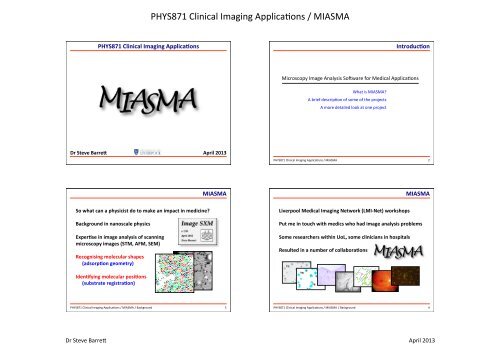
PHYS871 Clinical Imaging Applica4ons / <strong>MIASMA</strong> <br />
PHYS871 Clinical Imaging Applica>ons <br />
Introduc>on <br />
Microscopy Image <strong>Analysis</strong> SoDware for Medical Applica4ons <br />
What is <strong>MIASMA</strong>? <br />
A brief descrip4on <strong>of</strong> some <strong>of</strong> the projects <br />
A more detailed look at one project <br />
Dr Steve Barre* <br />
April 2013 <br />
PHYS871 Clinical Imaging Applica4ons / <strong>MIASMA</strong> <br />
1 <br />
PHYS871 Clinical Imaging Applica4ons / <strong>MIASMA</strong> <br />
2 <br />
<strong>MIASMA</strong> <br />
<strong>MIASMA</strong> <br />
So what can a physicist do to make an impact in medicine? <br />
Background in nanoscale physics <br />
Exper>se in image analysis <strong>of</strong> scanning <br />
microscopy images (STM, AFM, SEM) <br />
Recognising molecular shapes <br />
(adsorp>on geometry) <br />
Liverpool Medical Imaging Network (LMI-‐Net) workshops <br />
Put me in touch with medics who had image analysis problems <br />
Some researchers within UoL, some clinicians in hospitals <br />
Resulted in a number <strong>of</strong> collabora>ons <br />
Iden>fying molecular posi>ons <br />
(substrate registra>on) <br />
PHYS871 Clinical Imaging Applica4ons / <strong>MIASMA</strong> / Background <br />
3 <br />
PHYS871 Clinical Imaging Applica4ons / <strong>MIASMA</strong> / Background <br />
4 <br />
Dr Steve Barre?<br />
April 2013
What is <strong>MIASMA</strong>?<br />
PHYS871 Clinical Imaging Applica4ons / <strong>MIASMA</strong> <br />
Microscopy Image <strong>Analysis</strong> S<strong>of</strong>tware for Medical Applications<br />
<strong>MIASMA</strong> is the collective name for a number <strong>of</strong> projects involving image analysis in which I am collaborating with medics. Since about 1993 I have been<br />
writing s<strong>of</strong>tware for image analysis <strong>of</strong> scanning microscopy images, principally for applications in nanoscience and related disciplines. The s<strong>of</strong>tware that I<br />
have written, and continue to develop and expand, is Image SXM. Although written for scanning microscopy applications, I have found that Image SXM is<br />
an excellent platform on which to develop specialist image analysis solutions for the specific needs <strong>of</strong> users, including those who obtain images from light<br />
microscopes. <strong>MIASMA</strong> is the result <strong>of</strong> a number <strong>of</strong> these specialist applications having some common ground and so benefiting from being considered as<br />
part <strong>of</strong> a larger, overarching project.<br />
What are the <strong>MIASMA</strong> projects?<br />
<strong>MIASMA</strong> <br />
Intracellular Air Pollution Particulates<br />
Collaborators<br />
Aims<br />
SPM Image Display <br />
Projects include… <br />
Carbon par>culate ma*er in lung cells (lung cancer) <br />
Parasite analysis (malaria) <br />
Blood flow veloci>es in capillary networks (meningi>s) <br />
Re>nal image analysis (diabetes) <br />
Parasite morphology and development (leishmania) <br />
Assessing an>bio>c treatments (tuberculosis) <br />
Malaria Parasites<br />
<strong>Microcirculation</strong> Flow<br />
Dr Stephen Gordon<br />
Liverpool School <strong>of</strong> Tropical Medicine<br />
Dr Duncan Fullerton<br />
Liverpool School <strong>of</strong> Tropical Medicine<br />
Collaborator<br />
Pr<strong>of</strong>essor Alister Craig<br />
Liverpool School <strong>of</strong> Tropical Medicine<br />
i) To identify particulate matter and differentiate<br />
it from cell cytoplasm.<br />
ii) To measure the area <strong>of</strong> particulate matter<br />
relative to that <strong>of</strong> the cell cytoplasm.<br />
Documentation <strong>MIASMA</strong>-PMA-v7.pdf<br />
Aim<br />
To identify malaria parasites and differentiate<br />
them from background features.<br />
Documentation <strong>MIASMA</strong>-PCA-v5.pdf<br />
Collaborators<br />
Aims<br />
Dr Enitan Carrol<br />
Institute <strong>of</strong> Child Health, UoL<br />
i) To identify capillaries in videos <strong>of</strong> capillary<br />
networks and measure capillary vessel density.<br />
Dr Richard Sarginson<br />
Alder Hey Children's Hospital<br />
ii) To measure blood flow speed as a function <strong>of</strong><br />
capillary diameter.<br />
Dr Fauzia Paize<br />
UoL and Liverpool Women's Hospital<br />
Documentation <strong>MIASMA</strong>-MCA-v5.pdf<br />
Retinal Imaging<br />
Collaborators<br />
Aims<br />
Pr<strong>of</strong>essor Simon Harding<br />
Ophthalmology Research Unit, UoL<br />
Dr Yalin Zheng<br />
Ophthalmology Research Unit, UoL<br />
To identify specific features such as:<br />
Blood vessel network<br />
Optic disc<br />
Haemorrhages<br />
Exudates<br />
PHYS871 Clinical Imaging Applica4ons / <strong>MIASMA</strong> / Background 5 <br />
PHYS871 Clinical Imaging Applica4ons / <strong>MIASMA</strong> Documentation Not yet available<br />
6 <br />
http://www.liv.ac.uk/~sdb/<strong>MIASMA</strong>/<br />
Page 1 <strong>of</strong> 3<br />
<strong>MIASMA</strong><br />
14/05/12 12:20<br />
Parasite Morphology<br />
Collaborators<br />
Aims<br />
SPM Image Display <br />
Microcircula>on <strong>Analysis</strong> <br />
Lymphocyte Flow<br />
Dr Rod Dillon<br />
Liverpool School <strong>of</strong> Tropical Medicine<br />
Mr Hector Diaz<br />
Liverpool School <strong>of</strong> Tropical Medicine<br />
i) To identify leishmaniasis parasites.<br />
ii) To identify the developmental stage <strong>of</strong> the<br />
parasites by the shape and size <strong>of</strong> the parasite<br />
bodies and flagella.<br />
Documentation Not yet available<br />
Take one <strong>MIASMA</strong> project as an example… <br />
Blood flow veloci>es in capillary networks (meningi>s) <br />
Collaborator<br />
Aims<br />
Dr Carlo Laudanna<br />
Department <strong>of</strong> Pathology<br />
<strong>University</strong> <strong>of</strong> Verona<br />
i) To identify lymphocyte cells flowing through a<br />
glass capillary.<br />
ii) To measure the length <strong>of</strong> time that cells are<br />
arrested by or rolling along the capillary wall.<br />
Documentation <strong>MIASMA</strong>-LFA-v4.pdf<br />
Bacilli Lipid Bodies<br />
Collaborator<br />
Aim<br />
Dr Derek Sloan<br />
Clinical Sciences, UoL<br />
To measure the number <strong>of</strong> bacilli that contain<br />
lipid bodies.<br />
Documentation Not yet available<br />
Fibrillin Micr<strong>of</strong>ibrils<br />
Collaborator<br />
Aim<br />
Dr Riaz Akhtar<br />
Ocular Biomechanics Group<br />
School <strong>of</strong> Engineering, UoL<br />
To speed up the analysis <strong>of</strong> micr<strong>of</strong>ibrils by semiautomating<br />
the process <strong>of</strong> identifying micr<strong>of</strong>ibril<br />
beads and calculating their xy coordinates.<br />
PHYS871 Clinical Imaging Applica4ons / <strong>MIASMA</strong> <br />
Documentation <strong>MIASMA</strong>-MFA-v2.pdf<br />
7 <br />
PHYS871 Clinical Imaging Applica4ons / <strong>MIASMA</strong> / Microcircula4on <br />
8 <br />
Who is working on <strong>MIASMA</strong> projects?<br />
Apart from me, I recruite project students from Physics degree programmes and Summer students<br />
Name Status Project<br />
Martin Renvoize F303 MPhys Year 4 student <strong>Microcirculation</strong> <strong>Analysis</strong><br />
Fergus Dunn F350 BSc Year 3 student Retinal Imaging (diabetic retinopathy)<br />
Dr Steve Barre?<br />
Simon Blaen F350 BSc Year 3 student Parasite Morphology<br />
Ben Wadsworth Nuffield Bursary student Malaria Parasites<br />
Hannah Burton Nuffield Bursary student Retinal Imaging (optic disc recognition)<br />
James Dorman Nuffield Bursary student Parasite Morphology<br />
April 2013 <br />
Paul Mulligan F303 MPhys Year 4 student Parasite Morphology<br />
Kathryn Rose F300 BSc Year 3 student Lipid Body <strong>Analysis</strong>
PHYS871 Clinical Imaging Applica4ons / <strong>MIASMA</strong> <br />
Sepsis <br />
Magnifica>on <br />
Video Stability <br />
PHYS871 Clinical Imaging Applica4ons / <strong>MIASMA</strong> / Microcircula4on / Sepsis 9 <br />
PHYS871 Clinical Imaging Applica4ons / <strong>MIASMA</strong> / Microcircula4on / Video Stability <br />
10 <br />
Video Stability <br />
Video Stability <br />
Rota>on <br />
More likely, all <strong>of</strong> these…<br />
…plus ven>la>on, heartbeat <br />
PHYS871 Clinical Imaging Applica4ons / <strong>MIASMA</strong> / Microcircula4on / Video Stability <br />
11 <br />
PHYS871 Clinical Imaging Applica4ons / <strong>MIASMA</strong> / Microcircula4on / Video Stability <br />
12 <br />
Dr Steve Barre?<br />
April 2013
PHYS871 Clinical Imaging Applica4ons / <strong>MIASMA</strong> <br />
Microcircula>on <strong>Analysis</strong> <br />
Microcircula>on <strong>Analysis</strong> <br />
What informa>on can be extracted? <br />
How should the microcircula>on be quan>fied? <br />
What (manual) scoring systems exist? <br />
Percentage <strong>of</strong> perfused vessels (PPV) <br />
( Perfused = flow exists for > 50% <strong>of</strong> the >me ) <br />
Microcircula>on Flow Index (MFI) <br />
( Is the flow ‘intermi*ent’ or ‘sluggish’ or OK? ) <br />
Calcula>on <strong>of</strong> blood flow speeds <br />
• Stabilisa>on <strong>of</strong> the video <br />
• Iden>fica>on <strong>of</strong> the blood vessels (which are invisible) <br />
• Isola>on <strong>of</strong> each capillary vessel <br />
• <strong>Analysis</strong> <strong>of</strong> the movement <strong>of</strong> the blood cells <br />
Quan>fica>on <strong>of</strong> the flow distribu>on (PPV and MFI) <br />
• Flow speed as a func>on <strong>of</strong> >me <br />
• Flow speed as a func>on <strong>of</strong> vessel diameter <br />
• Varia>ons in flow speeds across the vessel network <br />
PHYS871 Clinical Imaging Applica4ons / <strong>MIASMA</strong> / Microcircula4on / Scores <br />
13 <br />
PHYS871 Clinical Imaging Applica4ons / <strong>MIASMA</strong> / Microcircula4on / Scores <br />
14 <br />
Microcircula>on <strong>Analysis</strong> <br />
Calcula>on <strong>of</strong> blood flow speeds <br />
• Stabilisa>on <strong>of</strong> the video <br />
fica>on <strong>of</strong> the blood vessels (which on <strong>of</strong> each capillary vessel cle <strong>Analysis</strong> <br />
• <strong>Analysis</strong> <strong>of</strong> the movement <strong>of</strong> the blood cells
PHYS871 Clinical Imaging Applica4ons / <strong>MIASMA</strong> <br />
Microcircula>on <strong>Analysis</strong> <br />
Microcircula>on <strong>Analysis</strong> <br />
!"#$%&'()#*+,-%./-*+0+- 01-234-*+0*<br />
!"#$%&$!'()$*+'',<br />
651<br />
!"#$%&'()*&$&+),&<br />
!"#$%&'()#*+,-./01-2-304- -.&/01&2%-3<br />
56-789-*+5:<br />
!"#$%&'()%*+"",-<br />
!"#$%&'()%*+"",-<br />
65+<br />
2%<br />
*;<br />
-4<br />
*+<br />
*51<br />
5;<br />
*5+<br />
-%<br />
9:&<br />
5+<br />
051<br />
4<br />
;<br />
05+<br />
%<br />
% -%% 2%% 3%% 5%% 4%% 6%% .%% $%% 7%%<br />
+8)9&"088:&;'?<br />
+<br />
+ 5++ *++ :++ 0?-)8>>@-ABCD.E<br />
+51<br />
Control<br />
Sepsis <br />
+<br />
+ 0++ *++ 6++ 7++ 1++ ,++ 8++<br />
9.;-?@ABCD<br />
PHYS871 Clinical Imaging Applica4ons / <strong>MIASMA</strong> / Microcircula4on <br />
17 <br />
PHYS871 Clinical Imaging Applica4ons / <strong>MIASMA</strong> / Microcircula4on <br />
18 <br />
Microcircula>on <strong>Analysis</strong> <br />
Microcircula>on <strong>Analysis</strong> <br />
!"#$%&'()*&$&+),&<br />
!"#$%&&#'()$*+*,-).((/#0() -.&/01&2%-2<br />
01)234).(0.<br />
!"#$%&'()#*+,-./01-2-304- 56-789-*+5:<br />
!"#$%&$''(<br />
2%<br />
.(<br />
!"#$%&'()%*+"",-<br />
!"#$%&'()%*+"",-<br />
:;+<br />
-.<br />
01<br />
*;<<br />
-%<br />
0(<br />
3@A9BC0.ADE09-FEBG-&HIJK-L3F&M<br />
*;+<br />
5;<<br />
5;+<br />
.<br />
%<br />
% -%% 2%% 3%% 4%% .%% 5%% 6%% $%% 7%%<br />
+8)9&":88;&?'@<br />
1<br />
(<br />
( 0(( .(( 5(( 6(( 1(( 7(( '(( 8(( /(( 0(((<br />
$9*:)&;99?@AB<br />
+;<<br />
+<br />
+= *+= >+= ,+= ?+= 5++=<br />
NJ9AJH/0OJ-BP-NJ9PD.JI-QJ..JE.-LNNQM<br />
PHYS871 Clinical Imaging Applica4ons / <strong>MIASMA</strong> / Microcircula4on <br />
19 <br />
PHYS871 Clinical Imaging Applica4ons / <strong>MIASMA</strong> / Microcircula4on <br />
20 <br />
Dr Steve Barre?<br />
April 2013
PHYS871 Clinical Imaging Applica4ons / <strong>MIASMA</strong> <br />
Terahertz Imaging <br />
Terahertz Imaging <br />
The latest <strong>MIASMA</strong> project… <br />
Imaging <strong>of</strong> biopsies with THz radia>on (cancer) <br />
Is there a pre-‐cancer signature in the infrared absorp>on? <br />
Is there a pre-‐cancer signature in the infrared absorp>on? <br />
ALICE is an accelerator at <br />
Daresbury Laboratory <br />
that is an intense source <br />
<strong>of</strong> infrared light with a <br />
Topography IR λ = 7.3 µm IR λ = 8.6 µm <br />
frequency <strong>of</strong> THz. <br />
This research shows poten>al but is s>ll at an early stage. <br />
PHYS871 Clinical Imaging Applica4ons / <strong>MIASMA</strong> / THz Imaging <br />
21 <br />
PHYS871 Clinical Imaging Applica4ons / <strong>MIASMA</strong> / THz Imaging <br />
22 <br />
Dr Steve Barre?<br />
April 2013