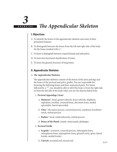
The Appendicular Skeleton
The Appendicular Skeleton
The Appendicular Skeleton

You also want an ePaper? Increase the reach of your titles
YUMPU automatically turns print PDFs into web optimized ePapers that Google loves.
3<br />
C H A P T E R<br />
E X E R C I S E<br />
<strong>The</strong> <strong>Appendicular</strong> <strong>Skeleton</strong><br />
I. Objectives<br />
A. To identify the bones of the appendicular skeleton and some of their<br />
prominent features.<br />
B. To distinguish between the bones from the left and right side of the body<br />
for the bones marked with a *.<br />
C. To learn to distinguish between a typical female and male pelvis.<br />
D. To learn the functional classification of joints.<br />
E. To learn the general structure of long bones.<br />
II. <strong>Appendicular</strong> <strong>Skeleton</strong><br />
A. <strong>The</strong> <strong>Appendicular</strong> <strong>Skeleton</strong><br />
<strong>The</strong> appendicular skeleton consists of the bones of the arms and legs and<br />
the bones of the pectoral and pelvic girdles. You are responsible for<br />
knowing the following bones and their anatomical parts. For bones<br />
followed by a “*”, you should be able to tell if the bone is from the right side<br />
or from the left side of the body when you see the disarticulated bone.<br />
1. Pectoral Appendage (Arm)<br />
a. Humerus*: head, greater tubercle, lesser tubercle, diaphysis,<br />
capitulum, trochlea, coronoid fossa, olecranon fossa, medial<br />
epicondyle, lateral epicondyle.<br />
b. Ulna*: olecranon process, coronoid process, semilunar (trochlear)<br />
notch, styloid process.<br />
c. Radius*: head, radial tuberosity, styloid process.<br />
d. Bones of the Hand: carpals, metacarpals, phalanges.<br />
2. Pectoral Girdle<br />
a. Scapula*: acromion, coracoid process, subscapular fossa,<br />
infraspinous fossa, supraspinous fossa, glenoid cavity, spine, lateral<br />
border, medial border.<br />
b. Clavicle: acromial end, sternal end.<br />
3-11
A Laboratory Manual of Human Anatomy and Physiology<br />
3. Pelvic Appendage (Leg)<br />
a. Femur*: head, neck, greater trochanter, lesser trochanter, medial<br />
condyle, lateral condyle, medial epicondyle, lateral epicondyle.<br />
b. Patella<br />
c. Tibia*: lateral condyle, medial condyle, tibial tuberosity, medial<br />
malleolus.<br />
d. Fibula: head, lateral malleolus.<br />
e. Bones of the Foot: tarsals (identify talus and calcaneus), metatarsals,<br />
and phalanges.<br />
4. Pelvic Girdle: Coxal (Innominate) bone*, acetabulum, obturator<br />
foramen.<br />
a. Ilium: iliac crest, anterior superior iliac spine, anterior inferior<br />
iliac spine, posterior superior iliac spine, posterior inferior iliac<br />
spine, greater sciatic notch, articular surface with sacrum.<br />
b. Ishium: ischial spine, ischial tuberosity.<br />
c. Pubis: pubic symphysis, pubic arch.<br />
d. Comparison of Male and Female Pelvis: a typical male and a typical<br />
female pelvis should be available in the lab for you to compare.<br />
Using your textbook as a guide, locate some of the structural<br />
differences. What obvious function underlies these differences?<br />
B. <strong>The</strong> Joints (Articulations)<br />
Joints are classified according to the amount of movement which they<br />
permit. Based on this criterion, three general types of joints can be<br />
identified. Examples of various joints are available in the laboratory. Using<br />
your textbook as a reference, you should be able to classify all joints into<br />
one of the three groups listed below. In the space provided define and give<br />
an example of each.<br />
1. Synarthroses –<br />
2. Amphiarthroses –<br />
3. Diarthroses –<br />
3-12
<strong>The</strong> <strong>Appendicular</strong> <strong>Skeleton</strong><br />
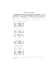
C. Long Bone in Longitudinal Section<br />
In addition to looking at various bones of the appendicular skeleton, you<br />
should take a look at a longitudinal section of the humerus or other long<br />
bone. This section will enable you to see the medullary (marrow) cavity,<br />
spongy and compact bone, and other features. On Figure 3–1, match<br />
the numbers to the appropriate parts of the bone. Some of the features (e.g.<br />
marrow and cartilage) can only be seen in fresh bone, and are not present<br />
in the dry bones that are available in the lab.<br />
1<br />
9<br />
8<br />
7<br />
5<br />
2<br />
3<br />
6<br />
A. Match the labels with the<br />
following terms:<br />
___ Articular cartilage<br />
___ Compact bone<br />
___ Diaphysis<br />
___ Distal epiphysis<br />
___ Endosteum<br />
___ Medullary cavity<br />
___ Periosteum<br />
___ Proximal epiphysis<br />
___ Spongy bone<br />
4<br />
Figure 3-1. PARTS OF A LONG BONE<br />
3-13
3-14<br />
Notes