The Appendicular Skeleton
The Appendicular Skeleton
The Appendicular Skeleton
Create successful ePaper yourself
Turn your PDF publications into a flip-book with our unique Google optimized e-Paper software.
3<br />
C H A P T E R<br />
E X E R C I S E<br />
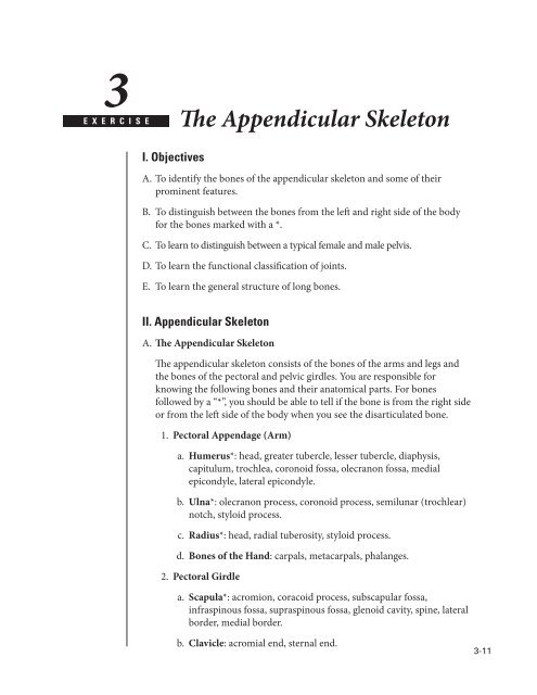
<strong>The</strong> <strong>Appendicular</strong> <strong>Skeleton</strong><br />
I. Objectives<br />
A. To identify the bones of the appendicular skeleton and some of their<br />
prominent features.<br />
B. To distinguish between the bones from the left and right side of the body<br />
for the bones marked with a *.<br />
C. To learn to distinguish between a typical female and male pelvis.<br />
D. To learn the functional classification of joints.<br />
E. To learn the general structure of long bones.<br />
II. <strong>Appendicular</strong> <strong>Skeleton</strong><br />
A. <strong>The</strong> <strong>Appendicular</strong> <strong>Skeleton</strong><br />
<strong>The</strong> appendicular skeleton consists of the bones of the arms and legs and<br />
the bones of the pectoral and pelvic girdles. You are responsible for<br />
knowing the following bones and their anatomical parts. For bones<br />
followed by a “*”, you should be able to tell if the bone is from the right side<br />
or from the left side of the body when you see the disarticulated bone.<br />
1. Pectoral Appendage (Arm)<br />
a. Humerus*: head, greater tubercle, lesser tubercle, diaphysis,<br />
capitulum, trochlea, coronoid fossa, olecranon fossa, medial<br />
epicondyle, lateral epicondyle.<br />
b. Ulna*: olecranon process, coronoid process, semilunar (trochlear)<br />
notch, styloid process.<br />
c. Radius*: head, radial tuberosity, styloid process.<br />
d. Bones of the Hand: carpals, metacarpals, phalanges.<br />
2. Pectoral Girdle<br />
a. Scapula*: acromion, coracoid process, subscapular fossa,<br />
infraspinous fossa, supraspinous fossa, glenoid cavity, spine, lateral<br />
border, medial border.<br />
b. Clavicle: acromial end, sternal end.<br />
3-11
A Laboratory Manual of Human Anatomy and Physiology<br />
3. Pelvic Appendage (Leg)<br />
a. Femur*: head, neck, greater trochanter, lesser trochanter, medial<br />
condyle, lateral condyle, medial epicondyle, lateral epicondyle.<br />
b. Patella<br />
c. Tibia*: lateral condyle, medial condyle, tibial tuberosity, medial<br />
malleolus.<br />
d. Fibula: head, lateral malleolus.<br />
e. Bones of the Foot: tarsals (identify talus and calcaneus), metatarsals,<br />
and phalanges.<br />
4. Pelvic Girdle: Coxal (Innominate) bone*, acetabulum, obturator<br />
foramen.<br />
a. Ilium: iliac crest, anterior superior iliac spine, anterior inferior<br />
iliac spine, posterior superior iliac spine, posterior inferior iliac<br />
spine, greater sciatic notch, articular surface with sacrum.<br />
b. Ishium: ischial spine, ischial tuberosity.<br />
c. Pubis: pubic symphysis, pubic arch.<br />
d. Comparison of Male and Female Pelvis: a typical male and a typical<br />
female pelvis should be available in the lab for you to compare.<br />
Using your textbook as a guide, locate some of the structural<br />
differences. What obvious function underlies these differences?<br />
B. <strong>The</strong> Joints (Articulations)<br />
Joints are classified according to the amount of movement which they<br />
permit. Based on this criterion, three general types of joints can be<br />
identified. Examples of various joints are available in the laboratory. Using<br />
your textbook as a reference, you should be able to classify all joints into<br />
one of the three groups listed below. In the space provided define and give<br />
an example of each.<br />
1. Synarthroses –<br />
2. Amphiarthroses –<br />
3. Diarthroses –<br />
3-12
<strong>The</strong> <strong>Appendicular</strong> <strong>Skeleton</strong><br />
C. Long Bone in Longitudinal Section<br />
In addition to looking at various bones of the appendicular skeleton, you<br />
should take a look at a longitudinal section of the humerus or other long<br />
bone. This section will enable you to see the medullary (marrow) cavity,<br />
spongy and compact bone, and other features. On Figure 3–1, match<br />
the numbers to the appropriate parts of the bone. Some of the features (e.g.<br />
marrow and cartilage) can only be seen in fresh bone, and are not present<br />
in the dry bones that are available in the lab.<br />
1<br />
9<br />
8<br />
7<br />
5<br />
2<br />
3<br />
6<br />
A. Match the labels with the<br />
following terms:<br />
___ Articular cartilage<br />
___ Compact bone<br />
___ Diaphysis<br />
___ Distal epiphysis<br />
___ Endosteum<br />
___ Medullary cavity<br />
___ Periosteum<br />
___ Proximal epiphysis<br />
___ Spongy bone<br />
4<br />
Figure 3-1. PARTS OF A LONG BONE<br />
3-13
3-14<br />
Notes


