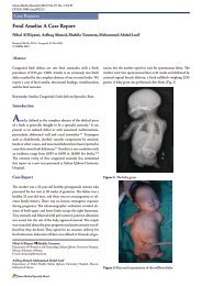
Malignant Epithelioid Hemangioendothelioma: A Case Report - OMJ
Malignant Epithelioid Hemangioendothelioma: A Case Report - OMJ
Malignant Epithelioid Hemangioendothelioma: A Case Report - OMJ

You also want an ePaper? Increase the reach of your titles
YUMPU automatically turns print PDFs into web optimized ePapers that Google loves.
Oman Medical Journal (2013) Vol. 28, No. 2:135-137<br />
DOI 10. 5001/omj.2013.36<br />
<strong>Malignant</strong> <strong>Epithelioid</strong> <strong>Hemangioendothelioma</strong>: A <strong>Case</strong> <strong>Report</strong><br />
Khouloud Bouslama, Fatma Houissa, Majd Ben Rejeb, Slim Bouzaidi, Leila Moualhi, Haifa Mekki,<br />
Radhouane Dabbeche, Mohamed Salem, Taoufik Najjar<br />
Received: 30 Nov 2012 / Accepted: 15 Jan 2013<br />
© OMSB, 2013<br />
Abstract<br />
<strong>Malignant</strong> epithelioid hemangioendothelioma (EH) is a rare tumor<br />
of vascular origin. We report a case of a woman who was found<br />
to have multiple hepatic masses in the right lobe of the liver on<br />
radiologic investigations, initially misdiagnosed as a metastatic<br />
carcinoma. The diagnosis of EH was made on histopathological<br />
study and confirmed by immunohistochemistry, which showed<br />
diffuse response for CD34 marker and no response to tissue CEA,<br />
HMB-45 or S-100 protein. Partial hepatectomy was made with<br />
good results.<br />
Keywords: <strong>Epithelioid</strong>; Liver; Histopathology; Immunohistochemistry.<br />
the abdomen. Systemic examination was unremarkable. Routine<br />
hematological and biochemical investigations, including serum<br />
bilirubin, transaminases, alkaline phosphatase and proteins were<br />
within reference ranges. Serological tests for hepatitis B and C were<br />
negative. Serum carcinoembryonic antigen, CA 19-9 and alphafetoprotein<br />
were also normal. Colonoscopy and upper endoscopy<br />
were normal.<br />
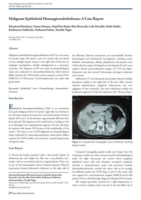
Abdominal CT scan during the portal phase showed multiple<br />
hypodense nodules in the right lobe of the liver. After contrast<br />
material administration, peripheral enhancement was seen<br />
suggestive of liver metastasis. The most voluminous nodule was<br />
localized in segment VI of the liver (diameter: 40 × 30 mm). (Fig. 1)<br />
Introduction<br />
<strong>Epithelioid</strong> hemangioendothelioma (EH) is an uncommon<br />
low-grade malignant tumor of vascular origin that may develop in<br />
the soft tissue, lung, bone, brain, liver, and small intestine. Primary<br />
hepatic EH is rare. 1 In the literature, approximately 200 cases have<br />
been reported. The diagnosis can be made both on cytology as well<br />
as on histology. Liver transplantation appears to be the only hope<br />
for patients with hepatic EH because of the multifocality of the<br />
tumor. 1,2 We report a case of EH diagnosed on histopathological<br />
study, confirmed by immunohistochemistry, which shows diffuse<br />
response for CD34 marker and treated by a partial hepatectomy<br />
with good results.<br />
<strong>Case</strong> <strong>Report</strong><br />
A 49-year-old female presented with a four-month history of<br />
abdominal pain and weight loss. She was a non-alcoholic, nonsmoker<br />
with no co-morbid medical or surgical ailment. There was<br />
no use of oral contraceptives and no chemical exposure. Physical<br />
examination showed abdominal tenderness on the right side of<br />
Khouloud Bouslama , Fatma Houissa, Majd Ben Rejeb, Slim<br />
Bouzaidi, Leila Moualhi, Haifa Mekki, Radhouane Dabbeche,<br />
Mohamed Salem, Taoufik Najjar<br />
Department of Hepatology and Gastroenterolog,. Charles nicolle’s hospital. Tunis-<br />
Tunisia.<br />
E-mail: khouloud13@voila.fr<br />
Figure 1: Computerized tomography scan of abdomen showing<br />
hepatic nodule.<br />
Computed tomography guided needle core biopsy from the<br />
voluminous nodule was taken and processed for histopathological<br />
study. On light microscope, the section shows polygonal<br />
epithelioid tumor cells with abundant vacuolated cytoplasm,<br />
vesicular to hyperchromatic nuclei and prominent nucleoli.<br />
Immunohistochemistry revealed the tumor cells to be strongly<br />
and diffusely positive for CD34 (Figs. 2 and 3). The tumor cells<br />
were negative for carcinoembryonic antigen, HMB-45 and S-100<br />
protein. Hence, a final histologic diagnosis of hepatic EH was made.<br />
The surgical procedure consisted of a right hepatectomy in<br />
order to insure complete tumor removal. At the last follow-up (2<br />
Oman Medical Specialty Board
Oman Medical Journal (2013) Vol. 28, No. 2:135-137<br />
years after diagnosis), the patient appeared well with no recurrence<br />
diagnosed on the remaining liver.<br />
Figure 2: Histopathology of EH (H&E × 200).<br />
Figure 3: Immunohistochemistry of EH revealing diffuse response<br />
to CD34 marker (CD34 × 200).<br />
Discussion<br />
<strong>Epithelioid</strong> hemangioendothelioma is a distinctive vascular<br />
tumor occurring mostly in soft tissues of extremities and lungs. 1,2<br />
Involvement of liver may be seen as a metastasis or rarely as<br />
a primary tumor. 3 Primary hepatic EH was first reported by<br />
Ishak et al. in 1984. 3 Review of literature for hepatic epithelioid<br />
hemangioendothelioma, between 1984 and 2012 using PubMed,<br />
revealed approximately 200 reported cases.<br />
The etiology or risk factors are unknown, although it is most<br />
frequently seen in middle-aged women. Clinical manifestations<br />
are nonspecific. 4 Diagnosing hepatic EH is fairly difficult for the<br />
radiologist because of its non specific findings. Ultrasonography<br />
reveals multiple discrete hypoechoic nodules or a diffusely<br />
heterogeneous echotexture of the liver. Computed tomography<br />
(CT) scan shows multiple discrete nodules with target appearance<br />
or a confluent hypodense mass in the liver. Magnetic resonance<br />
136<br />
imaging (MRI) also shows low signal intensity lesions on T1W<br />
images and heterogeneous high signal intensity on T2W sequences.<br />
Peripheral enhancement and a thin non-enhancing rim are seen on<br />
gadolinium-enhanced MRI sequences. 5,6 These features suggest a<br />
diagnosis of metastatic carcinoma, as also seen in the present case.<br />
In the absence of classic radiologic features, pathologic<br />
examination remains the mainstay of diagnosis of this rare<br />
tumor. Currently, there is an increasing use of guided fine needle<br />
aspiration cytology (FNAC) in the diagnosis of accessible<br />
visceral lesions; especially hepatic masses. 7-9 In the present case,<br />
the diagnosis of hepatic EH was made on core biopsy assisted<br />
by immunohistochemistry. The various cytologic differential<br />
diagnoses in a smear from hepatic EH would include hepatocellular<br />
carcinoma, cholangiocarcinoma, metastatic carcinoma, malignant<br />
melanoma and angiosarcoma. 9<br />
In the histopathologic appearance, the tumor is composed of<br />
pleomorphic cells with rare multinucleate giant cells. The vascular<br />
nature of the tumor may be apparent only by an occasional cell<br />
showing intracytoplasmic lumina formation containing erythrocyte<br />
or leukocyte. The endothelial nature of tumor cells may be<br />
confirmed by the immunohistochemical detection of CD31, CD34,<br />
factor VIII-related antigen or Ulex europeus 1 lectin. 7<br />
The prognosis may vary widely, with some patients exhibiting<br />
slow-growing lesions and others presenting a rapidly progressive<br />
form. 2 Orthotopic liver transplantation appears to be the only hope<br />
for patients with hepatic EH, since surgical resection is impossible<br />
due to the multifocality of the tumor. 4 Recently, some authors<br />
have reported a malignant EH of the liver successfully treated<br />
with pegylated liposomal doxorubicin and metastatic hepatic EH<br />
successfully treated with thalidomide. 10,11 In the present case, the<br />
choice of a right hepatectomy was done because the tumors were<br />
localized to the right liver, and also because liver transplantation is<br />
not available in Tunisia.<br />
Conclusion<br />
<strong>Malignant</strong> epithelioid hemangioendothelioma (EH) is a rare tumor<br />
of vascular origin which usually occurs in soft tissues. In case of liver<br />
localization, it presents as multiple hepatic nodules. Orthotopic liver<br />
transplantation appears to be the only hope for patients with hepatic<br />
EH because of the multifocality of the tumor. Partial resection of<br />
the liver is possible for the localized tumor with good results.<br />
Acknowledgements<br />
The authors reported no conflicts of interest and no funding was<br />
received for this work.<br />
References<br />
1. Liu YI, Brown SS, Elihu A, Bonham CA, Concepcion W, Longacre TA, et al.<br />
Hepatic epithelioid hemangioendothelioma. Dig Dis Sci 2011 Feb;56(2):303-<br />
306.<br />
Oman Medical Specialty Board
137<br />
Oman Medical Journal (2013) Vol. 28, No. 2:135-137<br />
2. Kim SG, Jung MK, Jeon SW, Cho CM, Tak WY, Kweon YO, et al. [A case<br />
of primary hepatic epithelioid hemangioendothelioma mimicking metastatic<br />
carcinoma]. Korean J Gastroenterol 2007 Jul;50(1):61-65.<br />
3. Ishak KG, Sesterhenn IA, Goodman ZD, Rabin L, Stromeyer FW. <strong>Epithelioid</strong><br />
hemangioendothelioma of the liver: a clinicopathologic and follow-up study of<br />
32 cases. Hum Pathol 1984 Sep;15(9):839-852.<br />
4. Nudo CG, Yoshida EM, Bain VG, Marleau D, Wong P, Marotta PJ, et al. Liver<br />
transplantation for hepatic epithelioid hemangioendothelioma: the Canadian<br />
multicentre experience. Can J Gastroenterol 2008 Oct;22(10):821-824.<br />
5. Chen Y, Yu RS, Qiu LL, Jiang DY, Tan YB, Fu YB. Contrast-enhanced multiplephase<br />
imaging features in hepatic epithelioid hemangioendothelioma. World J<br />
Gastroenterol 2011 Aug;17(30):3544-3553.<br />
6. Amin S, Chung H, Jha R. Hepatic epithelioid hemangioendothelioma: MR<br />
imaging findings. Abdom Imaging 2011 Aug;36(4):407-414.<br />
7. Soslow RA, Yin P, Steinberg CR, Yang GC. Cytopathologic features of hepatic<br />
epithelioid hemangioendothelioma. Diagn Cytopathol 1997 Jul;17(1):50-53.<br />
8. Gupta R, Mathur SR, Gupta SD, Durgapal P, Iyer VK, Das CJ, et al. Hepatic<br />
epithelioid hemangioendothelioma: A diagnostic pitfall in aspiration cytology.<br />
Cytojournal 2010;6:25.<br />
9. Manucha V, Sun CC. Cytologic findings and differential diagnosis in hepatic<br />
<strong>Epithelioid</strong> hemangioendothelioma: a case report. Acta Cytol 2008 Nov-<br />
Dec;52(6):713-717.<br />
10. Salech F, Valderrama S, Nervi B, Rodriguez JC, Oksenberg D, Koch A,<br />
et al. Thalidomide for the treatment of metastatic hepatic epithelioid<br />
hemangioendothelioma: a case report with a long term follow-up. Ann Hepatol<br />
2011 Jan-Mar;10(1):99-102.<br />
11. Grenader T, Vernea F, Reinus C, Gabizon A. <strong>Malignant</strong> epithelioid<br />
hemangioendothelioma of the liver successfully treated with pegylated liposomal<br />
doxorubicin. J Clin Oncol 2011 Sep;29(25):e722-e724.<br />
“Submit Proceedings of Conferences or<br />
Workshops As Supplements To Omj”<br />
v Proceedings of conferences or workshops will be considered as a supplement<br />
to the Oman Medical Journal.<br />
v Original work or abstracts will be accepted.<br />
v Material in supplements will be included with the regular issue of the journal<br />
or will be distributed separately as supplement.<br />
v Supplements will also be available on the Oman<br />
Medical Journal website.<br />
Oman Medical Specialty Board