An Under-recognized Complication: Diabetic Myonecrosis - OMJ
An Under-recognized Complication: Diabetic Myonecrosis - OMJ
An Under-recognized Complication: Diabetic Myonecrosis - OMJ

You also want an ePaper? Increase the reach of your titles
YUMPU automatically turns print PDFs into web optimized ePapers that Google loves.
<strong>An</strong> <strong>Under</strong>-<strong>recognized</strong> <strong>Complication</strong>: <strong>Diabetic</strong> <strong>Myonecrosis</strong><br />
Saif Khan, 1 *Dawood Al-Riyami, 1 Mohammed Al-Ghailani, 1<br />
Humoud Al-Dhuhli 2<br />
From the 1 Department of Medicine, 2 Radiology and Molecular Imaging; Sultan Qaboos University Hospital, Al Khod, Oman.<br />
Received:<br />
Accepted:<br />
*Address correspondence to: Dr. Dawood Al-Riyami, Department of Medicine,<br />
Sultan Qaboos University Hospital, P.O. Box 35, P.C. 123, Al Khod, Oman<br />
Email: dawood@squ.edu.om<br />
Khan S, et al. <strong>OMJ</strong>. 24, 228-230 (2009); doi:10.5001/omj.2009.46<br />
This report describes a case of diabetic myonecrosis in a patient<br />
with long standing diabetes mellitus (DM). This is a rare, possibly<br />
under-<strong>recognized</strong> complication of a common disease. This is a self<br />
limiting complication and requires only conservative management.<br />
Failure to recognize this condition can result in significant<br />
morbidity. Physicians should always consider this entity in the<br />
differential diagnosis of acute muscle pain in a patient with DM.<br />
Diabetes mellitus (DM) and its complications is a global<br />
challenge to health care systems. Ischemic heart disease, peripheral<br />
vascular disease, nephropathy and retinopathy are well <strong>recognized</strong><br />
complications. <strong>Diabetic</strong> myonecrosis or diabetic muscle infarct<br />
(DMI) is a rare complication of diabetes or possibly under<br />
diagnosed. It is described in longstanding diabetes. Although it<br />
was first reported in 1965, only case reports and a systemic review<br />
of these cases have been published. 1,2 It was also reported in some<br />
cases as the first manifestation of DM. Clinical presentation is<br />
with acute onset of muscle pain, commonly the thigh. Diabetes<br />
myonecrosis can be misdiagnosed as cellulitis, deep vein thrombosis<br />
or facsiatis. Magnetic resonance imaging (MRI) is sufficient, in the<br />
appropriate clinical context, to enable the diagosis, hence avoiding<br />
inappropriate treatment. 3<br />
Case Report<br />
A 57 year old female known to have past medical history of diabetes<br />
mellitus and hypertension for more then 20 years, on regular<br />
treatment with metformin, repaglinide, atenolol and amlodipine is<br />
described in this report. The patient developed severe excruciating<br />
left thigh pain and swelling for which she presented to a private<br />
clinic. The pain started 10 days before the presentation and<br />
worsened to the extent of limiting her activity to bed rest.<br />
At the private clinic, she had Doppler ultrasound of the<br />
thigh which reported no evidence of deep vein thrombosis. She<br />
was started on anticoagulation with Low Molecular Weight<br />
Heparin(LMWH) followed by warfarin for presumed superficial<br />
thrombophlebitis. In addition she was treated with an antibiotic<br />
(augmentin) and NSAID to control her pain.<br />
The pain persisted and she developed fever. The patient went<br />
back to the same clinic and was found to have a deterioration in<br />
renal function, with a creatinine of 590u/L, urea 30mmol/L,<br />
potassium 6.7mmol/L and bicarbonate of 13mmol/L. Her<br />
creatinine level was 135micromol/l at the first visit to the clinic.<br />
She was referred to the Sultan Qaboos University hospital for<br />
further evaluation and management of acute renal injury. The<br />
patient denied experiencing muscle trauma, skin breakdown or<br />
muscle weakness. Local examination revealed swelling and marked<br />
tenderness of left anteriomedial aspect of the thigh. The skin was<br />
warm but not erythematous. The patient had low grade fever of<br />
37.8°C and her blood pressure was 156/72 mmHg. There were no<br />
muco-cutaneous, joint or systemic abnormal signs.<br />
Laboratory studies for leukocyte, platelet, and eosinophil<br />
counts, C3, C4, ANA, hepatitis B surface antigen, and hepatitis<br />
C antibody yielded normal or negative findings. However, the<br />
patient had elevated ESR 116 and CK 168 U/L. Urine and blood<br />
cultures reported no growth. Chest x-ray was remarkable for mild<br />
cardiomegally. The patient remained oligouric despite initial fluid<br />
resuscitation and hemodialysis was started in addition to treatment<br />
with analgesics and antibiotic. The treatment with anticoagulants<br />
was stopped. MRI of the left thigh was performed and suggested<br />
myonecrosis of the vastus lateralis.<br />
The patient was diagnosed with diabetic myonecrosis. The<br />
pain gradually subsided with conservative measures and bed rest.<br />
Treatment with analgesics was continued but the antibiotic was<br />
discontinued 3 days after her admission when her cultures reported<br />
no growth. At the time of hospital discharge, the patient was able<br />
to walk and her thigh pain had improved significantly. Dialysis<br />
was discontinued before the hospital discharge. Renal function<br />
improved but not to her baseline creatinine. Her creatinine at<br />
her last follow up visit, 2 months following her admission was<br />
300micromol/L.<br />
Discussion<br />
The patient reported here had long standing DM with nephropathy<br />
evident by then elevated baseline creatinine. She was presented with<br />
clinical manifestation typical of what is described, in case reports,<br />
as diabetic myonecrosis or muscle infarction. This condition is<br />
described more in women. Although the exact pathogenesis is<br />
unknown, it is assumed to be the microvasculer complication of<br />
longstanding DM. The typical clinical presentation include abrupt<br />
onset of pain in involved muscle. The most commonly involved<br />
Oman Medical Journal 2009, Volume 24, Issue 3, July 2009
<strong>An</strong> <strong>Under</strong>-<strong>recognized</strong>... Khan et al.<br />
muscles are of the thigh, but calf muscles can also be affected. 4<br />
Often, there is palpable mass and swelling with or without systemic<br />
signs. The pain usually persists for weeks then spontaneously<br />
resolves. Recurrent episodes have also been reported.<br />
DMI needs to be differentiated from other common painful<br />
conditions that cause acute leg pain in a diabetic patient, including<br />
deep venous thrombosis, soft tissue abscess, haematoma,<br />
inflammatory myositis. Physician’s awareness should allow early<br />
diagnosis on the basis of clinical presentation, laboratory test and<br />
imaging, thereby avoiding biopsy and its potential complication.<br />
MRI plays an important role in the diagnosis. The main<br />
findings associated with <strong>Diabetic</strong> muscle Infarction, DMI, are<br />
increased signal intensity of the muscles involved on T2 weighted<br />
images. The affected muscles are either isointense or hypointense<br />
on T1 weighted images. Foci of increased signal intensity on T1<br />
weighted images may suggest focal hemorrhages. The affected<br />
muscles show modest amount of enhancement after administration<br />
of intravenous gadolinium. The enhancing muscles and fascia<br />
are almost always hyperintense on T2 weighted images. Areas<br />
within the muscles which show rim enhancement correspond to<br />
area of muscle infarction or necrosis within the ischemic muscle.<br />
Subcutaneous edema and subfascial fluid are commonly seen on<br />
the T2-weighted and inversion-recovery images. 5-7<br />
Sonographic findings of diabetic muscle infarction include<br />
internal linear echogenic structures coursing through the lesion;<br />
an absence of internal motion or swirling of fluid with transducer<br />
pressure; and a lack of a predominately anechoic area. These<br />
sonographic characteristics may help differentiate diabetic muscle<br />
infarction from abscess or necrotic<br />
tumor. 8<br />
<strong>Diabetic</strong> muscle infarction<br />
is frequently bilateral and the<br />
thigh is the most common site<br />
of involvement (81-87 %) of the<br />
reposted cases. Although any of<br />
the muscles of the thigh may be<br />
involved, the musculi vastus were<br />
the most frequently affected. 6 The<br />
second most commonly affected<br />
site is the calf muscles.<br />
The radiological differential<br />
diagnosis of diabetic muscle<br />
infarction includes soft-tissue<br />
abscess, pyomyositis, necrotizing<br />
fasciitis, and other causes of<br />
myositis. Necrotizing fasciitis is<br />
the major differential consideration<br />
that may not be distinguishable from diabetic muscle infarction on<br />
the basis of only MR imaging findings. Patients with necrotizing<br />
fasciitis are more likely to have fever, cellulitis, and an elevated<br />
peripheral white blood cell count. Severe pain is very characteristic<br />
of diabetic muscle infarction and may not be seen with necrotizing<br />
fasciitis or pyomyositis. 9 Therefore, the diagnosis of diabetic<br />
muscle infarction remains a clinical and radiographic diagnosis.<br />
Diabetes myonecrosis is a self-limiting disease, full recovery is<br />
expected overtime in most of the cases. Only general management<br />
is required including bed rest and analgesics. As a possible<br />
microvasculer complication antiplatelets could theoretically be<br />
effective. Tight glycemic control and cessation of smoking is<br />
prudent. Excisional biopsy, early debridement and mobility have<br />
lead to complications and delayed recovery. 10<br />
Conclusion<br />
<strong>Diabetic</strong>-associated muscle necrosis is an uncommon clinical entity.<br />
However, the incidence is likely to increase because of increasing<br />
global prevalence of diabetes. Physicians should always consider<br />
this entity in differential diagnosis of acute muscle pain in a diabetic<br />
patient. <strong>An</strong> early noninvasive diagnosis is possible by performing<br />
an MRI. Subsequent to reporting this case at the hospital, two<br />
more cases of diabetic myonecrosis were readily diagnosed in less<br />
then three months. <strong>Diabetic</strong> myonecrosis requires conservative<br />
management and minimal intervention to avoid complications and<br />
prolonged hospitalization. Biopsy is rarely indicated and if possible<br />
better avoided.<br />
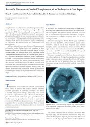
Figure 1: A) Axial fat-suppressed T2-weighted image shows enlargement and increased signal<br />
intensity of the vastus lateralis muscle (*) and rectus femoris (open white arrow). There is fluid in<br />
subcutaneous tissue and intermuscular fascia (black arrow). B) Axial fat-suppressed gadoliniumenhanced<br />
T1-wieghted image shows areas of diffuse enhancement of the vastus lateralis and rectus<br />
femoris. There is focal rim enhancement with a low–signal-intensity center within the vastus<br />
lateralis (white arrow) suggestive of muscle necrosis.<br />
Oman Medical Journal 2009, Volume 24, Issue 3, July 2009
<strong>An</strong> <strong>Under</strong>-<strong>recognized</strong>... Khan et al.<br />
Acknowledgements<br />
The authors reported no conflict of interest and no funding has<br />
been received on this work.<br />
References<br />
1. <strong>An</strong>gervall L, Stener B. Tumoriform focal muscular degeneration in two<br />
diabetic patients. Diabetologia 1965; 1:39-42.<br />
2. Trujillo–Santos A.J. <strong>Diabetic</strong> Muscle Infarction. <strong>An</strong> underdiagnosed<br />
complication of long-standing diabetes. Diabetes Care 2003 Jan 26: 211-15.<br />
3. Kattapuram TM, Suri R, Rosol MS, Rosenberg AE, Kattapuram SV.<br />
Idiopathic and diabetic skeletal muscle necrosis: evaluation by magnetic<br />
resonance imaging. Skeletal Radiol. 2005 Apr; 34:203-9.<br />
4. Umpierrez GE, Stiles RG, Kleinbart J, Krendal DA, Watts NB. <strong>Diabetic</strong><br />
muscle infarction. Am J Med 1996; 101:245<br />
5. Chason DP, Fleckenstein JL, Burns DK, et al. <strong>Diabetic</strong> muscle infarction:<br />
radiologic evaluation. Skeletal Radiol 1996; 127-132.<br />
6. Jelinek JS, Murphey MD, Aboulafia AJ, et al. Muscle infarction in patients<br />
with diabetes mellitus: MR imaging findings. Radiology1999; 211:241 –247<br />
7. Khoury NJ, El-Khoury GY, Kathol MH. MRI diagnosis of diabetic muscle<br />
infarction: report of two cases. Skeletal Radiol 1997; 26:122-7<br />
8. Delaney-Sathy LO; Fessell DP; Jacobson JA; Hayes CW. Sonography of<br />
diabetic muscle infarction with MR imaging, CT, and pathologic correlation.<br />
AJR Am J Roentgenol 2000 Jan;174:165-9.<br />
9. Schmid MR, Kossmann T, Duewell S. Differentiation of necrotizing fasciitis<br />
and cellulitis using MR imaging. AJR 1998; 170:615-620.<br />
10. Kapur S. and RJ McKendry. Treatment and outcomes of diabetic muscle<br />
infarction. J Clin Rheumatol. 2005 Feb; 11:8-12.<br />
Oman Medical Journal 2009, Volume 24, Issue 3, July 2009