Volume 9 Number 3 March 2012 - Oncology Practice Digital Network
Volume 9 Number 3 March 2012 - Oncology Practice Digital Network
Volume 9 Number 3 March 2012 - Oncology Practice Digital Network
Create successful ePaper yourself
Turn your PDF publications into a flip-book with our unique Google optimized e-Paper software.
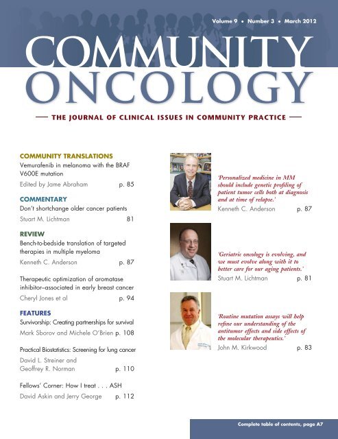
<strong>Volume</strong> 9 ● <strong>Number</strong> 3 ● <strong>March</strong> <strong>2012</strong><br />
COMMUNITY TRANSLATIONS<br />
Vemurafenib in melanoma with the BRAF<br />
V600E mutation<br />
Edited by Jame Abraham p. 85<br />
COMMENTARY<br />
Don’t shortchange older cancer patients<br />
Stuart M. Lichtman 81<br />
REVIEW<br />
Bench-to-bedside translation of targeted<br />
therapies in multiple myeloma<br />
Kenneth C. Anderson p. 87<br />
Therapeutic optimization of aromatase<br />
inhibitor–associated in early breast cancer<br />
‘Personalized medicine in MM<br />
should include genetic profiling of<br />
patient tumor cells both at diagnosis<br />
and at time of relapse.’<br />
Kenneth C. Anderson p. 87<br />
‘Geriatric oncology is evolving, and<br />
we must evolve along with it to<br />
better care for our aging patients.’<br />
Stuart M. Lichtman p. 81<br />
Cheryl Jones et al p. 94<br />
FEATURES<br />
Survivorship: Creating partnerships for survival<br />
Mark Sborov and Michele O’Brien p. 108<br />
Practical Biostatistics: Screening for lung cancer<br />
‘Routine mutation assays will help<br />
refine our understanding of the<br />
antitumor effects and side effects of<br />
the molecular therapeutics.’<br />
John M. Kirkwood p. 83<br />
David L. Streiner and<br />
Geoffrey R. Norman p. 110<br />
Fellows’ Corner: How I treat ...ASH<br />
David Askin and Jerry George p. 112<br />
Complete table of contents, page A7
Community <strong>Oncology</strong> <strong>March</strong> <strong>2012</strong><br />
<strong>Volume</strong> 9, <strong>Number</strong> 3 (pp 75–112)
<strong>March</strong> <strong>2012</strong><br />
VOLUME 9, NUMBER 3<br />
Editor-in-Chief<br />
Editors<br />
David H. Henry,<br />
MD, FACP<br />
Pennsylvania Hospital<br />
Philadelphia, PA<br />
Jame Abraham, MD<br />
West Virginia University<br />
Morgantown, WV<br />
Linda D. Bosserman,<br />
MD, FACP<br />
Wilshire <strong>Oncology</strong><br />
Medical Group<br />
La Verne, CA<br />
Editorial Board<br />
Johanna Bendell, MD<br />
Sarah Cannon Research Institute, Nashville, TN<br />
Charles L. Bennett, MD, PhD, MPP<br />
University of South Carolina, Columbia, SC<br />
Roy A. Beveridge, MD<br />
US <strong>Oncology</strong>, Houston, TX<br />
Ralph V. Boccia, MD<br />
Georgetown University, Washington, DC<br />
Matt Brow<br />
US <strong>Oncology</strong>, Washington, DC<br />
Michael J. Fisch, MD, MPH<br />
The University of Texas<br />
MD Anderson Cancer Center, Houston, TX<br />
John A. Fracchia, MD<br />
Lenox Hill Hospital, New York, NY<br />
James N. George, MD<br />
University of Oklahoma Health Sciences Center<br />
Oklahoma City, OK<br />
James Gilmore, PharmD<br />
Georgia Cancer Specialists, Atlanta, GA<br />
Patrick Grusenmeyer, ScD<br />
Helen F. Graham Cancer Center, Newark, DE<br />
David M.J. Hoffman, MD<br />
Tower Hematology <strong>Oncology</strong> Medical Group<br />
Beverly Hills, CA<br />
Jimmie Holland, MD<br />
Memorial Sloan-Kettering Cancer Center<br />
New York, NY<br />
Leslie Rodgers Laufman, MD<br />
Blood and Cancer Care of Ohio, Columbus, OH<br />
Stuart M. Lichtman, MD<br />
Memorial Sloan-Kettering Cancer Center, Commack, NY<br />
Charles Loprinzi, MD<br />
Mayo Medical School, Rochester, MN<br />
John L. Marshall, MD<br />
Lombardi Comprehensive Cancer Center, Washington, DC<br />
Cathy Maxwell, RN, OCN, CCRC<br />
Advanced Medical Specialties, LLC, Miami, FL<br />
Bradley J. Monk, MD, FACOG<br />
Creighton University School of Medicine at St. Joseph’s<br />
Hospital and Medical Center, Phoenix, AZ<br />
Anne Moore, MD<br />
Weill Medical College of Cornell University, New York, NY<br />
Deborah A. Nagle, MD<br />
Beth Israel Deaconess Medical Center, Boston, MA<br />
Geoffrey R. Norman, PhD<br />
McMaster University, Hamilton, Ontario, Canada<br />
Steven O’Day, MD<br />
The Angeles Clinic & Research Institute, Los Angeles, CA<br />
Theodore A. Okon, MBA<br />
Supportive <strong>Oncology</strong> Services, Memphis, TN<br />
Philip A. Philip, MD, PhD<br />
Barbara Ann Karmanos Cancer Institute, Detroit, MI<br />
Jondavid Pollock, MD, PhD<br />
Schiffler Cancer Center, Wheeling, WV<br />
Nicholas J. Robert, MD<br />
US <strong>Oncology</strong>, Fairfax, VA<br />
Peter J. Rosen, MD<br />
Roy & Patricia Disney Family<br />
Cancer Research Center, Burbank, CA<br />
Myrna R. Rosenfeld, MD, PhD<br />
University of Pennsylvania School of Medicine, Philadelphia, PA<br />
Philip Schulman, MD<br />
Memorial Sloan-Kettering Cancer Center, Commack, NY<br />
Lee S. Schwartzberg, MD, FACP<br />
The West Clinic, Memphis, TN<br />
David Streiner, PhD, CPsych<br />
University of Toronto, Toronto, Ontario, Canada<br />
Debu Tripathy, MD<br />
University of Southern California/ Norris Comprehensive<br />
Cancer Center, Los Angeles, CA<br />
Steven Tucker, MD<br />
Pacific Cancer Centre, Singapore, Malaysia<br />
<strong>Volume</strong> 9/<strong>Number</strong> 3 <strong>March</strong> <strong>2012</strong> COMMUNITY ONCOLOGY A5
<strong>March</strong> <strong>2012</strong><br />
VOLUME 9, NUMBER 3<br />
contents<br />
IMNG, LLC<br />
60B Columbia Road<br />
Morristown, NJ 07960<br />
973.290.8200 tel ● 631.424.8905 fax<br />
Alan Imhoff, President and Publisher<br />
Mary Jo Dales, Editorial Director<br />
Reneé Matthews, Managing Editor<br />
Elizabeth Mechcatie, Matt Stenger,<br />
Contributing Writers<br />
Virginia Ingram-Wells, Copy Editor<br />
Peter Murphy, Stuart Williams, National<br />
Accounts Managers<br />
Yvonne Evans, Production Manager<br />
Devin Gregorie, National Accounts<br />
Manager—<strong>Oncology</strong> Projects<br />
FROM THE EDITOR<br />
75 Assessing for physiologic not chronologic age in the elderly<br />
David H. Henry, MD, FACP<br />
COMMENTARY<br />
81 Don’t shortchange older cancer patients<br />
Stuart M. Lichtman, MD, FACP<br />
83 Vemurafenib’s companion assay refines use of the<br />
targeted therapy<br />
John M. Kirkwood, MD<br />
COMMUNITY TRANSLATIONS<br />
85 Vemurafenib in melanoma with the BRAF V600E mutation<br />
Edited by Jame Abraham, MD; report prepared by Matt Stenger, MS<br />
REVIEW<br />
87 Bench-to-bedside translation of targeted therapies in<br />
multiple myeloma<br />
Kenneth C. Anderson, MD<br />
94 Therapeutic optimization of aromatase inhibitor–associated<br />
arthralgia: etiology, onset, resolution, and symptom<br />
management in early breast cancer<br />
Cheryl Jones, MD, James Gilmore, PharmD, Mansoor Saleh, MD,<br />
Bruce Feinberg, DO, Michelle Kissner, RPh, PharmD, and Stacey J. Simmons, MD<br />
LETTERS<br />
Community <strong>Oncology</strong> (ISSN 1548-5315) is<br />
published monthly by IMNG, LLC, 60B<br />
Columbia Road, Morristown, NJ 07960.<br />
Periodicals postage paid at Morristown, NJ,<br />
and additional mailing offices.<br />
Change of Address<br />
Postmaster: send address changes to Community<br />
<strong>Oncology</strong>, Circulation, IMNG, LLC, 60B<br />
Columbia Road, Morristown, NJ 07960.<br />
Recipient: to change your address, contact<br />
b.cavallaro@elsevier.com, telephone:<br />
973.290.8253, or mail to Community <strong>Oncology</strong>,<br />
Circulation, IMNG, LLC, 60B Columbia Road,<br />
Morristown, NJ 07960.<br />
Case Letters<br />
102 Extramedullary BCR-ABL positive T-lymphoblastic leukemia<br />
in a patient with chronic myelogenous leukemia<br />
Mylene Go, MD, Le Wang, MD, PhD, JinMing Song, MD, and Rene Rubin, MD<br />
106 Decitabine–induced acute lung injury<br />
Monica Marwaha, MD, and Huzefa Bahrain, DO<br />
FEATURES<br />
Survivorship<br />
108 Creating partnerships for survival<br />
Mark Sborov, MD, and Michele O’Brien, RN, MSN, ACNS-BC, BA<br />
Practical Biostatistics<br />
110 Moving up in the world: screening for lung cancer<br />
David L. Streiner, PhD, CPsych, and Geoffrey R. Norman, PhD<br />
Fellows’ Corner<br />
112 How I treat ...ASH<br />
David Askin, DO, and Jerry George, DO<br />
<strong>Volume</strong> 9/<strong>Number</strong> 3 <strong>March</strong> <strong>2012</strong> COMMUNITY ONCOLOGY A7
<strong>March</strong> <strong>2012</strong><br />
VOLUME 9, NUMBER 3<br />
Information for Authors and Advertisers<br />
Aims and Scope<br />
COMMUNITY ONCOLOGY is an independent journal that publishes peerreviewed<br />
research, review articles and commentary on all aspects of<br />
clinical oncology practice. Article types include original clinical studies in<br />
practice-based settings, state-of-the-art review papers, peer viewpoints,<br />
commentaries, and letters to the editor.<br />
For a full and complete guide for authors, go to ees.elsevier.com/co/<br />
For further information, contact the Managing Editor, Renée Matthews,<br />
at 240-221-2461 or e-mail, renee.matthews@elsevier.com.<br />
Correspondence<br />
For general, noneditorial enquiries, write to COMMUNITY ONCOLOGY,<br />
60B Columbia Road, Morristown, NJ 07960; tel: 973-290-8200; fax:<br />
973-290-8250.<br />
Letters to the Editor should be addressed to the Editor-in-Chief, David<br />
H. Henry, MD, FACP, e-mail: c.oncology@elsevier.com.<br />
Advertising<br />
For information regarding advertising rates, contact Peter Murphy<br />
(tel: 201-529-4020; e-mail: pmurphy@braveheart-group.com) or Stuart<br />
Williams (tel: 201-529-4004; e-mail: swilliams@braveheart-group.com);<br />
for information regarding supplements and projects, contact Devin<br />
Gregorie (tel: 516-381-8613; e-mail: d.gregorie@elsevier.com).<br />
CME Supplements<br />
For information on CME supplements to COMMUNITY ONCOLOGY,<br />
contact Sylvia Reitman of Global Academy for Medical Education,<br />
LLC, at e-mail: s.reitman@globalacademycme.com.<br />
Annual Subscription Rates<br />
For 12 issues (in US$): Individual $380, Canada $413, International<br />
$413; Institutional $380, Canada $413, International, $413; Single<br />
copy $45.<br />
For further information regarding subscriptions, contact Barbara<br />
Cavallaro, e-mail: b.cavallaro@elsevier.com<br />
COMMUNITY ONCOLOGY (ISSN 1548-5315) is published monthly<br />
by International Medical News Group, LLC, 60B Columbia Road,<br />
Morristown, NJ 07960.<br />
Copyright<br />
Copyright © <strong>2012</strong> by Elsevier Inc. All rights reserved. No part of this<br />
publication may be reproduced or transmitted in any form or by any<br />
means, electronic or mechanical, including photocopy, recording, or any<br />
information storage and retrieval system, without written permission<br />
from the Publisher.<br />
Disclaimer<br />
Discussions, views, opinions, and recommendations as to medical<br />
procedures, products, choice of drugs, and drug dosages are the<br />
responsibility of the authors or advertisers. No responsibility is assumed<br />
by the Publisher, Editor, or Editorial Board for any injury and/or<br />
damage to persons or property as a matter of product liability,<br />
negligence, or otherwise or from any use or operation of any methods,<br />
products, instructions, or ideas contained in the material herein. Because<br />
of rapid advances in the medical sciences, independent verification of<br />
diagnoses and drug dosages should be made. Advertiser and advertising<br />
agency recognize, accept, and assume liability for all content (including<br />
text, representations, illustrations, opinions, and facts) of advertisements<br />
printed and also assume responsibility for any claims made against the<br />
Publisher arising from or related to such advertisements.<br />
In the event that legal action or a claim is made against the Publisher<br />
arising from or related to such advertisements, advertiser and advertising<br />
agency agree to fully defend, indemnify, and hold harmless the Publisher<br />
and to pay any judgment, expenses, and legal fees incurred by the<br />
Publisher as a result of said legal action or claim. The Publisher reserves<br />
the right to reject any advertising that he feels is not in keeping with the<br />
publication’s standards.<br />
The Publisher is not liable for delays in delivery and/or nondelivery in<br />
the event of Act of God, action by any government or quasigovernmental<br />
entity, fire, flood, insurrection, riot, explosion, embargo,<br />
strikes (whether legal or illegal), labor or material shortage, transportation<br />
interruption of any kind, work slowdown, or any condition beyond the<br />
control of the Publisher that affects production or delivery in any<br />
manner.<br />
This journal is printed on paper meeting the requirements of ANSI/<br />
NISO Z39.48-1992 (Permanence of Paper) effective with <strong>Volume</strong> 1,<br />
Issue 1, 2004.<br />
Community <strong>Oncology</strong> is indexed by EMBASE and the Cumulative Index to Nursing and Allied Health Literature (CINAHL)<br />
A8 COMMUNITY ONCOLOGY <strong>March</strong> <strong>2012</strong> www.Community<strong>Oncology</strong>.net
From the Editor<br />
Assessing for physiologic not chronologic<br />
age in the elderly<br />
Commun Oncol <strong>2012</strong>;9:75<br />
doi:10.1016/j.cmonc.<strong>2012</strong>.02.012<br />
© <strong>2012</strong> Published by Elsevier Inc.<br />
In recent years, increasing evidence has<br />
shown that older patients with cancer can<br />
tolerate standard cancer treatments and that<br />
the benefits they derive from those therapies are<br />
comparable with the benefits seen in younger<br />
patients. Yet despite such progress, some less<br />
encouraging trends about cancer care in the<br />
elderly persist—for example, that elderly nursing<br />
home residents are often diagnosed at a<br />
more advanced disease stage. 1 In a Commentary<br />
on page 81, Stuart M. Lichtman sends us an<br />
important wake-up call: don’t short-change<br />
older cancer patients. He urges us not to pull<br />
back from using standard therapies<br />
and dose densities simply because a<br />
patient is elderly. When doing an<br />
oncologic work-up of an elderly<br />
patient with cancer, it is more important<br />
to evaluate the patient’s<br />
physiologic age through routine<br />
geriatric assessment than it is to<br />
rely solely on the patient’s chronologic<br />
age. Dr. Lichtman goes on to<br />
note that elderly patients tend to be<br />
routinely excluded from clinical trials<br />
and he urges physicians in community-based<br />
practices to recruit<br />
their patients into clinical trials if appropriate.<br />
What could be more exciting than the recent<br />
developments in therapies for multiple myeloma<br />
and metastatic melanoma? On page 87,<br />
Kenneth C. Anderson brings us a riveting review<br />
on the bench-to-bedside translation of targeted<br />
therapies in multiple myeloma, which he originally<br />
presented at ASCO last year. 2 Dr. Anderson<br />
describes how bortezomib and lenalidomide, for<br />
example, target the tumor as well as multiple myeloma<br />
cells in the bone marrow microenvironment,<br />
and in doing so, double the median survival.<br />
He also writes that agents that drive bone biology<br />
can inhibit multiple melanoma cell growth, such<br />
as DKK-1 and MoAb; how certain combination<br />
therapies can be effective against multiple myeloma,<br />
even in some refractory cases of the disease;<br />
and that genomics is being used to define multiple<br />
myeloma heterogeneity, for new target discovery,<br />
and the development of personalized therapy.<br />
Continuing on the theme of targeted therapies,<br />
the approval last year of vermurafenib for metastatic<br />
melanoma and a companion diagnostic assay for<br />
determining whether a patient’s melanoma cells<br />
carry the activating mutation, BRAF V600E, has<br />
transformed the evaluation and treatment of the disease.<br />
This is especially important given the high<br />
number of patients who carry the V600E mutation,<br />
according to our Community Translations report on<br />
page 85. In an accompanying Commentary<br />
(p. 83), John Kirkwood is optimistic<br />
about the search for the therapy’s<br />
key mechanisms of action, the<br />
attempts to abolish treatment resistance,<br />
and improving the durability of<br />
clinical benefits for melanoma<br />
therapy.<br />
New to our pages this month is a<br />
feature called Fellows’ Corner, in<br />
which we hope to run brief articles<br />
by fellows, for fellows. The first feature<br />
(p. 112) is by David Askin and<br />
Jerry George, hem-onc fellows at<br />
the Lenox Hill Hospital in New York, who write<br />
about attending the American Society of Hematology<br />
for the first time and offer some useful<br />
advice for streamlining the process, from registration<br />
to planning your schedule—and more.<br />
David H. Henry, MD, FACP<br />
References<br />
1. Sara Freeman Cancer often goes untreated in nursing<br />
home residents Commun Oncol. 2011;8(12):557.<br />
2. Anderson, KC. The 39th David A. Karnofsky Lecture:<br />
bench-to-bedside translation of targeted therapies in multiple<br />
myeloma. J Clin Oncol. <strong>2012</strong>;30(4):445-452.<br />
<strong>Volume</strong> 9/<strong>Number</strong> 3 <strong>March</strong> <strong>2012</strong> COMMUNITY ONCOLOGY 75
Commentary<br />
Don’t shortchange older cancer patients<br />
Stuart M. Lichtman, MD, FACP<br />
Department of Medicine, Memorial Sloan-Kettering Cancer Center, New York and Commack, New York<br />
Enough data has accumulated in the past 10 years<br />
to show that our older patients not only can<br />
tolerate standard chemotherapies and other cancer<br />
treatments, but that they often obtain as much benefit<br />
as their younger counterparts. Of course, the tolerability<br />
of such treatments needs to be carefully evaluated and<br />
balanced against the likely therapeutic gains, but on the<br />
whole, increasing evidence shows that age alone does not,<br />
and should not, preclude treatment in even the most<br />
senior of our patients.<br />
Yet despite such progress, elderly patients still tend to<br />
be routinely excluded from clinical trials, are sometimes<br />
inadequately evaluated, and as a result, most probably are<br />
unnecessarily undertreated. Data presented at the 11th<br />
meeting of the International Society of Geriatric <strong>Oncology</strong><br />
(Société Internationale d’Oncologie, SIOG) clearly<br />
show, however, that clinical trials in more elderly individuals<br />
are possible and that geriatric assessment is evolving<br />
into a very practical and predictable activity.<br />
SIOG provides an important forum for everyone involved<br />
in geriatric oncologic care to meet and to encourage<br />
collaborative research between oncologists, geriatricians,<br />
nurses, and physical and occupational therapists,<br />
among others. We are slowly getting better at conducting<br />
clinical trials in more elderly individuals and that is partly<br />
due to better trial design. If you design a trial that is<br />
impractical or where the eligibility criteria are too restrictive,<br />
of course people will not go into the trial.<br />
Oncologists also influence patient accrual. Another<br />
factor is how we, as physicians, “sell” the trial to our<br />
patients. Slow patient accrual into the OMEGA study<br />
run by the Dutch Breast Cancer Trialists’ Group, for<br />
example, eventually resulted in the trial’s closure, with just<br />
74 out of a target 154 elderly women with metastatic<br />
breast cancer recruited. 1 Eligibility criteria were age 65<br />
years or older, frailty—a factor that can be hard to define<br />
but that geriatric assessment is helping to refine—and<br />
suitability for first-line treatment with pegylated doxorubicin<br />
or capecitabine. The trial’s investigators looked at<br />
reasons for the low recruitment and found that the most<br />
Correspondence to: Stuart M. Lichtman, MD, FACP, Memorial<br />
Sloan-Kettering Cancer Center, 650 Commack Road, Commack,<br />
New York 11725; e-mail: lichtmas@mskcc.org.<br />
Disclosures: Dr. Lichtman has no disclosures to make.<br />
common reported reasons were patient refusal of chemotherapy<br />
or unwillingness to be randomized.<br />
Data from the Cancer and Leukemia Group B have<br />
shown that a prime reason why eligible elderly patients do<br />
not enter clinical trials is that investigators simply do not<br />
ask patients if they would like to enter a trial. Even if a<br />
patient is asked, perhaps there is not enough encouragement<br />
or evaluation of exactly why patients decide not to<br />
actually participate.<br />
Study participation should be encouraged and physicians<br />
in community-based practices in particular should<br />
be empowered to recruit their patients into clinical trials.<br />
Ultimately, it is down to us as physicians to offer the best<br />
care to our patients, which for many more of our more<br />
elderly patients is to encourage their participation in a<br />
clinical trial if appropriate.<br />
On a more practical level, geriatric assessment needs to<br />
be an integral part of the oncologic work-up of an elderly<br />
individual with cancer and this can be easily done by the<br />
physician or, more likely in these resource-limited times,<br />
the oncology nurse. By geriatric assessment I do not mean<br />
a complicated battery of tests and questionnaires that<br />
need to be filled in with not much clue as to what to do<br />
with the results.<br />
Although the complete geriatric assessment is a defined<br />
tool that is used essentially to triage patients, geriatric<br />
assessment in more general terms can be as simple as<br />
asking the patient a few everyday questions (eg, ‘Can you<br />
get up by yourself?’, ‘Can you go to the bathroom unaided?’),<br />
or watching the patient walk to assess their level<br />
of mobility.<br />
Indeed, self-rated health and walking ability at diagnosis<br />
has been found to be predictive of 10-year overall<br />
survival in women aged over 65 with invasive breast cancer.<br />
2 Eng and colleagues reported at the SIOG meeting<br />
that women who were unable to walk several blocks and<br />
had poor self-rated health had a worse overall survival at<br />
10 years than did those with no walking limitations or<br />
higher self-rated health. But how many of us actually<br />
watch a patient walk?<br />
The various geriatric assessment tools that are currently<br />
available do not improve survival in themselves, but<br />
Commun Oncol <strong>2012</strong>;9:81-82<br />
doi:10.1016/j.cmonc.<strong>2012</strong>.02.008<br />
© <strong>2012</strong> Elsevier Inc. All rights reserved.<br />
<strong>Volume</strong> 9/<strong>Number</strong> 3 <strong>March</strong> <strong>2012</strong> COMMUNITY ONCOLOGY 81
Commentary<br />
they can help identify patient risks that influence outcome<br />
and thereby help us evaluate the suitability of treatments.<br />
Other research presented at SIOG showed that the various<br />
components of the geriatric assessments may have<br />
different influences on outcome, with sociodemographic<br />
domain deficits having the greatest effects. 3 Assessing a<br />
patient’s level of social support can be crucial in helping<br />
determine the outcome of treatment. Does the patient<br />
live alone? With family? Is there a friend or neighbor<br />
close by who could help if needed? How will they visit you<br />
in an emergency, or if they are experiencing side effects of<br />
treatment?<br />
A variety of geriatric assessment tools are available or<br />
in development, each with its own set of nuances. There<br />
are also new tools being developed to help determine<br />
older patients’ suitability for chemotherapy and likelihood<br />
of experiencing toxicity. The Cancer and Aging Research<br />
Group (CARG) Predictive Model for Chemotherapy<br />
Toxicity 4 is one example; the CRASH (Chemotherapy<br />
Risk Assessment Scale for High-Age Patients) 5 score is<br />
another. What we do with the results of these and other<br />
geriatric assessment tools is still being determined. Perhaps<br />
we will use the findings to be more cautious about<br />
the use of certain chemotherapies in older patients at<br />
particular risk of neuropathy; we may use lower doses to<br />
initiate therapy and titrate upwards until the balance between<br />
therapeutic benefit and toxicity is reached.<br />
Geriatric oncology is evolving and we must evolve<br />
along with it to better care for our aging patients. The<br />
bottom line is that geriatric assessment does not need to<br />
be time-consuming for the physician and it may help in<br />
managing the increasing number of elderly patients we<br />
see in our clinics every day.<br />
The important thing to remember perhaps is that it<br />
may not be what or how we do it, but that we do it as a<br />
matter of routine and use the findings to improve the care<br />
our patients receive.<br />
References<br />
1. Hamaker ME, Seynaeve C, Wymenga ANM, et al. Reasons for<br />
low accrual of elderly patients with metastatic breast cancer in the<br />
OMEGA study of the Dutch Breast cancer Trialists’ Group (BOOG)<br />
[SIOG abstract O10]. J Geriatr Oncol. 2011;2[suppl 1]:S23-S24.<br />
2. Eng JA, Clough-Gorr KM, Silliman RA. Self-rated health and<br />
walking ability predicts 10-year mortality in older women with breast<br />
cancer [SIOG abstract 03]. J Geriatr Oncol. 2011;2[suppl 1]:S21.<br />
3. Thwin SS, Silliman RR, Clough-Gorr KM. Cancer-Specific Geriatric<br />
Assessment (C-SGA): The relative importance of deficits in<br />
individual assessment domains on cancer outcomes [SIOG abstract O9].<br />
J Geriatr Oncol. 2011;2[suppl 1]:S23.<br />
4. Hurria A, Togawa K, Mohile SG, Owusu C, Klepin HD, Gross<br />
CP, et al. Predicting chemotherapy toxicity in older adults with cancer:<br />
A prospective multicenter study. J Clin Oncol. 2011;29(25):3457-3465.<br />
5. Extermann M, Boler I, Reich RR, Lyman GH, Brown RH,<br />
DeFelice J, et al. Predicting the risk of chemotherapy toxicity in older<br />
patients: The chemotherapy risk assessment scale for high-age patients<br />
(CRASH) score [published online ahead of print November 9, 2011].<br />
Cancer. doi:10.1002/cncr.26646.<br />
82 COMMUNITY ONCOLOGY <strong>March</strong> <strong>2012</strong> www.Community<strong>Oncology</strong>.net
Commentary<br />
Vemurafenib’s companion assay refines<br />
use of the targeted therapy<br />
John M. Kirkwood, MD<br />
University of Pittsburgh School of Medicine, PA<br />
See Community Translations on page 85.<br />
The advent of a molecularly targeted therapy for<br />
melanoma has revolutionized our approach to the<br />
evaluation and treatment of this disease. It is an<br />
especially important advance for community oncologists<br />
across the country, given that more than half of patients with<br />
cutaneous melanoma will have tumors with activating mutations<br />
of the BRAF gene. When melanoma patients have<br />
tumors showing the activating mutation (generally V600E),<br />
treatment with vemurafenib, can yield symptomatic benefit<br />
within days of therapy initiation and benefits may last a<br />
median of 6-7 months. 1 Early results also indicated that the<br />
therapy prolongs the lives of patients, delaying recurrence in<br />
a clinically and statistically significant manner.<br />
In August 2011, the Food and Drug Administration<br />
approved vermurafenib for metastatic melanoma and its<br />
companion diagnostic assay, the Cobas 4800 BRAF V600<br />
Mutation Test, for determining whether a patient’s melanoma<br />
cells carry the BRAF V600E mutation. Many<br />
large centers now perform mutation analysis for this and<br />
other increasingly relevant RAS, C-Kit, and other mutations<br />
as well as for PTEN loss, in the routine pathologic<br />
assessment of patients with metastatic melanoma. These<br />
may refine our understanding of the antitumor effects and<br />
side effects of the molecular therapeutics that are at hand.<br />
More mature survival data are needed to understand the<br />
durability of the BRAF inhibitors’ benefits, but regulatory<br />
approval of the first of these agents for melanoma adds a<br />
much-needed new approach for advanced inoperable melanoma<br />
therapy. Unfortunately, the development of resistance<br />
limits the duration of benefit from this therapy, and<br />
the fraction of patients who have durable benefit at 2 or<br />
more years appears to be small. Thus, for patients with<br />
asymptomatic metastatic melanoma, the question of how<br />
these agents may be combined with other molecular therapies<br />
(eg, MEK inhibitors) and whether immunotherapies<br />
will have benefits at longer horizons that may give<br />
them priority as initial therapy has yet to be answered:<br />
Correspondence to: John M Kirkwood, MD, University of Pittsburgh<br />
School of Medicine, M246 Scaife Hall, 3550 Terrace Street, Pittsburgh,<br />
PA 15261; e-mail: KirkwoodJM@upmc.edu.<br />
Disclosures Dr. Kirkwood has no disclosures to make.<br />
trials that will examine this question are currently in<br />
planning in the national cooperative groups. 2<br />
The benefits of two immunotherapies for metastatic<br />
melanoma—high-dose interleukin-2 (IL-2), an immunotherapy<br />
approved by the FDA in 1998; and ipilimumab, an<br />
immunotherapy approved in <strong>March</strong> 2011 3,4 —need to be<br />
considered. It is not clear whether the benefits of immunotherapy<br />
can be improved by previous or subsequent administration<br />
with BRAF inhibitors, and this will hopefully be<br />
tested in an intergroup trial recently proposed for study.<br />
The other obvious question regards the role of vemurafenib<br />
and other BRAF inhibitors, as well as combinations<br />
of molecularly targeted BRAF and MEK inhibitors,<br />
administered together with immunotherapies such as<br />
ipilimumab, IL-2, and interferon alfa-2. This question<br />
should be addressed through rigorous clinical trials over<br />
the next few years, and indeed, several trial proposals are<br />
in active discussion within the various investigative groups<br />
that focus upon melanoma. Whether vemurafenib alone<br />
will have a role in the adjuvant therapy of melanoma will<br />
be investigated in formal phase III adjuvant trials, and in<br />
neoadjuvant trial settings over the next several years.<br />
Altogether, the past year has been momentous for its<br />
addition of two new modalities of therapy for advanced<br />
inoperable melanoma—ipilimumab, an anti-CTLA4<br />
blocking antibody with durable survival benefits in inoperable<br />
advanced melanoma; and vemurafenib, a molecularly<br />
targeted therapy that has unprecedented capacities to<br />
rapidly remit metastatic disease. It has also been a year in<br />
which a new formulation of IFN has reached regulatory<br />
approval in the US—giving patients additional scheduling<br />
options for the adjuvant therapy of operable melanoma.<br />
Ultimately, we may hope that these new agents<br />
lead to new more rational, combined-modality regimens<br />
that improve the overall survival benefits of adjuvant therapy.<br />
Presently, the search for the key mechanisms of<br />
action, and means to abrogate treatment resistance as well<br />
as to improve the durability of clinical benefits for melanoma<br />
therapy are a bright prospect for the future.<br />
Commun Oncol <strong>2012</strong>;9:83-84<br />
doi:10.1016/j.cmonc.<strong>2012</strong>.02.010<br />
© <strong>2012</strong> Elsevier Inc. All rights reserved.<br />
<strong>Volume</strong> 9/<strong>Number</strong> 3 <strong>March</strong> <strong>2012</strong> COMMUNITY ONCOLOGY 83
Commentary<br />
References<br />
1. Su F, Viros A, Milagre C, et al. RAS mutations in cutaneous<br />
squamous-cell carcinomas in patients treated with BRAF inhibitors.<br />
N Engl J Med. <strong>2012</strong>; 366(3):207-215.<br />
2. Ribas A, Hersey P, Middleton MR, et al. New Challenges in<br />
Endpoints for Drug Development in Advanced Melanoma. Clin Cancer<br />
Res. <strong>2012</strong>;18(2):336-341.<br />
3. Hodi FS, O’Day SJ, McDermott DF, et al. Improved survival<br />
with ipilimumab in patients with metastatic melanoma. N Engl<br />
J Med. 2010;363(8):711-723. Erratum in: N Engl J Med. 2010;<br />
363(13):1290.<br />
4. Robert C, Thomas L, Bondarenko I, et al. Ipilimumab plus<br />
dacarbazine for previously untreated metastatic melanoma. Engl J Med.<br />
2011;364(26):2517-2526.<br />
84 COMMUNITY ONCOLOGY <strong>March</strong> <strong>2012</strong> www.Community<strong>Oncology</strong>.net
Community Translations<br />
Edited by Jame Abraham, MD<br />
Vemurafenib in melanoma with the<br />
BRAF V600E mutation<br />
See related Commentary on page 83<br />
What’s new, what’s important<br />
The treatment of refractory metastatic melanoma is one of<br />
the most frustrating challenges oncologists face in the clinic.<br />
But over the past 12 months, two new FDA-approved<br />
drugs, ipilimumab, an anti-CTLA4 blocking antibody, and<br />
more recently, vemurafenib, for patients with the BRAF<br />
V600E mutation, have boosted our treatment possibilities<br />
and present promising options for these patients.<br />
In August last year, the FDA approved vemurafenib for<br />
patients with the BRAF mutation as detected by the accompanying<br />
FDA-approved Cobas 4800 BRAF V600 Mutation<br />
Test. An interim analysis in the pivotal trial, comparing<br />
vermurafenib and dacarbazine, showed that both overall<br />
survival (OS) and progression-free survival (PFS) were significantly<br />
improved with vemurafenib, and patients in the<br />
dacarbazine arm were permitted to cross over to receive<br />
vemurafenib. Follow-up at 2 months showed that OS was<br />
84% in the vemurafenib group and 64% in the dacarbazine<br />
group. The estimated median progression-free survival durations<br />
were 5.3 months and 1.6 months, respectively. Superior<br />
PFS was observed for vemurafenib in all subgroups.<br />
The FDA-approved dose of vemurafenib is 960 mg,<br />
orally twice daily administered every 12 hours. Common<br />
side effects are joint pain, alopecia, fatigue, photosensitivity<br />
reaction, rash, and nausea. It is important to note that about<br />
24% of the patients who were treated with vermurafenib<br />
developed cutaneous squamous cell carcinomas. Patients<br />
who develop these lesions can have them excised and continue<br />
to be treated with vemurafenib.<br />
Long-term benefit from this drug is still limited due to<br />
the emergence of resistance. Better understanding of the<br />
mechanism of resistance and development of novel drugs to<br />
overcome the resistance will be looked at future trials.<br />
— Jame Abraham, MD<br />
Vemurafenib, an oral inhibitor of some mutated<br />
forms of the BRAF serine threonine kinase,<br />
was recently approved for the treatment of patients<br />
with unresectable or metastatic melanoma with<br />
the BRAF V600E mutation as detected by an FDAapproved<br />
test. 1,2 It is not recommended for use in<br />
patients with wild-type BRAF melanoma. The clinical<br />
trial supporting approval of vemurafenib (PLX4032)<br />
was performed in treatment-naïve patients with the<br />
V600E mutation as detected by the Cobas 4800 BRAF<br />
V600 Mutation Test. About 40%-60% of cutaneous<br />
melanomas have BRAF mutations that result in constitutive<br />
activation of downstream signaling through the<br />
MAPK pathway; about 90% of those carry the V600E<br />
mutation.<br />
In a phase III trial, 675 patients with unresectable<br />
previously untreated stage IIIC or IV melanoma positive<br />
for the BRAF V600E mutation were randomized to receive<br />
vemurafenib 960 mg orally twice daily (337 patients)<br />
or dacarbazine 1,000 mg/m 2 (338 patients) via IV infusion<br />
every 3 weeks. 1 Patients were excluded if they had a<br />
history of cancer within the previous 5 years (except for<br />
basal or squamous cell carcinoma of the skin or carcinoma<br />
of the cervix) or metastases to the central nervous system,<br />
unless such metastases had been definitively treated more<br />
than 3 months previously with no progression and no<br />
requirement for continued glucocorticoid therapy. Concomitant<br />
treatment with any other cancer therapy was<br />
not permitted. For the vemurafenib and dacarbazine<br />
groups, respectively, median ages were 56 and 52 years,<br />
59% and 52% of patients were men, 99% and 100%<br />
were white, 68% and 68% had ECOG performance<br />
status of 0, 66% and 65% had M1c extent of metastatic<br />
disease and 6% and 4% had unresectable stage IIIC<br />
disease, and 58% and 58% had lactate dehydrogenase<br />
above the upper limit of normal. Coprimary endpoints<br />
of the trial were overall survival (OS) and progressionfree<br />
survival (PFS).<br />
At interim analysis, both OS and PFS were significantly<br />
improved with vemurafenib, and patients in the<br />
dacarbazine arm were subsequently permitted to cross<br />
over to receive vemurafenib. At that time, median follow-up<br />
durations were 3.8 months in the vemurafenib<br />
group and 2.3 months in the dacarbazine group. Among<br />
672 patients evaluated for OS, vemurafenib treatment was<br />
associated with a 63% reduction in risk for death (hazard<br />
ratio [HR], 0.37; P .001), with a survival benefit being<br />
observed in all prespecified subgroups according to age,<br />
Report prepared by Matt Stenger, MS<br />
Commun Oncol <strong>2012</strong>;9:85-86<br />
doi:10.1016/j.cmonc.<strong>2012</strong>.02.013<br />
© <strong>2012</strong> Elsevier Inc. All rights reserved.<br />
<strong>Volume</strong> 9/<strong>Number</strong> 3 <strong>March</strong> <strong>2012</strong> COMMUNITY ONCOLOGY 85
Community Translations<br />
TABLE 1 Adverse events of grade 2 or higher in<br />
patients receiving vemurafenib or dacarbazine<br />
Vemurafenib<br />
(n 336)<br />
% of patients<br />
Dacarbazine<br />
(n 282)<br />
Arthralgia<br />
Grade 2 18 1<br />
Grade 3 3 1<br />
Rash<br />
Grade 2 10 0<br />
Grade 3 8 0<br />
Fatigue<br />
Grade 2 11 12<br />
Grade 3 2 2<br />
Cutaneous squamous cell<br />
carcinoma<br />
Grade 3 12 1<br />
Keratoacanthoma<br />
Grade 2 2 0<br />
Grade 3 6 0<br />
Nausea<br />
Grade 2 7 11<br />
Grade 3 1 2<br />
Alopecia<br />
Grade 2 8 0<br />
Pruritus<br />
Grade 2 6 0<br />
Grade 3 1 0<br />
Hyperkeratosis<br />
Grade 2 5 0<br />
Grade 3 1 0<br />
Diarrhea<br />
Grade 2 5 1<br />
Grade 3 1 1<br />
Headache<br />
Grade 2 4 2<br />
Grade 3 1 0<br />
Vomiting<br />
Grade 2 3 5<br />
Grade 3 1 1<br />
Neutropenia<br />
Grade 2 1 1<br />
Grade 3 0 5<br />
Grade 4 1 3<br />
Grade 5 0 1<br />
From Chapman et al [Chapman 2011].<br />
sex, ECOG performance status, tumor stage, lactate dehydrogenase<br />
level, and geographic region. At the time of<br />
interim analysis, the number of patients with follow-up<br />
greater than 7 months was inadequate to provide reliable<br />
estimates for Kaplan-Meier survival curves. At 6 months,<br />
OS was 84% in the vemurafenib group and 64% in the<br />
dacarbazine group. Follow-up for OS is ongoing. In 549<br />
patients who were evaluated for PFS, vemurafenib was<br />
associated with a 74% reduction in risk for tumor progression<br />
(HR, 0.26; P .001). Estimated median PFS<br />
durations were 5.3 months for vermurafenib, compared<br />
with 1.6 months for dacarbazine, and superior PFS was<br />
observed for vemurafenib in all subgroups examined.<br />
Among 439 patients evaluated for tumor response, response<br />
rates were 48% in the vemurafenib group (104<br />
partial and 2 complete responses), compared with 5% (all<br />
partial responses) in the dacarbazine group (P .001).<br />
Most patients in the vemurafenib group had a detectable<br />
decrease in tumor size.<br />
Adverse events of grade 2 or higher among 618 patients<br />
included in the safety analysis are shown in the<br />
Table 1. The most common adverse events in the vemurafenib<br />
group were cutaneous events, arthralgia, and fatigue.<br />
Photosensitivity reactions of grade 2 or 3 were<br />
observed in 12% of vemurafenib patients; grade 3 reactions<br />
were characterized by blistering that could be<br />
prevented with sun block. Cutaneous squamous cell<br />
carcinoma or keratoacanthoma or both developed in 61<br />
vemurafenib patients (18%), with all lesions being<br />
treated by simple excision. Pathological analysis of skin<br />
biopsies from these patients is under way. The most<br />
common adverse events in dacarbazine patients were fatigue,<br />
nausea, vomiting, and neutropenia. Adverse events<br />
required dose modification or interruption in 38% of<br />
vemurafenib patients, compared with 16% of dacarbazine<br />
patients.<br />
The safety and efficacy of vemurafenib have not been<br />
investigated in melanoma with wild-type BRAF. The<br />
labeling for vemurafenib carries warnings and precautions<br />
for cutaneous squamous cell carcinomas, serious hypersensitivity<br />
reactions (including anaphylaxis), severe dermatologic<br />
reactions (including Stevens-Johnson syndrome<br />
and toxic epidermal necrolysis), QT interval<br />
prolongation, liver function abnormalities, photosensitivity,<br />
serious ophthalmologic reactions (including uveitis,<br />
iritis, and retinal vein occlusion), new primary malignant<br />
melanomas, and use in pregnancy.<br />
References<br />
1. Chapman PB, Hauschild A, Robert C, Haanen JB, Ascierto P,<br />
Larkin J, et al. Improved survival with vemurafenib in melanoma with<br />
BRAF V600E mutation. N Engl J Med. 2011;364(26):2507-2516.<br />
2. Zelboraf (vemurafenib) [package insert]. San Francisco, CA: Genentech<br />
USA Inc; 2011.<br />
86 COMMUNITY ONCOLOGY <strong>March</strong> <strong>2012</strong> www.Community<strong>Oncology</strong>.net
Review<br />
Bench-to-bedside translation of targeted<br />
therapies in multiple myeloma<br />
Kenneth C. Anderson, MD<br />
Jerome Lipper Multiple Myeloma Center, Dana-Farber Cancer Institute, Harvard Medical School, Cambridge, Mass<br />
This article is an edited version of the 39th David A. Karnofsky Lecture, which was presented by Dr. Anderson at the American<br />
Society of Clinical <strong>Oncology</strong> annual meeting in Chicago, last year. It is published here with permission from ASCO, and was<br />
published in the Journal of Clinical <strong>Oncology</strong> (<strong>2012</strong>;30:445-452).<br />
Manuscript received July 9, 2011; accepted July 11, 2011.<br />
Correspondence to: Kenneth C. Anderson, MD, Dana-<br />
Farber Cancer Institute, 450 Brookline Avenue Boston, MA<br />
02115-5450; e-mail: kenneth_anderson@dfci.harvard.edu<br />
Disclosures: Dr. Anderson has served as an advisor or consultant<br />
for Bristol-Myers Squibb, Millennium Pharmaceuticals,<br />
Celgene, Novartis Pharmaceuticals, Merck, and Onyx<br />
Pharmaceuticals.<br />
Multiple myeloma (MM) is characterized by excess<br />
monoclonal plasma cells in the bone marrow<br />
(BM), in most cases associated with monoclonal<br />
protein in blood or urine. Nearly 50 years ago, the<br />
use of combined melphalan and prednisone was<br />
shown to extend median survival of patients with<br />
MM to 2-3 years. In an approach pioneered by<br />
Prof. Tim McElwain in the 1970s, high-dose<br />
melphalan followed by BM transplantation in the<br />
1980s and peripheral blood stem cell rescue in the<br />
1990s further increased median survival to 3-4<br />
years. Since 1998, MM has represented a new<br />
paradigm in drug development due to the remarkable<br />
therapeutic efficacy of targeting tumor cells in<br />
their microenvironment 1,2 —an approach perhaps<br />
best exemplified by the use of the proteasome<br />
inhibitor bortezomib and immunomodulatory<br />
drugs (IMiDs) thalidomide and lenalidomide to<br />
target the MM cell in the BM microenvironment.<br />
This approach has rapidly translated from bench<br />
to bedside, producing six new Food and Drug<br />
Administration (FDA)-approved treatments in<br />
the past 7 years and a doubling of patient survival<br />
from 3-4 to 7-8 years as a direct result. 3 My<br />
colleagues and I have made contributions in the<br />
areas of identifying novel targets in the tumor and<br />
microenvironment, confirming the activity of inhibitors<br />
directed at these targets, and then leading<br />
clinical trials assessing the efficacy and safety of<br />
these agents. These collaborative efforts have included<br />
basic and clinical investigators, the pharmaceutical<br />
industry, the National Cancer Institute,<br />
FDA regulators, and patient advocacy<br />
groups, with the common focus and sole goal of<br />
improving MM treatments. 4 Indeed, the use of<br />
novel targeted inhibitors in relapsed refractory<br />
MM, relapsed MM, newly diagnosed MM and,<br />
most recently, consolidation and maintenance<br />
therapies has totally transformed MM therapy and<br />
patient outcome.<br />
I have been carrying out bench-to-bedside research<br />
in MM now for 38 years, initially inspired<br />
by my mentor Dr. Richard L. Humphrey, who<br />
taught me the two most important lessons that<br />
have shaped my research and clinical practice ever<br />
since. When I was a medical student at Johns<br />
Hopkins, he instilled in me the opportunity in<br />
MM to “make science count for patients” by developing<br />
laboratory and animal models of disease<br />
and then rapidly translating promising leads from<br />
the bench to the bedside in clinical trials. Moreover,<br />
he showed me the importance of treating<br />
patients as family. He has served as my inspiration<br />
and role model ever since.<br />
Monoclonal antibodies and immunebased<br />
therapies<br />
After an introduction to MM in both the laboratory<br />
and the clinic at Johns Hopkins during my<br />
medical school and internal medicine training, I<br />
moved to the Dana-Farber Cancer Institute for<br />
training in medical oncology, hematology, and tumor<br />
immunology. There, Drs. George Canellos and<br />
Robert Mayer showed me the importance of clinical<br />
investigation. Under the tutelage of Drs. Lee Nadler<br />
and Stuart Schlossman, I was part of a team that<br />
developed monoclonal antibodies (MoAbs) directed<br />
at B-cell malignancies, including MM. 5,6 It was an<br />
extraordinary time, since these MoAbs allowed for<br />
Commun Oncol <strong>2012</strong>;9:87-93 © <strong>2012</strong> Elsevier Inc. All rights reserved.<br />
doi:10.1016/j.cmonc.<strong>2012</strong>.02.009<br />
<strong>Volume</strong> 9/<strong>Number</strong> 3 <strong>March</strong> <strong>2012</strong> COMMUNITY ONCOLOGY 87
Review<br />
identification of the lineage and stage of differentiation of<br />
B-cell malignancies, as well as permitting comparisons of the<br />
neoplastic B-cell to its normal cellular counterpart. A panel<br />
of B-cell MoAbs was very useful for complementing histopathologic<br />
diagnosis and identifying non-T-acute lymphoblastic<br />
leukemia, chronic lymphocytic leukemia and lymphomas,<br />
and MM as tumors corresponding to pre-B cells,<br />
isotype diversity B differentiative stages, and plasma cells,<br />
respectively. 5<br />
From the outset, these MoAbs were also used in innovative<br />
treatment strategies in MM, and our efforts to<br />
develop immune-based MoAb and immunotoxin therapies,<br />
tumor vaccines, and mechanisms to abrogate host<br />
immunosuppression continue to the present. For example,<br />
given that high-dose therapy and autologous BM<br />
transplantation achieved remarkable extent and frequency<br />
of response, we early on examined whether cocktails of<br />
MoAbs (CD 10, CD20, PCA-1) could purge MM cells<br />
from autografts ex vivo prior to autologous BM transplantation.<br />
7 Although effective at purging 2–3 logs of<br />
MM cells, this strategy had little impact on overall outcome,<br />
likely due to residual systemic tumor burden. T-cell<br />
(CD6)-directed MoAbs were used to purge T cells from<br />
allogeneic BM grafts to abrogate graft-versus-host disease.<br />
8 However, the transplant-related mortality of allotransplantation<br />
in MM remains unacceptably high to the<br />
present, and we continue to carry out studies to identify<br />
targets of allogeneic graft-versus-myeloma effect (GVM) 9<br />
and develop clinical protocols of nonmyeloablative allografting<br />
in order to exploit GVM while avoiding attendant<br />
toxicity.<br />
Over many years, we have continued to carry out<br />
preclinical and clinical studies of MoAbs targeting MM<br />
cells, tumor-host interactions, and cytokines, as well as<br />
evaluating MoAb-based immunotoxin therapies. 1,10,11<br />
For example, we found CS-1 to be highly and uniformly<br />
expressed at the gene and protein levels in patient MM<br />
cells, and then showed that targeting this antigen with<br />
elotuzumab was effective in preclinical models of MM in<br />
the BM milieu both in vitro and in vivo. 12 These promising<br />
data in turn motivated a clinical trial of elotuzumab,<br />
which showed that the agent achieved stable disease in<br />
relapsed refractory MM but did not induce responses<br />
sufficient to warrant new drug development. However,<br />
our preclinical studies showed that lenalidomide enhanced<br />
antibody-dependent cellular cytotoxicity triggered<br />
by elotuzumab, 12 providing the rationale for a combination<br />
clinical trial with very promising results. This<br />
bedside-to-bench-and-back iterative process illustrates<br />
our translational focus. An example of an immunotoxin<br />
clinical trial is that of CD138 linked to maytansinoid<br />
toxin DM, which is currently ongoing based upon our<br />
promising data both in vitro and in xenograft models of<br />
human MM in mice. 13<br />
Our more recent focus in immune therapies has been<br />
on the development of vaccines. Vasair and colleagues<br />
have shown in murine MM 14 and Rosenblatt and colleagues<br />
in human MM 15 that vaccination with fusions of<br />
dendritic cells (DC) with tumor cells allows for generation<br />
of T- and B-cell tumor-specific responses in vitro<br />
and in vivo in preclinical models. Recent clinical trials of<br />
MM-DC vaccinations to treat minimal residual disease<br />
after transplantation show that these vaccinations are triggering<br />
host antitumor T and humoral responses associated<br />
with high rates of complete response. An alternative<br />
strategy is the use of cocktails of peptides for vaccination.<br />
Specifically, we have shown that CS-1, XBP-1, and<br />
CD138 are functionally significant targets in MM cells, and<br />
we have gone on to derive peptides from these antigens that<br />
can be presented to trigger cytotoxic T-lymphocyte responses<br />
in HLA-A2-positive patients. 16 Ongoing clinical<br />
trials are evaluating vaccination with cocktails of these peptides<br />
in patients most likely to respond, with the goal of<br />
triggering clinically significant immune responses.<br />
We have also characterized the underlying immunodeficiency<br />
in MM patients in order to design strategies to<br />
overcome it. 17 Our studies have demonstrated decreased<br />
help, increased suppression, pro-MM growth cytokines,<br />
and dysregulated immune-homeostasis. And, for example,<br />
the demonstration of increased TH-17 cytokines<br />
promoting MM cell growth has set the stage for a related<br />
clinical trial of anti-IL-17 MoAb in MM. 17 In our studies<br />
of host accessory cells, we have shown that plasmacytoid<br />
DCs (pDCs) in MM patients do not induce immune<br />
effector cells as do normal pDCs, but instead promote<br />
tumor growth, survival, and drug resistance. 18 In preclinical<br />
studies, maturation of pDCs with CpG oligonucleotides<br />
both restores immune stimulatory function of pDCs<br />
and abrogates their tumor, promoting activity, setting the<br />
stage for a related clinical trial.<br />
The tumor in its microenvironment<br />
Therapies targeting MM<br />
From the 1990s to the present, we have developed in vitro<br />
and in vivo models to define the role of MM-BM interactions<br />
in pathogenesis, identify novel targets, and validate<br />
novel targeted therapies. As a result, we have been<br />
able to take multiple single and combination agents targeting<br />
the tumor and microenvironment from bench to<br />
bedside in clinical trials. We have also used oncogenomics<br />
to characterize pathogenesis, identify novel targets, predict<br />
response, and inform the designs of single-agent and<br />
combination treatment clinical trials.<br />
88 COMMUNITY ONCOLOGY <strong>March</strong> <strong>2012</strong> www.Community<strong>Oncology</strong>.net
Anderson<br />
Specifically, we have developed models of MM in the<br />
BM microenvironment that have been useful in defining<br />
the roles of tumor cell-BM accessory cell contact as well<br />
as cytokines in the BM milieu, in conferring growth,<br />
survival, and drug resistance in MM. 1,19,20 These models<br />
have allowed for the identification of agents that can<br />
overcome cell adhesion-mediated drug resistance to conventional<br />
therapies. The proteasome inhibitor bortezomib,<br />
for example, triggers MM cell cytotoxicity in the<br />
BM, whereas the anti-tumor activity of dexamethasone is<br />
attenuated. 21 At both the gene transcript and proteasome<br />
activity levels, the ubiquitin proteasome cascade is upregulated<br />
by MM-BM binding, perhaps contributing to its enhanced<br />
activity in this context. 22 Bortezomib directly targets<br />
chymotryptic proteasome activity, inhibits growth and survival,<br />
induces apoptosis, upregulates heat shock proteins,<br />
inhibits DNA damage repair, and induces endoplasmic reticulum<br />
stress in MM cells; downregulates adhesion molecules<br />
on the tumor and in BM, thereby abrogating adhesion;<br />
and targets the microenvironment to trigger anti-angiogenesis,<br />
as well as triggering apoptosis of osteoclasts while promoting<br />
osteoblast differentiation. 21,23-27 This drug was rapidly<br />
translated from the bench to the bedside and received accelerated<br />
FDA approval in 2003 for treatment of relapsed<br />
refractory MM, followed by approval for relapsed MM and<br />
as initial therapy based upon its superiority in randomized<br />
phase III clinical trials. 28-30 Most recently, very promising<br />
data on the use of bortezomib as consolidation and maintenance<br />
therapy are emerging.<br />
However, not all MMs respond to bortezomib, and<br />
some tumors ultimately develop resistance. From the outset,<br />
we have therefore tried to identify gene signatures of<br />
response versus resistance to bortezomib in MM 33 as well<br />
as to develop functional assays to better predict whose<br />
cancer is most likely to respond. For example, we developed<br />
a predictive model in which tumors like MM with<br />
high proteasome load and low proteasome capacity have<br />
high proteasome stress and are therefore susceptible to<br />
proteasome inhibition, whereas solid tumors with high<br />
proteasome capacity and low proteasome load are relatively<br />
resistant to proteasome inhibitors. 32 It is remarkable<br />
that bortezomib has opened a whole new area of preclinical<br />
and clinical experimentation in cancer targeting the<br />
ubiquitin proteasome cascade; the strategies include targeting<br />
deubiquitinating enzymes upstream of the proteasome,<br />
selective and broad targeting of proteasome activity,<br />
and targeting the immunoproteasome. For example,<br />
our preclinical studies show that inhibitors of deubiquitinating<br />
enzymes upstream of the proteasome, such as<br />
USP-7 inhibitor P5091, inhibit human MM cell growth<br />
and prolong host survival in a murine xenograft model.<br />
Carfilzomib, a next-generation, more potent intravenous<br />
inhibitor of chymotryptic activity, has overcome bortezomib<br />
resistance in preclinical and early clinical trials.<br />
Oral proteasome inhibitors targeting chymotryptic activity<br />
that have translated from the bench to bedside in<br />
phase I clinical trials include Onx 0912, which triggers<br />
cytotoxicity against MM cell lines and patient cells, and<br />
MLN2238/9708, which demonstrates more potent preclinical<br />
activity against MM cells in vivo than bortezomib.<br />
33-38 NPI-0052 targets chymotryptic, tryptic-like,<br />
and caspase-like activities, and similarly shows clinical<br />
promise. 37 Finally, inhibitors of the immunoproteasome,<br />
such as the PR-924 inhibitor of the LMP-7 immunoproteasome<br />
subunit, also block MM growth in vitro and<br />
in vivo. 39<br />
Since the empiric observation that thalidomide had<br />
anti-MM activity in 1998, we have studied the IMiDs<br />
thalidomide, lenalidomide, and pomalidomide in our<br />
models of MM in the BM microenvironment. These<br />
agents directly trigger caspase 8-mediated apoptosis; decrease<br />
binding of tumor cells to BM; inhibit constitutive<br />
and MM cell binding-induced secretion of cytokines from<br />
BM; inhibit angiogenesis; and stimulate autologous NK,<br />
T, and NK-T cell immunity to MM cells. 40-43 Like<br />
bortezomib, lenalidomide was rapidly translated from the<br />
bench to the bedside. Our preclinical studies demonstrated<br />
increased responses when lenalidomide (triggers<br />
caspase 8-mediated apoptosis) was combined with dexamethasone<br />
(induces caspase 9-mediated apoptosis); our<br />
phase I and II clinical trials established the maximumtolerated<br />
dose and confirmed the enhanced clinical efficacy<br />
of combined lenalidomide and dexamethasone, informing<br />
the design of phase III clinical trials leading to its<br />
FDA and European Medicines Agency approvals to treat<br />
relapsed MM. 28,29,43-47 Trials of lenalidomide as initial<br />
therapy in both the transplant candidate and elderly populations,<br />
as well as in consolidation and maintenance<br />
therapy, have yielded very promising results. 48,49 For<br />
example, maintenance lenalidomide has been shown to<br />
add years of progression-free survival (PFS) in both<br />
newly diagnosed transplant and nontransplant candidates.<br />
We and others recently have shown that the secondgeneration<br />
IMiD pomalidomide produces remarkable and<br />
durable responses, with a favorable side effect profile, even<br />
in the setting of MM resistant to lenalidomide and<br />
bortezomib. 50,51<br />
Targeting the tumor in the microenvironment<br />
Bortezomib and lenalidomide are examples of targeting<br />
the tumor and also impacting the microenvironment,<br />
since both have a positive impact on bone disease in<br />
MM. 27,52 We have also had a long-term interest in targeting<br />
the MM BM microenvironment with the goal of<br />
<strong>Volume</strong> 9/<strong>Number</strong> 3 <strong>March</strong> <strong>2012</strong> COMMUNITY ONCOLOGY 89
Review<br />
triggering MM responses. For example, MM cells secrete<br />
DKK-1, which downregulates osteoblast function via an<br />
effect on Wnt signaling. In our preclinical murine xenograft<br />
models of human MM, the neutralizing anti-<br />
DKK-1 BHQ880 MoAb not only triggers new bone<br />
formation, but also inhibits MM cell growth; 53 a clinical<br />
trial of BHQ880 MoAb is ongoing. We have also shown<br />
that B-cell activating factor (BAFF) is elevated in the BM<br />
plasma of patients with MM and mediates osteoclastogenesis,<br />
as well as tumor cell survival and drug resistance;<br />
anti-BAFF MoAb can neutralize these effects, 54 and a<br />
clinical trial of this MoAb is ongoing. Most recently, we<br />
have shown that targeting BTK in our preclinical models<br />
not only blocks osteoclast formation and growth, thereby<br />
maintaining bone integrity, but also inhibits MM cell<br />
growth. These studies illustrate the principle that targeting<br />
cytokines or accessory cells in the tumor microenvironment<br />
can also impact MM cell growth, further validating<br />
the utility of our in vitro and in vivo model<br />
systems.<br />
Preclinical studies to identify combination<br />
targeted therapies<br />
We have used functional oncogenomics to inform the<br />
design of novel combination therapies. For example, bortezomib<br />
was shown to inhibit DNA damage repair in<br />
vitro, 27 providing the rationale for its combination with<br />
DNA damaging agents to enhance or overcome drug<br />
resistance. Indeed, a large randomized phase III trial of<br />
bortezomib versus bortezomib with pegylated doxorubicin<br />
showed prolonged PFS and overall survival and increased<br />
extent and frequency of response with the combination,<br />
55 leading to FDA approval of bortezomib with<br />
pegylated doxorubicin to treat relapsed MM.<br />
In a second example, we found heat shock protein 27<br />
(Hsp 27) to be increased at transcript and protein levels in<br />
patient MM cells in the setting of bortezomib refractoriness.<br />
Our bedside-back-to-bench studies showed that<br />
overexpression of Hsp 27 conferred bortezomib resistance,<br />
whereas knockdown of Hsp 27 in bortezomibresistant<br />
MM cells restored sensitivity. 56 Hideshima and<br />
colleagues then showed that p38MAPK inhibitor decreased<br />
downstream Hsp 27 and thereby overcame bortezomib<br />
resistance in MM cell lines and patient cells, 57<br />
providing the rationale for a clinical trial of bortezomib<br />
and p38MAPK inhibitor.<br />
In another example, based upon hallmark cyclin D<br />
abnormalities in MM, Raje and colleagues have studied<br />
cyclin D kinase inhibitors alone and in combination in<br />
MM. 58,59 In addition, Ghobrial and colleagues have<br />
translated promising preclinical data on an mTOR inhibitor<br />
and bortezomib into clinical trials. 60 We also have<br />
shown that bortezomib triggers activation of Akt, and<br />
that bortezomib with the Akt inhibitor perifosine can<br />
overcome resistance to bortezomib in preclinical models.<br />
61 Our phase I and II trials of this combination therapy<br />
showed durable responses even in the setting of bortezomib<br />
resistance, and a phase III trial of bortezomib<br />
versus bortezomib with perifosine in relapsed MM is<br />
ongoing.<br />
Finally, we believe that protein homeostasis represents<br />
one of the most attractive novel therapeutic targets in<br />
MM. Specifically, we have shown that inhibition of the<br />
proteasome upregulates aggresomal degradation of protein,<br />
and, conversely, that blockade of aggresomal degradation<br />
induces compensatory upregulation of proteasomal<br />
activity. 62 Most important, blockade of aggresomal and<br />
proteasomal degradation of proteins by histone deacetylase<br />
(HDAC) inhibitors (vorinostat, panobinostat, tubacin)<br />
and proteasome inhibitors (bortezomib, carfilzomib),<br />
respectively, triggers synergistic MM cell cytotoxicity in<br />
preclinical studies. 62-64 We are leading international<br />
phase I/II trials combining the HDAC inhibitors vorinostat<br />
or panobinostat with bortezomib, which have thus<br />
far shown that responses are achieved in the majority of<br />
patients with relapsed bortezomib-refractory MM, as well<br />
as phase III trials for FDA registration of these combinations.<br />
A very promising finding is that an HDAC6-<br />
selective inhibitor causes acetylation of tubulin and more<br />
potently and selectively blocks aggresomal protein degradation,<br />
providing synergistic MM cytotoxicity when combined<br />
with bortezomib. This combination has rapidly<br />
translated from our laboratory to the bedside in clinical<br />
trials aimed at determining whether clinical efficacy can<br />
be achieved without the side effect profile of fatigue,<br />
diarrhea, thrombocytopenia, and cardiac abnormalities<br />
associated with the more broad type HDAC1 or 2<br />
inhibitors.<br />
To date, the most exciting combination emerging from<br />
our preclinical studies is that of lenalidomide and bortezomib,<br />
with the respective caspase 8-mediated apoptosis<br />
and caspase 9-mediated apoptosis inducing synergistic<br />
cytotoxicity in models of MM cells in the BM milieu. 65<br />
Richardson and colleagues led efforts to translate these<br />
findings to clinical trials in advanced MM, which showed<br />
that lenalidomide, bortezomib, and dexamethasone<br />
achieved a response rate of 58% in relapsed MM that was<br />
often refractory to either agent. 66 Most important, our<br />
center has shown that lenalidomide, bortezomib, and<br />
dexamethasone combination therapy achieves a response<br />
rate of 100% in newly diagnosed MM, with 74% of<br />
patients having at least very good partial response and<br />
52% having complete or near complete response. 45 Given<br />
these unprecedented results, a clinical trial is now evalu-<br />
90 COMMUNITY ONCOLOGY <strong>March</strong> <strong>2012</strong> www.Community<strong>Oncology</strong>.net
Anderson<br />
ating whether high-dose chemotherapy and stem cell<br />
transplantation adds value in the context of this high<br />
extent and frequency of response to combined novel<br />
therapies.<br />
The integration of novel combination therapy, predicated<br />
upon scientific rationale, has transformed and continues<br />
to transform the treatment of MM. Going forward<br />
and based upon these exciting results, we are now carrying<br />
out high throughput drug screening to identify novel<br />
agents active against MM cells bound to BM stromal cells<br />
reflective of their microenvironment.<br />
Oncogenomic studies<br />
From the 1990s to the present, we have used oncogenomics<br />
to characterize MM pathogenesis, identify novel targets,<br />
predict response, and inform the design of singleagent<br />
and combination therapy clinical trials. Our earliest<br />
studies profiled transcriptional changes occurring with<br />
transition from normal plasma cells to monoclonal gammopathy<br />
of undetermined significance to MM, as well as<br />
identifying gene and protein changes distinguishing patient<br />
MM cells from normal plasma cells in a syngeneic<br />
twin. 67 We have repeatedly used transcript profiling to<br />
identify signatures of response, initially with bortezomib<br />
and subsequently with multiple other single-agent and<br />
combination therapies, 31 and most recently showed that<br />
microRNA profiling can also identify prognostic subgroups.<br />
Our DNA-based array comparative genomic hybridization<br />
studies have identified copy number alterations<br />
(CNAs) and suggested novel MM oncogenes or<br />
suppressor genes; once validated using knock in and<br />
knock down experiments in our models of MM cells in<br />
the BM milieu, these may serve as potential therapeutic<br />
targets. 68<br />
Single nucleotide polymorphism (SNP) arrays have<br />
also identified CNAs and allowed for the development of<br />
novel prognostic models. 69 For example, recent SNP<br />
analyses of clinically annotated samples identified CNAs<br />
that may predict clinical outcome, including increased 1q<br />
and 5q as sites for putative MM oncogenes and decreased<br />
12p as a site of putative MM suppressor genes. 69 Most<br />
important, as one of the founding centers of the Multiple<br />
Myeloma Research Consortium, we have participated in<br />
MM genome sequencing studies that have revealed mutated<br />
genes involved in protein homeostasis, NF-B signaling,<br />
IRF4 and Blimp-1, and histone methylating enzymes,<br />
all consistent with MM biology. 70 These studies<br />
also identified unexpected mutations, such as those in<br />
BRAF observed in melanoma, and these discoveries may<br />
have clinical application in the near future. Finally, we<br />
have now shown that there is continued evolution of<br />
genetic changes with progressive MM, strongly supporting<br />
the view that personalized medicine in MM must<br />
include profiling patient tumor cells not only at diagnosis,<br />
but also at time of relapse.<br />
Future directions and conclusions<br />
Our ongoing efforts include identification and development<br />
of immune strategies (vaccines and adoptive immunotherapy),<br />
novel agents targeting the MM cell in the<br />
BM microenvironment, and rational multi-agent combination<br />
therapies and use of genomics to improve patient<br />
classification and allow for personalized medicine in MM.<br />
With continued rapid progress, MM will become a<br />
chronic illness with sustained complete responses in a<br />
significant proportion of patients.<br />
Acknowledgments<br />
Supported by NIH grants RO1-50947, PO-1 78378, and P50-100707.<br />
KCA is an American Cancer Society Clinical Research Professor.<br />
References<br />
1. Hideshima T, Mitsiades C, Tonon G, et al. Understanding multiple<br />
myeloma pathogenesis in the bone marrow to identify new therapeutic<br />
targets. Nat Rev Cancer. 2007;7:585-598.<br />
2. Laubach JP, Richardson PG, Anderson KC. The evolution and<br />
impact of therapy in multiple myeloma. Med Oncol. 2010;27(suppl<br />
1):S1-S6.<br />
3. Kumar SK, Rajkumar SV, Dispenzieri A, et al. Improved survival<br />
in multiple myeloma and the impact of novel therapies. Blood. 2008;<br />
111:2516-2520.<br />
4. Anderson KC, Pazdur R, Farrell AT. Development of effective<br />
new treatments for multiple myeloma. J Clin Oncol. 2005;28:7207-7211.<br />
5. Anderson KC, Bates MP, Slaughenhoupt B, et al. Expression of<br />
human B-cell associated antigens on leukemias and lymphomas: a model<br />
of human B-cell differentiation. Blood. 1984;63:1424-1433.<br />
6. Anderson KC, Bates MP, Slaughenhoupt B, et al. A monoclonal<br />
antibody with reactivity restricted to normal and neoplastic plasma cells.<br />
J Immunol. 1984;32:3172-3179.<br />
7. Anderson KC, Barut BA, Ritz J, et al. Monoclonal antibody<br />
purged autologous bone marrow transplantation therapy for multiple<br />
myeloma. Blood. 1991;77:712-720.<br />
8. Anderson KC, Anderson J, Soiffer R, et al. Monoclonal antibodypurged<br />
bone marrow transplantation therapy for multiple myeloma.<br />
Blood. 1993;82:2568-2576.<br />
9. Belucci R, Alyea E, Chiaretti S, et al. Graft versus tumor response<br />
in patients with multiple myeloma is associated with antibody response<br />
to BCMA, a plasma cell membrane receptor. Blood. 2005;105:3945-<br />
3950.<br />
10. Rabb M, Podar K, Breitreutz I, et al. Recent advances in the<br />
biology and treatment of multiple myeloma. Lancet. 2009;374:324-339.<br />
11. Palumbo A, Anderson K. Multiple myeloma. N Engl J Med.<br />
2011;364:1046-1060.<br />
12. Tai YT, Anderson KC. Antibody-based therapies in multiple<br />
myeloma. Bone Marrow Res. [Epub ahead of print]. doi:10.1155/2011/<br />
924058.<br />
13. Ikeda H, Hideshima T, Fulciniti M, et al. The monoclonal<br />
antibody nBT062 conjugated to cytotoxic maytansinoids has selective<br />
cytotoxicity against CD138-positive multiple myeloma cells in vitro and<br />
in vivo. Clin Cancer Res. 2009;15:4028-4037.<br />
14. Vasir B, Borges V, Wu Z, et al. Fusion of dendritic cells with<br />
multiple myeloma cells results in maturation and enhanced antigen<br />
presentation. Br J Haematol. 2005;129:687-700.<br />
15. Rosenblatt J, Vasir B, Uhl L, et al. Vaccination with dendritic<br />
cell/tumor fusion cells results in cellular and humoral antitumor immune<br />
responses in patients with multiple myeloma. Blood. 2011;117:393-402.<br />
<strong>Volume</strong> 9/<strong>Number</strong> 3 <strong>March</strong> <strong>2012</strong> COMMUNITY ONCOLOGY 91
Review<br />
16. Bae J, Carrasco R, Sukhdeo K, et al. Identification of novel<br />
myeloma-specific XBP-1 peptides able to generate cytotoxic T lymphocytes:<br />
potential therapeutic application in multiple myeloma. Leukemia.<br />
2011;doi: 10.1038/leu.2011.120.<br />
17. Prabhala RH, Pelluru D, Fulciniti M, et al. Elevated IL-17<br />
produced by TH17 cells promotes myeloma cell growth and inhibits<br />
immune function in multiple myeloma. Blood. 2010;115:5385-5392.<br />
18. Chauhan D, Singh AV, Brahmandam M, et al. Functional<br />
interaction of plasmacytoid dendritic cells with multiple myeloma cells:<br />
a therapeutic target. Cancer Cell. 2009;16:309-323.<br />
19. Laubach J, Richardson P, Anderson K. Multiple myeloma. Ann<br />
Rev Med. 2011;62:249-264.<br />
20. Anderson KC, Carrasco RD. Pathogenesis of myeloma. Ann Rev<br />
Pathol. 2011;6:249-274.<br />
21. Hideshima T, Richardson P, Chauhan D, et al. The proteasome<br />
inhibitor PS-341 inhibits growth, induces apoptosis, and overcomes<br />
drug resistance in human multiple myeloma cells. Cancer Res. 2011;61:<br />
3071-3076.<br />
22. McMillin DW, Delmore J, Weisberg E, et al. Tumor cellspecific<br />
bioluminescence platform to identify stroma-induced changes to<br />
anticancer drug activity. Nat Med. 2010;16:483-489.<br />
23. Mitsiades N, Mitsiades C, Poulaki V, et al. Molecular sequelae<br />
of proteasome inhibition in human multiple myeloma cells. Proc Natl<br />
Acad Sci USA. 2002;99:14374-14379.<br />
24. LeBlanc R, Catley L, Hideshima T, et al. Proteasome inhibitor<br />
PS-341 inhibits human myeloma cell growth in vivo and prolongs<br />
survival in a murine model. Cancer Res. 2002;62:4996-5000.<br />
25. Hideshima T, Mitsiades C, Akiyama M, et al. Molecular mechanisms<br />
mediating anti-myeloma activty of proteasome inhibitor PS-341.<br />
Blood. 2003;101:1530-1534.<br />
26. Chauhan D, Hideshima T, Anderson KC. Targeting proteasomes<br />
as therapy in multiple myeloma. Adv Exp Med Biol. 2008;615:<br />
251-260.<br />
27. Mukherjee S, Raje N, Schoonmaker JA, et al. Pharmacologic<br />
targeting of a stem/progenitor population in vivo is associated with<br />
enhanced bone regeneration in mice. J Clin Invest. 2008;118:491-504.<br />
28. Richardson P, Schlossman R, Weller E, et al. The IMiD CC<br />
5013 overcomes drug resistance and is well tolerated in patients with<br />
relapsed multiple myeloma. Blood. 2002;100:3063-3067.<br />
29. Richardson P, Blood E, Mitsiades CS, et al. A randomized phase<br />
2 trial of lenalidomide therapy for patients with relapsed or relapsed and<br />
refractory multiple myeloma. Blood. 2006;15:3458-3464.<br />
30. San Miguel JF, Schlag R, Khuageva NK, et al. Bortezomib plus<br />
melphalan and prednisone for initial treatment of multiple myeloma.<br />
N Engl J Med. 2008;359:906-917.<br />
31. Mulligan G, Mitsiades C, Bryant B, et al. Gene expression<br />
profiling and correlation with outcome in clinical trials of the proteasome<br />
inhibitor bortezomib. Blood. 2007;109:3177-3188.<br />
32. Bianchi G, Oliva L, Cascio P, et al. The proteasome load versus<br />
capacity balance determines apoptotic sensitivity of multiple myeloma<br />
cells to proteasome inhibition. Blood. 2009;113:3040-3049.<br />
33. Parlati F, Lee SJ, Aujay M, et al. Carfilzomib can induce tumor<br />
cell death through selective inhibition of the chymotrypsin-like activity<br />
of the proteasome. Blood. 2009;114:3439-3447.<br />
34. Zhou HJ, Aujay MA, Bennett MK, et al. Design and synthesis<br />
of an orally bioavailable and selective peptide epoxyketone proteasome<br />
inhibitor (PR-047). J Med Chem. 2009;52:3028-3038.<br />
35. Chauhan D, Singh AV, Aujay M, et al. A novel orally active<br />
proteasome inhibitor ONX 0912 triggers in vitro and in vivo cytotoxicity<br />
in multiple myeloma. Blood. 2010;116:4906-4915.<br />
36. Chauhan D, Tian Z, Zou B, et al. In vitro and in vivo selective<br />
antitumor activity of a novel orally bioavailable proteasome inhibitor<br />
MLN9708 against multiple myeloma cells. Clin Cancer Res. 2011;17:<br />
5311-5321.<br />
37. Chauhan D, Catley L, Li G, et al. A novel orally active proteasome<br />
inhibitor induces apoptosis in multiple myeloma cells with mechanisms<br />
distinct from bortezomib. Cancer Cell. 2005;8:407-419.<br />
38. O’Connor OA, Stewart AK, Vallone M, et al. A phase 1 dose<br />
escalation study of the safety and pharmacokinetics of the novel proteasome<br />
inhibitor carfilzomib (PR-171) in patients with hematologic<br />
malignancies. Clin Cancer Res. 2009;15:7085-7091.<br />
39. Singh AV, Bandi M, Aujay MA, et al. PR-924, a selective<br />
inhibitor of the immunoproteasome subunit LMP-7, blocks multiple<br />
myeloma cell growth both in vitro and in vivo. Br J Haematol. 2011;<br />
152:155-163.<br />
40. Hideshima T, Chauhan D, Shima Y, et al. Thalidomide and its<br />
analogues overcome drug resistance of human multiple myeloma cells to<br />
conventional therapy. Blood. 2000;96:2943-2950.<br />
41. Lentzsch S, LeBlanc R, Podar K, et al. Immunodulatory analogs<br />
of thalidomide inhibit growth of HS sultan cells and angiogenesis in<br />
vivo. Leukemia. 2003;17:41-44.<br />
42. Görgün G, Calabrese E, Soydan E, et al. Immunomodulatory<br />
effects of lenalidomide and pomalidomide on interaction of tumor and<br />
bone marrow accessory cells in multiple myeloma. Blood. 2010;116:<br />
3227-3237.<br />
43. Weber DM, Chen C, Niesvizky R, et al. Lenalidomide plus<br />
dexamethasone for relapsed multiple myeloma in North America.<br />
N Engl J Med. 2007;357:2133-2142.<br />
44. Dimopoulos M, Spencer A, Attal M, et al. Lenalidomide plus<br />
dexamethasone for relapsed or refractory multiple myeloma. N Engl<br />
J Med. 2007;357:2123-2132.<br />
45. Richardson PG, Weller E, Lonial S, et al. Lenalidomide, bortezomib,<br />
and dexamethasone combination therapy in patients with newly<br />
diagnosed multiple myeloma. Blood. 2010;116:679-686.<br />
46. Palumbo A, Bringhen S, Rossi D, et al. Bortezomib-melphalanprednisone-thalidomide<br />
followed by maintenance with bortezomibthalidomide<br />
compared with bortezomib-melphalan-prednisone for initial<br />
treatment of multiple myeloma: a randomized controlled trial. J Clin<br />
Oncol. 2010;28:5101-5109.<br />
47. Palumbo A. First-line treatment of elderly multiple myeloma<br />
patients. Clin Adv Hematol Oncol. 2010;8:529-530.<br />
48. Rajkumar SV, Jacobus S, Callander NS, et al. Lenalidomide plus<br />
high-dose dexamethasone versus lenalidomide plus low-dose dexamethasone<br />
as initial therapy for newly diagnosed multiple myeloma: an<br />
open-label randomised controlled trial. Lancet Oncol. 2010;11:29-37.<br />
49. Palumbo A, Falco P, Musto P, et al. Oral revlimid plus melphalan<br />
and prednisone for newly diagnosed multiple myeloma. Blood.<br />
2005;106:231a.<br />
50. Lacy MQ, Hayman SR, Gertz MA, et al. Pomalidomide<br />
(CC4047) plus low-dose dexamethasone as therapy for relapsed multiple<br />
myeloma. J Clin Oncol. 2009;27:5008-5014.<br />
51. Lacy MQ, Hayman SR, Gertz MA, et al. Pomalidomide<br />
(CC4047) plus low dose dexamethasone (Pom/dex) is active and well<br />
tolerated in lenalidomide refractory multiple myeloma (MM). Leukemia.<br />
2010;24:1934-1939.<br />
52. Breitkreutz I, Raab MS, Vallet S, et al. Lenalidomide inhibits<br />
osteoclastogenesis, survival factors and bone-remodeling markers in<br />
multiple myeloma. Leukemia. 2008;22:1925-1932.<br />
53. Fulciniti M, Tassone P, Hideshima T, et al. Anti-DKK1 mAb<br />
(BHQ880) as a potential therapeutic agent for multiple myeloma. Blood.<br />
2009;114:371-379.<br />
54. Neri P, Kumar S, Fulciniti MT, et al. Neutralizing B-cell activating<br />
factor antibody improves survival and inhibits osteoclastogenesis<br />
in a severe combined immunodeficient human multiple myeloma model.<br />
Clin Cancer Res. 2007;13:5903-5909.<br />
55. Orlowski RZ, Nagler A, Sonneveld P, et al. Randomized phase<br />
III study of pegylated liposomal doxorubicin plus bortezomib compared<br />
with bortezomib alone in relapsed or refractory multiple myeloma:<br />
combination therapy improves time to progression. J Clin Oncol. 2007;<br />
25:3892-3901.<br />
56. Chauhan D, Li G, Hideshima T, et al. Hsp27 overcomes<br />
bortezeomib/proteasome inhibitor PS-341 resistance in lymphoma cells.<br />
Cancer Res. 2003;63:6174-6177.<br />
57. Hideshima T, Podar K, Chauhan D, et al. p38MAPK inhibition<br />
enhances PS-341 (bortezomib)-induced cytotoxicity against multiple<br />
myeloma cells. Oncogene. 2004;23:8766-8776.<br />
58. Raje N, Kumar S, Hideshima T, et al. Seliciclib (CYC 202 or<br />
R-roscovitine), a small molecule cyclin dependent kinase inhibitor, me-<br />
92 COMMUNITY ONCOLOGY <strong>March</strong> <strong>2012</strong> www.Community<strong>Oncology</strong>.net
Anderson<br />
diates activity via downregulation of Mcl-1 in multiple myeloma. Blood.<br />
2005;106:1042-1047.<br />
59. Raje N, Hideshima T, Mukherjee S, et al. Preclinical activity of<br />
P276-00, a novel small-molecule cyclin-dependent kinase inhibitor in<br />
the therapy of multiple myeloma. Leukemia. 2009;23:961-970.<br />
60. Ghobrial IM, Weller E, Vij R, et al. Weekly bortezomib in<br />
combination with temsirolimus in relapsed or relapsed and refractory<br />
multiple myeloma: a multicentre, phase 1/2, open-label, dose-escalation<br />
study. Lancet Oncol. 2011;12:263-272.<br />
61. Hideshima T, Catley L, Yasui H, et al. Perifosine, an oral<br />
bioactive novel alkyl-lysophospholipid, inhibits Akt and induces in vitro<br />
and in vivo cytotoxicity in human multiple myeloma cells. Blood. 2006;<br />
107:4053-4062.<br />
62. Hideshima T, Bradner J, Wong J, et al. Small molecule inhibition<br />
of proteasome and aggresome function induces synergistic antitumor<br />
activity in multiple myeloma: therapeutic implications. Proc Natl<br />
Acad Sci USA. 2005;102:8567-8572.<br />
63. Mitsiades CS, Mitsiades NS, MuMullan CJ, et al. Transcriptional<br />
signature of histone deacetylase inhibition in multiple myeloma:<br />
biological and clinical implications. Proc Natl Acad Sci USA. 2004;101:<br />
540-545.<br />
64. Catley L, Weisberg E, Kiziltepe T, et al. Aggresome induction<br />
by proteasome inhibitor bortezomib and a-tubulin hyperacetylation by<br />
tubulin deacetylase (TDAC) inhibitor LBH589 are synergistic in myeloma<br />
cells. Blood. 2006;108:3441-3449.<br />
65. Mitsiades N, Mitsiades CS, Poulaki V, et al. Apoptotic signaling<br />
induced by immunomodulatory thalidomide analogs in human multiple<br />
myeloma cells: therapeutic implications. Blood. 2002;99:4525-4530.<br />
66. Richardson PG, Weller E, Jagannath S, et al. Multicenter, phase<br />
I, dose-escalation trial of lenalidomide plus bortezomib for relapsed and<br />
relapsed/refractory multiple myeloma. J Clin Oncol. 2009;27:5713-5719.<br />
67. Munshi N, Hideshima T, Carrasco R, et al. Identification of<br />
genes modulated in multiple myeloma using genetically identical twin<br />
samples. Blood. 2004;103:1799-1806.<br />
68. Carrasco DR, Tonon G, Huang Y, et al. High-resolution<br />
genomic profiles define distinct clinico-pathogenetic subgroups of multiple<br />
myeloma patients. Cancer Cell. 2006;9:313-325.<br />
69. Avet-Loiseau H, Li C, Magrangeas F, et al. Prognostic significance<br />
of copy-number alterations in multiple myeloma. J Clin Oncol.<br />
2009;27:4585-4590.<br />
70. Chapman MA, Lawrence MS, Keats JJ, et al. Initial genome<br />
sequencing and analysis of multiple myeloma. Nature. 2011;471:<br />
467-472.<br />
<strong>Volume</strong> 9/<strong>Number</strong> 3 <strong>March</strong> <strong>2012</strong> COMMUNITY ONCOLOGY 93
Review<br />
Therapeutic optimization of aromatase<br />
inhibitor–associated arthralgia: etiology,<br />
onset, resolution, and symptom<br />
management in early breast cancer<br />
Cheryl Jones, MD, 1 James Gilmore, PharmD, 1 Mansoor Saleh, MD, 1<br />
Bruce Feinberg, DO, 1 Michelle Kissner, RPh, PharmD, 2 and Stacey J. Simmons, MD 2<br />
1 Georgia Cancer Specialists, Macon, GA; 2 Pfizer Inc., New York, NY<br />
Third-generation aromatase inhibitors (AIs) used in the treatment of hormone-responsive breast cancer are associated with arthralgia,<br />
which is the most common reason for treatment discontinuation. This review characterizes the observed arthralgia and describes its<br />
variable definitions in key clinical trials; its typical onset and duration; symptom management strategies; and symptom resolution. The<br />
symptomatic manifestations of AI-associated arthralgia are highly variable, with typical onset occurring 2-6 months after treatment<br />
initiation. Aromatase inhibitor-associated arthralgia is most often bilateral and symmetrical, involving hands and wrists. Other common<br />
locations include knees, hips, lower back, shoulders, and feet. To improve standardization of care as well as patient quality of life, we<br />
propose a diagnostic algorithm for the management of patients who receive AIs and who develop arthralgia or worsening symptoms<br />
from preexisting joint pain. We conclude that although arthralgia is often associated with AI therapy, prompt diagnosis and<br />
management of musculoskeletal symptoms may ensure continued AI treatment and improve quality of life.<br />
The use of third-generation aromatase inhibitors<br />
(AIs) such as anastrozole, letrozole,<br />
and exemestane for the treatment of<br />
postmenopausal women with hormone-sensitive<br />
breast cancer has increased steadily since 2000,<br />
and AIs have been incorporated into many clinical<br />
practice guidelines as an effective therapeutic option.<br />
1,2 In the adjuvant setting, AIs reduce the risk<br />
of recurrence by 20%-29% relative to tamoxifen. 3,4<br />
Increased use of AIs has led to broader awareness<br />
of their side-effect profiles, leading clinicians to<br />
consider proactive management of some symptoms<br />
with the intent to improve adherence to<br />
therapy.<br />
Anastrozole and letrozole reversibly block the<br />
cytochrome P450 enzyme aromatase, while exemestane<br />
irreversibly blocks aromatase, but a re-<br />
Manuscript received May 19, 2011; accepted February 7, <strong>2012</strong>.<br />
Correspondence: Cheryl Jones, MD, 308 Coliseum Drive,<br />
Suite 120, Macon, GA 31217; e-mail: Cheryl.Jones@<br />
gacancer.com.<br />
Disclosures: Ms. Kissner and Dr. Simmons are employees and<br />
stockholders of Pfizer Inc.; they contributed to the study’s concept<br />
and design; Ms. Kissner was also involved in the collection,<br />
analysis, and interpretation of the data. Dr. Saleh has received<br />
clinical research funding from Pfizer Inc. Drs. Jones, Gilmore,<br />
and Feinberg report no potential conflicts of interest.<br />
Funding: Pfizer Inc. provided support for medical editorial<br />
assistance.<br />
view of the major adjuvant studies has shown that<br />
the three AIs have similar safety profiles and<br />
disease-free survival rates. 5 One of the commonly<br />
reported adverse events (AEs) is arthralgia, which<br />
occurs in 18%-36% of patients 5-7 and is particularly<br />
important for postmenopausal women who<br />
have an increased incidence of joint complaints.<br />
Indeed, the reported arthralgia incidence in the<br />
general population of postmenopausal women is<br />
as high as 74%. 8 In a recent survey of 416 breast<br />
cancer specialists, 92% graded AI-induced arthralgia<br />
as important or very important. 9<br />
Subsequent data analyses of AI adjuvant studies<br />
show that about 2%-20% of patients reporting arthralgia<br />
discontinue treatment. 7,10,11 In addition,<br />
retrospective analyses of survey data and medical<br />
records from either clinical practice or prescription<br />
refill databases have shown that adherence to AI<br />
regimens significantly decreased after 1 year of treatment<br />
(to 82%-88%) and continued to decrease<br />
through year 3 (to 62%-79%). 12-14 The reasons for<br />
treatment nonadherence were varied, but they included<br />
AEs, especially those events that decrease<br />
quality of life, such as arthralgia. 5,15 Reduced<br />
medication compliance may then lead to de-<br />
Commun Oncol <strong>2012</strong>;9:94-101 © <strong>2012</strong> Elsevier Inc. All rights reserved.<br />
doi:10.1016/j.cmonc.<strong>2012</strong>.02.005<br />
94 COMMUNITY ONCOLOGY <strong>March</strong> <strong>2012</strong> www.Community<strong>Oncology</strong>.net
Jones et al<br />
creased efficacy and increased rates of breast cancer recurrence.<br />
7 Although arthralgia-related symptoms may be<br />
severe and lead to discontinuation of AI therapy, data<br />
from the ATAC (Arimidex, Tamoxifen Alone or in<br />
Combination) study suggest that symptoms may improve<br />
within 6 months with continuous AI therapy. 8 It is therefore<br />
clinically relevant to differentiate between any comorbid<br />
arthralgia and the arthralgia/musculoskeletal<br />
symptoms (MSS) that are associated with AI therapy.<br />
Despite the uncertainty surrounding the establishment<br />
of an accurate incidence of AI-related arthralgia/MSS<br />
(partly due to the wide variability of symptoms and terms<br />
used to define arthralgia in clinical studies), several studies<br />
have attempted to evaluate and identify potential risk<br />
factors for developing arthralgia/MSS during AI therapy.<br />
A cross-sectional survey of patients who were treated in<br />
the community setting identified prior taxane chemotherapies<br />
as a risk factor, 16 and a retrospective analysis of<br />
ATAC identified prior chemotherapy, prior hormone<br />
therapy, positive hormone-receptor status, anastrozole<br />
treatment, and obesity as risk factors. 17 In addition, AIinduced<br />
arthralgia seems to be related inversely to the<br />
length of time since cessation of menstrual function, with<br />
incidence significantly lower in patients whose last menstrual<br />
period was more than 10 years ago. 18,19 Prompt<br />
diagnosis and management of MSS could ensure continued<br />
AI treatment and improve quality of life. 15<br />
This review characterizes arthralgia-related symptoms,<br />
discusses the temporal relationship between symptom onset<br />
and duration and the possible etiologies of arthralgiarelated<br />
symptoms in these patients, and presents diagnostic<br />
criteria for arthralgia as well as management strategies<br />
to ameliorate these symptoms.<br />
Methods<br />
Evidence was collected from a literature review of the<br />
PubMed database through December 2011. Search terms<br />
were aromatase inhibitors and breast cancer with arthralgia<br />
or musculoskeletal; the permutations included anastrozole,<br />
letrozole, or exemestane. Additional information<br />
was garnered from oncology conference Web sites.<br />
Arthralgia definition and diagnosis<br />
Arthralgia is commonly defined as pain in one or more<br />
joints, and is distinguished from arthritis by the absence<br />
of joint inflammation related to structural damage, infection,<br />
autoimmunity, or metabolic conditions.<br />
Clinical history and physical examination provide the<br />
best assessment tools; laboratory and radiographic analyses<br />
can provide additional information. 20 The medical<br />
history should include a brief assessment for any comorbidities<br />
or medication usage that may contribute to the<br />
presence of MSS. A focused physical examination should<br />
note any extra-articular features such as nodules, tophi,<br />
rashes, or joint effusion, as well as the number of affected<br />
joints and any pattern of joint symptoms. The joint pain’s<br />
location (eg, inside or surrounding the joint), time of<br />
onset (eg, morning or at rest), and duration (eg, intermittent<br />
or constant), as well as any associated symptoms, are<br />
important for determining the cause of arthralgia. 21 A<br />
baseline clinical assessment of MSS and the proactive<br />
treatment of preexisting joint symptoms are important<br />
before AI therapy is initiated.<br />
Typically, AI-associated arthralgia is reported as stiffness,<br />
achiness, or pain that is symmetrical, is most noticeable<br />
in the morning, and may improve with activity. 22<br />
It is most often bilateral, involving hands and wrists.<br />
Other common locations include knees, hips, lower back,<br />
shoulders, and feet. However, joint pain has also been<br />
reported in the feet, pelvis, arms, and back. 22 There may<br />
also be soft tissue thickening and/or fluid in the tendon<br />
sheaths. 20,22 Clinical evidence of joint changes has been<br />
reported in several small studies. 23-26<br />
Two studies evaluating musculoskeletal pain during AI<br />
therapy found fluid in the sheath surrounding the digital<br />
flexor tendons, as well as tendon sheath thickening and<br />
enhancement (tenosynovial changes); however, in one of<br />
these studies, the majority of patients had tenosynovial<br />
changes before initiating AI therapy. 24,25 A retrospective<br />
study in patients with AI-induced arthralgia identified a<br />
trend toward reduced incidence of arthralgia among patients<br />
receiving chronic diuretics, further suggesting the<br />
value of reducing fluid in the joints. 27 Another study<br />
found no association between tenosynovial changes and<br />
reports of new MSS; 23 still another study found no correlation<br />
between tenosynovitis and AI use, although MSS<br />
were more common among patients receiving AIs. 28<br />
Therefore, although there is some evidence of joint<br />
changes in patients receiving AIs, it remains unclear<br />
whether those changes are associated with AI therapy.<br />
The most recent National Cancer Institute Common<br />
Terminology Criteria for Adverse Events (CTCAE) for<br />
assessing arthralgia severity integrates both pain severity<br />
and its effect on physical functioning. 29 According to<br />
these criteria, arthralgia ranges from grade 1 (mild pain<br />
with no limitations on activities of daily living) to grade<br />
3 (severe pain that limits self-care and activities of daily<br />
living).<br />
Onset and duration of MSS<br />
The temporal relationship between MSS onset and the initiation<br />
of AI therapy is important in identifying possible<br />
etiologies. The most thorough assessment of time to first<br />
joint symptoms in patients with BC receiving AI therapy<br />
<strong>Volume</strong> 9/<strong>Number</strong> 3 <strong>March</strong> <strong>2012</strong> COMMUNITY ONCOLOGY 95
Review<br />
6-Month first event rate, %<br />
12<br />
11<br />
10<br />
9<br />
8<br />
7<br />
6<br />
5<br />
4<br />
3<br />
2<br />
1<br />
0<br />
Treatment first received<br />
Time to first event, months<br />
At risk:<br />
A:<br />
T:<br />
N = 3,092<br />
N = 3,094<br />
2,593<br />
2,701<br />
2,311<br />
2,429<br />
2,083<br />
2,184<br />
1,902<br />
2,023<br />
1,755<br />
1,874<br />
1,621<br />
1,724<br />
1,528<br />
1,603<br />
1,420<br />
1,481<br />
1,297<br />
1,345<br />
Anastrozole<br />
Tamoxifen<br />
0 6 12 18 24 30 36 42 48 54 60 66<br />
711<br />
772<br />
FIGURE 1 Time to onset of first joint symptom event in the ATAC trial.<br />
The 6-month first event rate D/S, where D is the decrement in the<br />
Kaplan-Meier event-free estimate over the previous 6 months and S is<br />
the Kaplan-Meier estimate 6 months before the time point. Reprinted<br />
with permission from Mackey J, Gelmon K. Adjuvant aromatase inhibitors<br />
in breast cancer therapy: significance of musculoskeletal complications.<br />
Curr Opin Oncol. 2007;19:S9-S18, 31 originally appeared in<br />
poster by Buzdar AU, presented at 2006 ASCO meeting.<br />
5<br />
13<br />
was conducted in the ATAC study (Figure 1). 10,30,31 This<br />
subanalysis showed that the event rate for joint symptoms<br />
peaked within 6 months after initiation of AI therapy and<br />
decreased thereafter. The majority of events (anastrozole,<br />
68%; tamoxifen, 59%) were reported within 24 months of<br />
AI therapy initiation. 10 Among patients reporting joint<br />
symptoms, 46% had exacerbation of an existing condition.<br />
10 In addition, patients who received prior chemotherapy<br />
had a higher incidence of joint symptoms and a<br />
shorter median time to onset (TTO). 31 A recent analysis<br />
of the Breast International Group (BIG) 1-98 study<br />
showed that the incidence of arthralgia/myalgias in patients<br />
who received letrozole was higher in years 1 and 2<br />
than in years 3-5 (26% vs. 14%, respectively). 32 A recent<br />
1-year, prospective, joint-symptom evaluation in a clinical<br />
practice involving 58 postmenopausal women who initiated<br />
AI therapy showed that MSS were increased from<br />
baseline at both 3 and 6 months after treatment began. 33<br />
Rheumatologic evaluation of the hands showed worsening<br />
function, stiffness, and pain at 3 months; however, only<br />
function continued to worsen at the 6-month evaluation,<br />
when a significant decrease in pinch grip strength was noted<br />
(P .05). The 1-year results were not available.<br />
Several AI studies have reported a median TTO of<br />
arthralgia, which yields more precise timing of onset. In a<br />
study involving 97 postmenopausal women who were<br />
randomized to either exemestane or letrozole for 1 year,<br />
44 women met the criteria for rheumatologic evaluation.<br />
34 Among those evaluated, the median TTO was 1.6<br />
months (range, 0.4-10 months). In another study involving<br />
24 patients who were referred for rheumatologic evaluation,<br />
the median TTO was 2.5 months. 5 The timing of<br />
arthralgia in these two AI studies was similar to what was<br />
observed in 102 premenopausal women receiving leuprolide,<br />
a drug that reduces hormone production to menopausal<br />
levels; the timing suggested that estrogen deprivation<br />
may be involved in the development of arthralgia. 35<br />
In fact, the prevalence of arthralgia peaks in women<br />
during menopause (age 50-59 years). 15,35 In this study,<br />
the AEs of estrogen deprivation (such as vaginal dryness)<br />
began 2 weeks after leuprolide initiation and corresponded<br />
to the decline of estradiol to menopausal levels.<br />
The development of arthralgias and myalgias began during<br />
weeks 3-7 of therapy, with 25% of patients experiencing<br />
symptoms. 35 Symptoms resolved at 2-12 weeks<br />
after discontinuing leuprolide treatment. In a separate<br />
study, conjugated estrogen therapy in postmenopausal<br />
women reduced the risk of developing MSS by up to 38%,<br />
compared with placebo. 35<br />
These studies suggest that a high percentage of postmenopausal<br />
women with breast cancer may be predisposed<br />
to develop joint symptoms, or may have a preexisting<br />
joint condition. Therefore, in general, arthralgia<br />
that is related to estrogen suppression may worsen or<br />
develop within the first few months of AI treatment<br />
initiation, and subside within a few months after treatment<br />
has been discontinued.<br />
Potential etiologies for MSS with AI use<br />
Despite increased awareness of the clinical importance of<br />
AI-associated MSS, the mechanisms underlying symptom<br />
development remain poorly understood. Given<br />
symptom variability, multiple etiologies likely can lead to<br />
the development of MSS in individual patients on AI<br />
therapy. 5 Several potential mechanisms have been discussed<br />
in recent reviews, including estrogen deprivation,<br />
inflammatory or autoimmune response, the direct offtarget<br />
effect of AIs or their metabolites, and vitamin D<br />
deficiency. 5,36,37 Identification of the mechanisms leading<br />
to MSS may facilitate the development of directed approaches<br />
for symptom management.<br />
A recent case-control, genomewide association study of<br />
patients in the National Cancer Institute of Canada Clinical<br />
Trials Group (NCIC CTG) MA.27 phase III study of<br />
anastrozole and exemestane identified four single-nucleotide<br />
polymorphisms that were related to the incidence of grade 3<br />
or 4 musculoskeletal symptoms. 38 Genetic polymorphisms<br />
in CYP19A1 (the final enzyme in estrogen synthesis) is also<br />
associated with patient-reported arthralgia. 39 Further investigation<br />
of these genetic variations may lead to a better<br />
understanding of the mechanism, more effective symptom<br />
management, and earlier identification of patients at risk of<br />
developing arthralgia.<br />
96 COMMUNITY ONCOLOGY <strong>March</strong> <strong>2012</strong> www.Community<strong>Oncology</strong>.net
Jones et al<br />
Estrogen deprivation is thought to be a crucial contributing<br />
factor for AI-associated MSS because estrogen<br />
is involved in various signaling pathways that are implicated<br />
in MSS etiology. 5,8,15,35,37 Current evidence suggests<br />
that estrogen is involved in bone and collagen maintenance,<br />
peripheral and central nervous system pain<br />
perception, and inflammation. 5,8,15,35,37 Accordingly, patients<br />
with osteoporosis have an increased risk for MSS<br />
during AI therapy. 40 Estrogen deprivation also seems to<br />
lower the pain threshold, and the increased pain perception<br />
may expose an underlying joint pathology. 15 Low<br />
estrogen levels may alter the natural melatonin cycle,<br />
leading to morning joint stiffness. 41 However, a definitive<br />
relationship between decreased estrogen levels and the<br />
development arthralgia has not been established.<br />
Several recent studies have reported conflicting results<br />
with regard to the involvement of inflammation in the<br />
development of MSS. 34,42,43 Currently, there is no consistent<br />
evidence based on inflammatory biomarkers, but a<br />
link may exist. Localized inflammation in the joint may<br />
activate nociceptive fibers that innervate the joint capsule<br />
and ligaments. 35,37 Joint inflammation may also lead to an<br />
expansion of the nociceptive fields, thereby sensitizing<br />
nociceptive receptors to pain signals that might otherwise<br />
be ignored, or to pain signals originating from other parts<br />
of the body. 35,37 Therefore, if inflammation surrounding<br />
the joints is promoted by AI-induced estrogen deprivation,<br />
then a peripheral nociceptive mechanism may explain<br />
the development of joint pain. One case-control<br />
study of 30 participants that was part of a larger, prospective,<br />
randomized clinical trial found no statistically significant<br />
changes in 36 inflammatory cytokines and lipid<br />
mediators that were assayed after AI treatment, compared<br />
with pretreatment levels. 44 However, further research is<br />
necessary to clearly define the role of inflammation in the<br />
development of AI-associated arthralgia.<br />
Although it is possible that AIs or their metabolites may<br />
affect the development of arthralgia through a direct offtarget<br />
mechanism, this is less likely than other proposed<br />
mechanisms, based on the observation that musculoskeletal<br />
AEs are common to steroidal and nonsteroidal AIs as well as<br />
to gonadotropin-releasing hormone antagonists. 5,45 Further<br />
studies investigating AI metabolism are necessary to elucidate<br />
whether this is a viable hypothesis.<br />
Another potential mechanism is exacerbation of an existing<br />
vitamin D deficiency. Vitamin D deficiency can lead<br />
to musculoskeletal pain and joint stiffness/discomfort, and a<br />
recent study reported that 88% of women with early breast<br />
cancer had vitamin D deficiency. 46 Nevertheless, the data<br />
supporting this hypothesis are inconsistent. An intervention<br />
study showed a significant inverse correlation between<br />
arthralgia symptoms and vitamin D levels, and<br />
lowered pain scores were reported in a randomized study<br />
with high-dose vitamin D supplementation (50,000 IU<br />
weekly), compared with placebo. 47-49 Another prospective<br />
study suggested that a vitamin D target concentration<br />
of 40 ng/mL may prevent the development of AI-induced<br />
arthralgia. 50 In contrast, a study in postmenopausal<br />
women receiving anastrozole or placebo reported no effect<br />
of baseline vitamin D levels on arthralgia incidence. 51<br />
Interestingly, one prospective study noted that vitamin D<br />
levels increased significantly from baseline during 6<br />
months of AI treatment (P .004), although this study<br />
also found no association between AI-associated symptoms<br />
and vitamin D concentration. 52 In summary, several<br />
of these potential mechanisms may play a role, but further<br />
study is needed.<br />
Limitations to determining etiology<br />
Analysis of MSS etiology during AI therapy in postmenopausal<br />
women with breast cancer is complicated by<br />
a variety of factors, including prior or concomitant anticancer<br />
therapy and/or comorbidities. For example, MSS<br />
may occur as a result of chemotherapy. 53 In a neoadjuvant<br />
docetaxel study involving 45 patients with operable breast<br />
cancer, 6.7% developed grade 3 myalgia/arthralgia during<br />
chemotherapy. 54 Among 18 patients who received chemotherapy<br />
for a variety of tumor types and then developed<br />
arthralgia, their joint symptoms arose about 6<br />
months after the first chemotherapy session and lasted for<br />
a mean of 3 months with treatment. 55 Therefore, MSS<br />
may overlap between treatments, or may arise during<br />
subsequent treatment but be related to the prior treatment.<br />
Arthralgias are also known to occur after treatment<br />
with certain antihypertensives, statins, and vaccines. 20,53<br />
To add to the complexity, there are 41 preferred terms<br />
for MSS in the CTCAE (version 4.0). 29 In Common<br />
Toxicity Criteria (version 2.0), there are just seven preferred<br />
terms for MSS (arthralgia, arthritis, muscle weakness,<br />
myalgia, myositis, osteonecrosis, and other). 56 Indeed,<br />
arthralgia arising during AI therapy is difficult to<br />
distinguish from bone diseases, inflammatory and degenerative<br />
arthropathies, and secondary pain from other<br />
causes. 20 Clearly, a uniform assessment of arthralgia/MSS<br />
in AI-treated patients is lacking. 35 To facilitate the identification<br />
of AI-induced arthralgia, we propose a diagnostic<br />
algorithm (Figure 2) rather than a more comprehensive<br />
rheumatologic evaluation, which may not be applicable.<br />
Optimal management<br />
Management of MSS is usually palliative, with patients<br />
primarily receiving nonsteroidal anti-inflammatory drugs<br />
(NSAIDs), cyclooxygenase-2 inhibitors (coxibs), and opioids<br />
(for severe symptoms). 15 Interventions to reduce ar-<br />
<strong>Volume</strong> 9/<strong>Number</strong> 3 <strong>March</strong> <strong>2012</strong> COMMUNITY ONCOLOGY 97
Review<br />
No<br />
Assess Baseline Characteristics & Pre-existing Arthralgia<br />
– Other medications (eg, statins, antihypertensive)<br />
– Obesity, body mass index > 30<br />
– Depression<br />
– Social history<br />
– Pre-existing (rheumatologic diagnosis, carpal tunnel syndrome,<br />
morning stiffness, and arthralgia complaint)<br />
1. Baseline exam<br />
2. Education<br />
3. Vitamin D (monitor and replete if low)<br />
Initiate Aromatase Inhibitor Therapy<br />
New or Worsening Arthralgia Symptoms<br />
•Consider switching to tamoxifen if appropriate<br />
•Re-address baseline risk for intervention<br />
•Exam: joints and grip strength b<br />
•Hypothyroid: TSH<br />
Arthralgia Management<br />
1. Baseline exam<br />
2. Education<br />
3. Vitamin D (monitor and replete if low)<br />
4. Diet<br />
5. Exercise (eg, yoga, stretching)<br />
6. Antidepressants, stress management<br />
7. Anti-inflammatories<br />
8. Refer to rheumatologist, if appropriate a<br />
thralgia symptoms during AI treatment have not been<br />
formally studied; they have been extrapolated from the<br />
current management of arthritis and other related entities.<br />
Treatment of MSS symptoms should be individualized<br />
based on symptoms, differential diagnoses, and concomitant<br />
therapies. Furthermore, arthralgia symptoms could be an<br />
early sign of rheumatoid arthritis, requiring referral to a<br />
rheumatologist (Figure 2). 53<br />
Published recommendations from experts in the field<br />
and an arthralgia working group for the management of<br />
arthralgia symptoms in patients receiving AI therapy suggest<br />
a sequential use of lifestyle changes and pharmacologic<br />
interventions, depending on symptom severity (Figure<br />
2). 6,15 The recommendations also stress patient<br />
Yes<br />
• Behavior modification: diet, exercise, and stress reduction<br />
• Pharmacologic: anti-inflammatory c<br />
• Complementary treatments: acupuncture, TENS, patellar taping, biofeedback, and/or probiotics<br />
• Trial wash-out and switch to another aromatase inhibitor<br />
FIGURE 2 Algorithm for diagnosis and management of AI-associated arthralgia. TSH indicates<br />
thyroid-stimulating hormone; TENS indicates transcutaneous electrical nerve stimulation. a Criteria for<br />
referral include 3 swollen joints, metatarso-/metacarpophalangeal involvement, and morning<br />
stiffness that lasts 30 minutes. b Joint examination is for absence of effusion and pain (mild tenderness<br />
permissible), with no associated joint changes (if positive, refer to rheumatologist); grip strength should<br />
be normal. c Treatment is with a nonsteroidal anti-inflammatory agent, ibuprofen, naproxen, or diclofenac,<br />
or a cyclo-oxygenase-2–specific inhibitor such as celecoxib, if not contraindicated.<br />
counseling and education as important<br />
components of arthralgia management.<br />
Advising patients that arthralgia<br />
is common with AI<br />
treatment—and that symptoms can<br />
be managed—may increase the likelihood<br />
that patients will report these<br />
events. This, in turn, promotes appropriate<br />
symptom management and<br />
discourages AI therapy discontinuation<br />
or nonadherence.<br />
Lifestyle changes—including dietary<br />
changes, weight loss, and<br />
exercise—are suggested for patients<br />
with either preexisting or new-onset<br />
symptoms. 15 Weight loss may decrease<br />
the risk for joint symptoms, as<br />
obese women (BMI 30 kg/m 2 )in<br />
the ATAC study were more likely to<br />
report joint symptoms than were overweight<br />
or normal-weight women<br />
(BMI 30). 17 In a prospective<br />
study of tenosynovial changes in patients<br />
who were treated with an AI or<br />
tamoxifen, a regression analysis suggested<br />
that grip strength decreased<br />
more for patients with high or low<br />
body mass index (BMI). 57 Yoga, exercise<br />
(especially weight-bearing exercise)<br />
with regular stretching, and<br />
physical therapy with joint-mobility<br />
exercises have been suggested for<br />
the management of mild arthralgia<br />
pain. 5,53,58 Such measures may also<br />
support breast cancer treatment<br />
goals, for example, in patients without<br />
hot flashes who were effectively<br />
treated for breast cancer, dietary<br />
changes that were maintained over 4 years were also<br />
shown to reduce the risk of breast cancer. 59 Exercise<br />
may also contribute to improved survival after adjuvant<br />
breast cancer treatment. 60 Other nonpharmacologic<br />
approaches to MSS management include heat (eg, hot<br />
packs), footwear with lateral-wedge insoles (for kneeassociated<br />
symptoms), massage therapy, and acupressure<br />
(Figure 2). 5,6,53<br />
Pharmacologic treatment options for AI-associated arthralgia<br />
that reportedly provide symptom relief include<br />
conventional NSAIDs (eg, ibuprofen), 11,16 analgesics (eg,<br />
acetaminophen), 16 coxibs (eg, celecoxib), tramadol, glucosamine<br />
plus chondroitin sulfate, 16 opioids, 16 probiotics<br />
98 COMMUNITY ONCOLOGY <strong>March</strong> <strong>2012</strong> www.Community<strong>Oncology</strong>.net
Jones et al<br />
(eg, VSL#3), bisphosphonates, vitamin D supplements,<br />
antidepressants, sleep aids, nerve-pain medication, and<br />
topical capsaicin plus methylsalicylate. 5,15,20,53 Recently,<br />
testosterone undecanoate was reported to reduce joint<br />
symptom morbidity. 61 According to one recommendation,<br />
conventional NSAIDs and coxibs should be started<br />
at a high dose to provide rapid symptom relief, followed<br />
by titration down to the minimum effective dose. 15 Duloxetine<br />
(a selective serotonin norepinephrine reuptake<br />
inhibitor that has been used to treat chronic pain) has<br />
recently been tested in a randomized phase II study for 29<br />
patients with AI-induced MSS. 62 Results were promising;<br />
72% of patients receiving duloxetine had at least 30%<br />
reduction in average pain. 62 By allowing normal joint<br />
function to resume quickly, these drugs may encourage<br />
patients to continue AI treatment.<br />
Switching to another AI may reduce arthralgia symptom<br />
severity. Although all three AIs reduce estrogen<br />
levels through inhibition of the aromatase enzyme, they<br />
differ in terms of their pharmacokinetics and their effects<br />
on lipid parameters, aldosterone levels, and cortisol levels.<br />
63 The dissimilarities among AIs may lead to variations<br />
in tolerability, and switching agents may allow patients<br />
to continue AI therapy. Several studies have<br />
evaluated this strategy. 64-66 Among 182 patients randomized<br />
to receive 12 weeks of letrozole followed by 12 weeks<br />
of anastrozole and vice versa, joint pain was reported by<br />
131 patients. 64 However, 56% of those who reported joint<br />
symptoms with upfront letrozole did not report these<br />
symptoms after they switched to anastrozole; similar results<br />
were observed with the opposite sequence. Patients<br />
who discontinued anastrozole because of grade 2 or 3<br />
arthralgia or myalgia and switched to letrozole after a<br />
1-month period without AI therapy experienced a significant<br />
improvement in pain and disability scores after 6<br />
months. 65 Another similarly designed study involved patients<br />
who discontinued anastrozole because of musculoskeletal<br />
pain; among those who switched to letrozole,<br />
about 30% fewer patients reported pain after 6 months. 66<br />
Therefore, switching to another AI may allow patients to<br />
continue treatment and maximize benefits. 43<br />
Resolving MSS<br />
Spontaneous resolution of arthralgia-related AEs associated<br />
with AI therapy occurs slowly during treatment, but<br />
resolution is common after cessation of AI therapy. 8,15 In<br />
one study, 53% (56 of 106 patients) with joint pain and/or<br />
stiffness reported use of oral medications for symptom<br />
relief (including NSAIDs, acetaminophen, and opiates),<br />
as well as oral supplements (eg, glucosamine, chondroitin<br />
sulfate, omega fish oils); 46% used a nonpharmacologic<br />
intervention (eg, exercise). 16 Among patients who used<br />
oral medications, 78% reported moderate to complete relief<br />
of joint symptoms. Among 34 patients reporting arthralgia<br />
and/or bone pain in a clinical setting, 50% stated that<br />
NSAIDs were effective for pain relief. 11 Testosterone and<br />
dehydroepiandrosterone-sulfate have each been reported<br />
to reduce the severity of pain and stiffness (as measured by<br />
visual analog score or questionnaire) in patients receiving<br />
AI therapy. 61,67<br />
Although there are few published clinical studies for<br />
nonpharmacologic interventions, three small studies in<br />
postmenopausal women with early breast cancer who reported<br />
MSS during AI therapy showed that acupuncture<br />
reduced pain severity, reduced joint symptoms, improved<br />
joint function, and was well tolerated. 68-70 However, supporting<br />
data from larger studies are necessary to establish<br />
benefits from acupuncture.<br />
Temporary discontinuation of an AI with or without<br />
initiation of tamoxifen may be useful to establish MSS<br />
causality and health care providers may then decide<br />
whether to switch to another AI or to tamoxifen. Completion<br />
of adjuvant endocrine therapy is important for the<br />
cancer patient to receive maximum treatment benefit. To<br />
that end, physicians may improve adherence to therapy<br />
through patient education about arthralgia and effective<br />
symptom management. 15<br />
Conclusion<br />
Adjuvant AI therapy is associated with arthralgia/MSS in<br />
approximately one-third of patients with hormone-sensitive<br />
early breast cancer. Although the reported symptoms are<br />
primarily mild to moderate, the development of more<br />
severe arthralgia does occur in approximately 2%-12% of<br />
patients treated with an AI.<br />
Because of variability in the definition of arthralgia, the<br />
limitations in the data establishing causality, and the high<br />
baseline incidence of MSS in postmenopausal women, an<br />
accurate estimate of the incidence and etiology of AIassociated<br />
arthralgia is difficult to establish. Nonetheless,<br />
the risk-benefit ratio favors adjuvant AI therapy. 32,71-73<br />
Therefore, steps to manage MSS should be taken in order<br />
for patients to complete AI therapy and receive its full<br />
clinical benefit. These steps include a baseline examination<br />
and patient education before initiating AI therapy, as<br />
well as lifestyle changes and pharmacologic treatment, if<br />
necessary, when arthralgia develops or worsens during AI<br />
therapy. Because the majority of patients who develop<br />
new or worsening arthralgia during AI therapy report<br />
ameliorated symptoms with palliative treatment—and algorithms<br />
to aid in optimal arthralgia management are<br />
available—arthralgia should not be a deterrent to using<br />
AIs in this patient population.<br />
<strong>Volume</strong> 9/<strong>Number</strong> 3 <strong>March</strong> <strong>2012</strong> COMMUNITY ONCOLOGY 99
Review<br />
Acknowledgments<br />
Medical editorial assistance was provided by Tamalette Loh, PhD, at<br />
Accuverus, Beachwood, OH.<br />
References<br />
1. National Comprehensive Cancer <strong>Network</strong>. NCCN Clinical <strong>Practice</strong><br />
Guidelines in <strong>Oncology</strong>: Breast Cancer v.2.2010. Available at:<br />
http://www.nccn.org/professionals/physician_gls/PDF/breast.pdf. Accessed<br />
January 10, 2010.<br />
2. Winer EP, Hudis C, Burstein HJ, et al. American Society of<br />
Clinical <strong>Oncology</strong> technology assessment on the use of aromatase inhibitors<br />
as adjuvant therapy for postmenopausal women with hormone<br />
receptor-positive breast cancer: status report 2004. J Clin Oncol. 2005;<br />
23(3):619-629.<br />
3. Ingle JN, Dowsett M, Cuzick J, Davies C. Aromatase inhibitors<br />
versus tamoxifen as adjuvant therapy for postmenopausal women with<br />
estrogen receptor positive breast cancer: meta-analyses of randomized<br />
trials of monotherapy and switching strategies. Cancer Res. 2009;69:12.<br />
doi:10.1158/0008-5472.SABCS-12.<br />
4. Dowsett M, Cuzick J, Ingle J, et al. Meta-analysis of breast cancer<br />
outcomes in adjuvant trials of aromatase inhibitors versus tamoxifen.<br />
J Clin Oncol. 2010;28(3):509-518.<br />
5. Henry NL, Giles JT, Stearns V. Aromatase inhibitor-associated<br />
musculoskeletal symptoms: etiology and strategies for management.<br />
<strong>Oncology</strong> (Williston Park). 2008;22(12):1401-1408.<br />
6. Younus J, LKligman L. Management of aromatase inhibitorinduced<br />
arthralgia. Curr Oncol. 2010;17(1):87-90.<br />
7. Gaillard S, Stearns V. Aromatase inhibitor–associated bone and<br />
musculoskeletal effects: new evidence defining etiology and strategies for<br />
management. Breast Cancer Res. 2011;13(2):205.<br />
8. Chlebowski RT. Aromatase inhibitor-associated arthralgias.<br />
J Clin Oncol. 2009;27(30):4932-4934.<br />
9. Din OS, Dodwell D, Winter MC, Mori S, Coleman RE. Current<br />
opinion of aromatase inhibitor–induced arthralgia in breast cancer in the<br />
UK. Clin Oncol (R Coll Radiol). 2011;23 (10):674-680.<br />
10. Buzdar AU; ATAC Trialist’ Group. Clinical features of joint<br />
symptoms observed in the ’Arimedex,’ Tamoxifen, Alone or in Combination<br />
(ATAC) trial [ASCO abstract 551]. J Clin Oncol. 2006;24(18S,<br />
pt 1)(suppl.).<br />
11. Presant CA, Bosserman L, Young T, et al. Aromatase inhibitorassociated<br />
arthralgia and/or bone pain: frequency and characterization in<br />
non-clinical trial patients. Clin Breast Cancer. 2007;7(10):775-778.<br />
12. Hadji P. Improving compliance and persistence to adjuvant tamoxifen<br />
and aromatase inhibitor therapy. Crit Rev Oncol Hematol.<br />
2010;73(2):156-166.<br />
13. Partridge AH, LaFountain A, Mayer E, Taylor BS, Winer E,<br />
Asnis-Alibozek A. Adherence to initial adjuvant anastrozole therapy<br />
among women with early-stage breast cancer. J Clin Oncol. 2008;26(4):<br />
556-562.<br />
14. Ziller V, Kalder N, Albert US, et al. Adherence to adjuvant<br />
endocrine therapy in postmenopausal women with breast cancer. Ann<br />
Oncol. 2009;20(3):431-436.<br />
15. Coleman RE, Bolten WW, Lansdown M, et al. Aromatase<br />
inhibitor-induced arthralgia: clinical experience and treatment recommendations.<br />
Cancer Treat Rev. 2008;34(3):275-282.<br />
16. Crew KD, Greenlee H, Capodice J, et al. Prevalence of joint<br />
symptoms in postmenopausal women taking aromatase inhibitors for<br />
early-stage breast cancer. J Clin Oncol. 2007;25(25):3877-3883.<br />
17. Sestak I, Cuzick J, Sapunar F, et al. Risk factors for joint<br />
symptoms in patients enrolled in the ATAC trial: a retrospective,<br />
exploratory analysis. Lancet Oncol. 2008;9(9):866-872.<br />
18. Mao JJ, Stricker C, Bruner D, et al. Patterns and risk factors<br />
associated with aromatase inhibitor-related arthralgia among breast cancer<br />
survivors. Cancer. 2009;115(16):3631-3639.<br />
19. Kanematsu M, Morimoto M, Honda J, et al. The time since last<br />
menstrual period is important as a clinical predictor for non-steroidal<br />
aromatase inhibitor-related arthralgia. BMC Cancer. 2011;11:436.<br />
20. Thorne C. Management of arthralgias associated with aromatase<br />
inhibitor therapy. Curr Oncol. 2007;14(suppl 1): S11-S19.<br />
21. Goodwin MA. Arthralgia, excerpt from: The 10-Minute Diagnosis<br />
Manual: Symptoms and Signs in the Time-Limited Encounter,<br />
R.B. Taylor (ed), Lippincott Williams & Wilkins: Philadelphia (2008;<br />
originally published 2000). Available at: http://www.wrongdiagnosis.<br />
com/a/arthritis/book-diseases-10a.htm. Accessed January 10, 2010.<br />
22. Burstein HJ, Winer EP. Aromatase inhibitors and arthralgias: a<br />
new frontier in symptom management for breast cancer survivors. J Clin<br />
Oncol. 2007;25(25):3797-3799.<br />
23. Henry NL, Jacobson J, Banerjee M, et al. Association of aromatase<br />
inhibitor-associated musculoskeletal symptoms with ultrasonographic<br />
changes at the wrist. Poster presented at: 32nd Annual San<br />
Antonio Breast Cancer Symposium; December 9-13, 2009; San Antonio,<br />
TX; Abstract 802.<br />
24. Morales L, Pans S, Paridaens R, et al. Debilitating musculoskeletal<br />
pain and stiffness with letrozole and exemestane: associated tenosynovial<br />
changes on magnetic resonance imaging. Breast Cancer Res<br />
Treat. 2007;104(1):87-91.<br />
25. Morales L, Pans S, Verschueren K, et al. Prospective study to<br />
assess short-term intra-articular and tenosynovial changes in the aromatase<br />
inhibitor-associated arthralgia syndrome. J Clin Oncol. 2008;<br />
26(19):3147-3152.<br />
26. Henry NL, Jacobson JA, Banerjee M, et al. A prospective study<br />
of aromatase inhibitor-associated musculoskeletal symptoms and abnormalities<br />
on serial high-resolution wrist ultrasonography. Cancer. 2010;<br />
116(18):4360-4367.<br />
27. Xepapadakis G, Ntasiou P, Koronarchis D, et al. New views on<br />
treatment of aromatase inhibitors induced arthralgia. Breast. 2010;19(3):<br />
249-250.<br />
28. Shanmugam VK, McCloskey J, Elston B, Allison SJ, Eng-Wong<br />
J. The CIRAS study: a case control study to define the clinical, immunologic,<br />
and radiographic features of aromatase inhibitor-induced musculoskeletal<br />
symptoms. Breast Cancer Res Treat. <strong>2012</strong>;131(2):699-708.<br />
29. National Cancer Institute. Common Terminology Criteria for<br />
Adverse Events, version 4.0. Available at: http://safetyprofiler-ctep.<br />
nci.nih.gov/CTC/CTC.aspx. Accessed January 10, 2010.<br />
30. Buzdar AU. Clinical features of joint symptoms observed in the<br />
‘Arimedex’, Tamoxifen, Alone or in Combination (ATAC) trial. Poster<br />
presented at: 42nd Annual Meeting of the American Society of Clinical<br />
<strong>Oncology</strong>; June 2-6, 2006; Atlanta, GA; Abstract 551.<br />
31. Mackey J, Gelmon K. Adjuvant aromatase inhibitors in breast<br />
cancer therapy: significance of musculoskeletal complications. Curr Opin<br />
Oncol. 2007;19:S9-S18.<br />
32. Mouridsen H, Giobbie-Hurder A, Goldhirsch A, et al; the BIG<br />
1-98 Collaborative Group. Letrozole therapy alone or in sequence with<br />
tamoxifen in women with breast cancer. N Engl J Med. 2009;361(8)766-<br />
776.<br />
33. Crew KD, Podolanczuk A, Brafman L, Awad D, Hershman<br />
DL. Prospective evaluation of joint symptoms in postmenopausal<br />
women initiating aromatase inhibitors for early breast cancer. Poster<br />
presented at: 32nd Annual San Antonio Breast Cancer Symposium;<br />
December 9-13, 2009; San Antonio, TX; Abstract 5044.<br />
34. Henry NL, Giles JT, Ang D, et al. Prospective characterization of<br />
musculoskeletal symptoms in early stage breast cancer patients treated with<br />
aromatase inhibitors. Breast Cancer Res Treat. 2008;111(2):365-372.<br />
35. Felson DT, Cummings SR. Aromatase inhibitors and the syndrome<br />
of arthralgias with estrogen deprivation. Arthritis Rheum. 2005;<br />
52(9):2594-2598.<br />
36. DinOS, Dodwell D, Wakefield RJ, Coleman RE. Aromatase<br />
inhibitor-induced arthralgia in early breast cancer: what do we know and<br />
how can we find out more? Breast Cancer Res Treat. 2010;120(3):525-538.<br />
37. Josse RG. Roles for estrogen in bone loss and arthralgia during<br />
aromatase inhibitor treatment. Curr Opin Oncol. 2007;19:S1-S8. doi:<br />
10.1097/01.cco.0000266465.24079.63.<br />
38. Ingle JN, Schaid DJ, Goss PE, et al. Genome-wide associations<br />
and functional genomic studies of musculoskeletal adverse events in<br />
women receiving aromatase inhibitors. J Clin Oncol. 2010;28(31):4674-<br />
4682.<br />
100 COMMUNITY ONCOLOGY <strong>March</strong> <strong>2012</strong> www.Community<strong>Oncology</strong>.net
Jones et al<br />
39. Mao JJ, Su HI, Feng R, et al. Association of functional polymorphisms<br />
in CYP19A1 with aromatase inhibitor associated arthralgia<br />
in breast cancer survivors. Breast Cancer Res. 2011;13(1):R8.<br />
40. Muslimani AA, Spiro TP, Chaudhry AA, Taylor HC, Jaivesimi<br />
I, Daw HA. Aromatase inhibitor-related musculoskeletal symptoms: is<br />
preventing osteoporosis the key to eliminating these symptoms? Clin<br />
Breast Cancer. 2009;9(1):34-38.<br />
41. Burk R. Aromatase inhibitor-induced joint pain: melatonin’s<br />
role. Med Hypotheses. 2008;71(6):862-867.<br />
42. Henry NL, Charles P, Pshezhetskiy D, et al. A distinct inflammatory<br />
marker pattern in patients with aromatase inhibitor (AI)-<br />
induced musculoskeletal symptoms. Poster presented at: 31st Annual<br />
San Antonio Breast Cancer Symposium; December 10-14, 2008; San<br />
Antonio, TX; Abstract 1129.<br />
43. Briot K, Tubiana-Hulin M, Bastit L, Kloos I, Roux C. Effect of<br />
a switch of aromatase inhibitors on musculoskeletal symptoms in postmenopausal<br />
women with hormone-receptor-positive breast cancer: the<br />
ATOLL (articular tolerance of letrozole) study. Breast Cancer Res Treat.<br />
2010;120(1):127-134.<br />
44. Henry NL, Pchejetski D, A’Hern R, et al. Inflammatory cytokines<br />
and aromatase inhibitor-associated musculoskeletal syndrome: a<br />
case-control study. Br J Cancer. 2010;103(3):291-296.<br />
45. Friedman AJ, Juneau-Norcross M, Rein MS. Adverse effects of<br />
leuprolide acetate depot treatment. Fertil Steril. 1993;59(2):448-450.<br />
46. Nogues X, Servitja S, Peña MJ, et al. Vitamin D deficiency and<br />
bone mineral density in postmenopausal women receiving aromatase<br />
inhibitors for early breast cancer. Maturitas. 2010;66(3):291-297.<br />
47. Ott CD, Waltman NL, Twiss JJ. Vitamin D insufficiency and<br />
musculoskeletal symptoms in breast cancer survivors on aromatase inhibitor<br />
therapy. Bone. 2007;40(suppl 2):S310. Abstract 347Th.<br />
48. Rastelli AL, Taylor ME, Villareal R. A double-blind, randomized,<br />
placebo-controlled trial of high dose vitamin D therapy on musculoskeletal<br />
pain and bone mineral density in anastrozole-treated breast<br />
cancer patients with marginal vitamin D status. Presented at: 32nd<br />
Annual San Antonio Breast Cancer Symposium; December 9-13, 2009;<br />
San Antonio, TX; Abstract 803.<br />
49. Rastelli AL, Taylor ME, Gao F, et al. Vitamin D and aromatase<br />
inhibitor-induced musculoskeletal symptoms (AIMSS): a<br />
phase II, double-blind, placebo-controlled, randomized trial. Breast<br />
Cancer Res Treat. 2011;129(1):107-116.<br />
50. Prieto-Alhambra D, Javaid MK, Servitja S, et al. Vitamin D<br />
threshold to prevent aromatase inhibitor-induced arthralgia: a prospective<br />
cohort study. Breast Cancer Res Treat. 2011;125(3):869-878.<br />
51. Singh S, Howell A, Cuzick J. Vit D levels among patients with<br />
arthralgia: results from IBIS-II breast cancer prevention study. Presented<br />
at: 27th Annual San Antonio Breast Cancer Symposium; December<br />
14–17, 2006; San Antonio, TX; Abstract 1068.<br />
52. Helzlsouer KJ, Gallicchio L, MacDonald R, Wood B, Ruchovich<br />
E. A prospective study of aromatase inhibitor therapy, vitamin D,<br />
C-reactive protein and musculoskeletal symptoms. Breast Cancer Res<br />
Treat. <strong>2012</strong>;131(1):277-285.<br />
53. Thorne C. Clinical management of arthralgia and bone health in<br />
women undergoing adjuvant aromatase inhibitor therapy. Curr Opin<br />
Oncol. 2007;19:S19-S28. doi:10.1097/01.cco.0000266467.69821.2a.<br />
54. Jacot W, Bibeau F, Gourgou-Bourgade S, et al. Phase II trial of<br />
dose dense docetaxel followed by FEC100 as neoadjuvant chemotherapy<br />
in patients with operable breast cancer. Am J Clin Oncol. 2010;33(6):<br />
544-549.<br />
55. Kim MJ, Ye YM, Park HS, Suh CH. Chemotherapy-related<br />
arthropathy. J Rheumatol. 2006;33(7):1364-1368.<br />
56. National Cancer Institute. Common Toxicity Criteria, version<br />
2.0. Available at: http://ctep.cancer.gov/protocoldevelopment/electronic_<br />
applications/docs/ctcv20_4-30-992.pdf. Accessed January 10, 2010.<br />
57. Lintermans A, Van Calster B, Van Hoydonck M, et al. Aromatase<br />
inhibitor-induced loss of grip strength is body mass index dependent:<br />
hypothesis-generating findings for its pathogenesis. Ann Oncol.<br />
2011;22(8):1763-1769.<br />
58. Galantino ML, Desai K, Greene L, Demichele A, Stricker CT,<br />
Mao JJ. Impact of yoga on functional outcomes in breast cancer survivors<br />
with aromatase inhibitor-associated arthralgias. Integr Cancer Ther.<br />
2011. [Epub ahead of print]. doi:10.1177/1534735411413270.<br />
59. Gold EB, Pierce JP, Natarajan L, et al. Dietary pattern influences<br />
breast cancer prognosis in women without hot flashes: the women’s<br />
healthy eating and living trial. J Clin Oncol. 2009;27(3):352-359.<br />
60. Pierce JP, Stefanick ML, Flatt SW, et al Greater survival after<br />
breast cancer in physically active women with high vegetable-fruit intake<br />
regardless of obesity. J Clin Oncol. 2007;25(17):2345-2351.<br />
61. Birrell SN, Tilley WD. Testosterone undecanoate treatment<br />
reduces joint morbidities induced by anastrozole therapy in postmenopausal<br />
women with breast cancer: results of a double-blind, randomized<br />
phase II trial. Presented at: 32nd Annual San Antonio Breast Cancer<br />
Symposium; December 9–13, 2009; San Antonio, TX; Abstract 804.<br />
62. Henry NL, Banerjee M, Wicha M, et a. Pilot study of duloxetine<br />
for treatment of aromatase inhibitor-associated musculoskeletal symptoms.<br />
Cancer. 2011; 117(24):5469-5475.<br />
63. Buzdar AU, Robertson JF, Eiermann W, Nabholtz JM. An<br />
overview of the pharmacology and pharmacokinetics of the newer generation<br />
aromatase inhibitors anastrozole, letrozole, and exemestane.<br />
Cancer. 2002;95(9):2006-2016.<br />
64. Renshaw L, McHugh M, Williams L, et al. Comparison of joint<br />
problems as reported by patients in a randomized adjuvant trial of<br />
anastrozole and letrozole. Breast Cancer Res Treat. 2007;106(suppl 1):<br />
S108. Abstract 2072.<br />
65. Yardley DA, Green NB, Papish S, et al. Rheumatologic evaluation<br />
of adjuvant letrozole in postmenopausal breast cancer patients<br />
discontinuing anastrozole due to grade 2-3 arthralgia or myalgia. Poster<br />
presented at: 32nd Annual San Antonio Breast Cancer Symposium;<br />
December 9-13, 2009; San Antonio, TX; Abstract 805.<br />
66. Briot K, Bastit L, Rotarsky M, et al. Effects of switching aromatase<br />
inhibitors on arthralgia: the ATOLL study. Poster presented at:<br />
31st Annual San Antonio Breast Cancer Symposium; December 10-14,<br />
2008; San Antonio, TX. Abstract 1142.<br />
67. Gallicchio L, MacDonald R, Wood B, Rushovich E, Helalsouer<br />
KJ. Androgens and musculoskeletal symptoms among breast cancer<br />
patients on aromatase inhibitor therapy. Breast Cancer Res Treat. 2011;<br />
130(2):569-577.<br />
68. Crew KD, Capodice JL, Greenlee H, et al. Pilot study of acupuncture<br />
for the treatment of joint symptoms related to adjuvant aromatase<br />
inhibitor therapy in postmenopausal breast cancer patients. J<br />
Cancer Surviv. 2007;1(4):283-291.<br />
69. Mao JJ, Bruner DW, Stricker C, et al. Feasibility trial of electroacupuncture<br />
for aromatase inhibitor–related arthralgia in breast cancer<br />
survivors. Integr Cancer Ther. 2009;8(2):123-129.<br />
70. Crew KD, Capodice JL, Greenlee H, et al. Randomized,<br />
blinded, sham-controlled trial of acupuncture for the management of<br />
aromatase inhibitor-associated joint symptoms in women with earlystage<br />
breast cancer. J Clin Oncol. 2010;28(7):1154-1160.<br />
71. Coates AS, Keshaviah A, Thürlimann B, et al. Five years of<br />
letrozole compared with tamoxifen as initial adjuvant therapy for postmenopausal<br />
women with endocrine-responsive early breast cancer: update<br />
of study BIG 1-98. J Clin Oncol. 2007;25(5):486-492.<br />
72. Coombes R, Kilburn L, Beare S, Snowdon C, Bliss J. Survival<br />
and safety post study treatment completion: an updated analysis of the<br />
Intergroup Exemestane Study (IES) – submitted on behalf of the IES<br />
investigators. Presented at: Joint ECCO 15–34th ESMO Multidisciplinary<br />
Congress; September 20–24, 2009; Berlin, Germany; Abstract<br />
O-5010.<br />
73. Howell A, Cuzick J, Baum M, et al. Results of the ATAC<br />
(Arimidex, Tamoxifen, Alone or in Combination) trial after completion<br />
of 5 years’ adjuvant treatment for breast cancer. Lancet. 2005;365(9453):<br />
60-62.<br />
<strong>Volume</strong> 9/<strong>Number</strong> 3 <strong>March</strong> <strong>2012</strong> COMMUNITY ONCOLOGY 101
Letters<br />
Case Letter<br />
Extramedullary BCR-ABL positive<br />
T-lymphoblastic leukemia in a patient<br />
with chronic myelogenous leukemia<br />
Mylene Go, MD, Le Wang, MD, PhD, JinMing Song, MD, and Rene Rubin, MD<br />
Department of Hematology and <strong>Oncology</strong>, Drexel University, Philadelphia, PA<br />
The introduction of imatinib has significantly<br />
improved outcomes in patients<br />
with chronic myelogenous leukemia<br />
(CML). Before the Food and Drug Administration<br />
(FDA) approved imatinib for leukemia in<br />
2001, progression from chronic phase to CML<br />
blast crisis was almost inevitable in the absence of<br />
allogeneic stem cell transplantation. However, in<br />
the recent update of the IRIS trial, 93% of patients<br />
who received imatinib defied the natural history of<br />
the disease progression from a relatively protracted,<br />
benign chronic phase to the accelerated<br />
phase, and then the terminal blast phase. 1 CML is<br />
an early stem cell disease, so its blast transformation<br />
may be myeloid, lymphoid, or of undifferentiated<br />
nature. Fifty percent of blast transformation<br />
has myeloid blast phenotype and 25% has lymphoid<br />
phenotype, of which most are of B-cell<br />
lineage. T-cell blast crisis is rare and associated<br />
with poor prognosis. To date, there are a few case<br />
reports on precursor T-cell blast crisis, but none<br />
are as interesting as the current case about the<br />
demonstration of predominantly extramedullary<br />
nodal T-cell blastic transformation while the bone<br />
marrow remained in chronic phase CML.<br />
Case report<br />
A previously healthy 59-year-old black man presented<br />
with a 1-month history of fatigue, weight<br />
loss, drenching night sweats, and enlarging axillary<br />
and cervical adenopathy. On admission, the<br />
results of a complete blood count test showed that<br />
he had profound leukocytosis, with a total white<br />
blood cell count (WBC) of 255,000 cells per<br />
Manuscript received January 7, <strong>2012</strong>; accepted February 7, <strong>2012</strong>.<br />
Correspondence to: Mylene Go, MD, Division of Hematology<br />
and <strong>Oncology</strong>, Drexel University College of Medicine,<br />
245 N 15th Street, MS 412, Philadelphia, Pennsylvania 19102;<br />
e-mail: mylenego88@yahoo.com.<br />
Disclosures: None of the authors had any disclosures to make.<br />
microliter, which was comprised predominantly of<br />
neutrophils in different stages of maturation and<br />
blasts accounting for less than 2% of the total<br />
WBC. Initial laboratory findings showed a hemoglobin<br />
count of 9.4 g/dL, normocytic anemia with<br />
a mean corpuscular volume of 92 fL, leukocytosis<br />
with 54% neutrophils, 18% basophils, 6% lymphocytes,<br />
2% monocytes, 3% metamyelocytes,<br />
15% myelocytes, and 2% blasts (Figure 1). The<br />
patient was treated with hydroxyurea and supportive<br />
care measures, and achieved a nice reduction in<br />
his white blood cell count.<br />
A physical examination, also at admission, revealed<br />
multiple 2-3 cm (diameter), palpable, fixed,<br />
nontender bilateral cervical, axillary, and inguinal<br />
adenopathy and hepatomegaly, and a markedly<br />
enlarged spleen. A computed tomography scan<br />
showed extensive lymphadenopathy in the patient’s<br />
neck, mediastinum, and hilum axillary, with<br />
bulky retrocrural and inguinal lymphadenopathy<br />
and moderate hepatosplenomegaly. The patient<br />
underwent a left inguinal lymph node excisional<br />
biopsy (Figure 2) and the results of a flow cytometry<br />
analysis showed extramedullary T-lymphoblastic<br />
transformation with 86% positive for CD34,<br />
CD13, CD2, CD5, CD7, and terminal deoxynucleotidyl<br />
transferase (TdT), and negative for<br />
myeloperoxidase. He had an abnormal male<br />
52,XY karyotype with the identification of<br />
the Philadelphia (Ph) chromosome translocation<br />
at t(9;22) in one out of four metaphases<br />
and numerous other chromosomal changes,<br />
5,7,8,-9,10,1319. No clonal T-cell<br />
or immunoglobulin heavy gene rearrangement<br />
was detected. A bone marrow biopsy revealed<br />
100% cellularity with occasional micromegakaryocytes.<br />
Fewer than 5% of blasts detected in<br />
the flow cytometry analysis were positive for<br />
Commun Oncol <strong>2012</strong>;9:102-105 © <strong>2012</strong> Elsevier Inc. All rights reserved.<br />
doi:10.1016/j.cmonc.<strong>2012</strong>.02.004<br />
102 COMMUNITY ONCOLOGY <strong>March</strong> <strong>2012</strong> www.Community<strong>Oncology</strong>.net
Case Letter<br />
FIGURE 1 Peripheral blood smear upon diagnosis demonstrating<br />
an increase in myeloid cells at different stages of maturation.<br />
FIGURE 2 Inguinal node excisional biopsy with diffuse effacement<br />
by leukemic cells.<br />
CD2, CD5, CD13, CD34, and TdT (Figure 3). A<br />
quantitative reverse transcription polymerase chain reaction<br />
of the patient’s peripheral blood identified a<br />
major breakpoint cluster region in intron b3 forming<br />
the fusion gene b3a2, which encodes for the 210 kDa<br />
BCR-ABL protein known as p210, accounting for<br />
130% units (130,000 cells out of 100,000 total cells)<br />
consistent with chronic myelogenous leukemia.<br />
The rest of the relevant laboratory results were as<br />
follows: iron level, 20 mg/dL; iron saturation, 14%; and<br />
elevated ferritin, 579 ng/mL. Except for an elevated level<br />
of lactate dehydrogenase (553 u/L), the rest of his<br />
chemistry panel was unremarkable: calcium (8.5 mg/<br />
dL), phosphorus (3 mg/dL), potassium (5.1 mEq/L),<br />
uric acid (8.9 mg/dL), and creatinine (1.4 mg/dL). An<br />
echocardiogram showed a well-preserved left ventricular<br />
function of 75%. The results of a baseline CSF<br />
analysis before therapy initiation were unremarkable.<br />
The patient was started on the ECOG-2993 protocol<br />
(an acute lymphoblastic leukemia [ALL] induction chemotherapy)<br />
and standard dose imatinib. During the first<br />
induction phase treatment (weeks 1-4), he received<br />
daunorubicin 60 mg/m 2 on days 1, 8, 15, and 22; vincristine<br />
1.4 mg/m 2 on days 1, 8, 15, and 22; prednisone 60<br />
mg/m 2 on days 1-28; PEG-asparaginase 2,500 u/m 2 on<br />
day 17, and intrathecal (IT) methotrexate 12 mg on day<br />
23, during week 1 to week 4. He achieved complete<br />
hematologic remission and substantial decrease in his<br />
adenopathy within 1 month after therapy initiation. Repeated<br />
CSF analysis was negative for CNS involvement.<br />
During the second induction phase (weeks 5- 8), he was<br />
treated with cyclophosphamide 650 mg/m 2 on days 1,<br />
15, and 29; cytarabine 75 mg/m 2 on days 1-4, 8-11,<br />
15-18, and 22-25; and methotrexate IT 12 mg on days 1,<br />
8, 15, and 22. After completion of the second induction<br />
phase, his bone marrow biopsy was remarkably hypocellular,<br />
consistent with postchemotherapy effect (Figure 4),<br />
and a flow cytometry analysis showed no evidence of<br />
residual disease. During the third month (weeks 9-12),<br />
conventional karyotype and FISH (fluorescence in-situ<br />
hybridization) cytogenetics identified no BCR-ABL rearrangement<br />
in 75 interphase cells that were examined,<br />
consistent with achievement of a complete cytogenetic<br />
response. A plan for nonmyeloablative allogeneic stem<br />
cell transplant with a haploidentical donor is in process.<br />
Discussion<br />
Numerous studies have demonstrated that the dysregulation<br />
of normal apoptotic process by BCR-ABL underlies<br />
the major mechanism providing a milieu for accumulation<br />
of genetic mutations. 2 As a result, clonal evolution becomes<br />
a common phenomenon rather than an exception<br />
during continued unperturbed BCR-ABL independence.<br />
Two other BCR-ABL proteins, p190 and p230, generated<br />
by variant fusion genes are occasionally detected in<br />
classic CML. Expression of p210 BCR-ABL, an oncoprotein<br />
with constitutive tyrosine kinase activity, is necessary<br />
for malignant transformation and has been strongly<br />
linked to the leukemogenesis in murine CML models.<br />
Gross cytogenetic abnormalities are commonly seen in<br />
the blast crisis, including duplication of the Ph chromosome,<br />
trisomy 8, and isochrome 17. 3 Alterations in p53<br />
genes and loss of p16 genes have also been reported in the<br />
lymphoid blast crisis. As many as 83% of patients with<br />
lymphoid blast crisis also develop gene amplification,<br />
which results in protein overexpression and/or point mutations<br />
in the ABL tyrosine-kinase domain. However, it<br />
is not known if these additional chromosomal changes<br />
alter the management of blast crisis, and there is intensive<br />
study underway in this area. The sudden onset of the blast<br />
<strong>Volume</strong> 9/<strong>Number</strong> 3 <strong>March</strong> <strong>2012</strong> COMMUNITY ONCOLOGY 103
Letters<br />
FIGURE 3<br />
Flow cytometry revealing 97% of mature myeloid cells with 2% CD34, CD2, CD5, CD13 positive T-cell myeloblasts.<br />
FIGURE 4 Bone marrow biopsy showing decreased cellularity at<br />
5% consistent with postchemotherapy effect.<br />
phase has been cited as a reason for advocating early<br />
allogeneic transplant, despite the inherent high mortality<br />
rate in the first year after transplant. Most lymphoid blast<br />
crises are associated with a favorable prognosis, and the<br />
achievement of second remission through intensive chemotherapy<br />
is possible and critical for better outcomes<br />
after stem cell transplantation. Front-line therapy with a<br />
high-dose combination ALL chemotherapy regimen produced<br />
a high response rate of 70%, but the duration of the<br />
response was disappointing. Addition of imatinib to the<br />
above regimen, however, could result in a significant improvement<br />
in both hematologic and cytogenetic response<br />
rate and better durable disease control. 4<br />
Furthermore, the second-generation, multitargeted kinase<br />
inhibitor, dasatinib, has a 325-fold greater potency<br />
attributed to its binding to BCR-ABL in its active and<br />
inactive conformations.<br />
In the START-L trial, 42 patients with lymphoid<br />
blast crisis who either progressed through imatinib or who<br />
were intolerant of imatinib were started on standard-dose<br />
dasatinib and maintained until disease progression. In this<br />
cohort, 79% of patients had received previous chemotherapy.<br />
At the 8-month follow-up, 31% of the patients<br />
achieved a major hematologic response and 50%, a major<br />
cytogenetic response (MCyR). 5 In a phase II study of<br />
another second-generation, multikinase TKI, nilotinib,<br />
13% of patients achieved complete hematologic response<br />
(CHR) with a median duration of 3.6 months and a 52%<br />
major cytogenetic response. However, nilotinib has not<br />
been approved by the FDA approved for the treatment of<br />
patients with blast crisis for CML. Although a significant<br />
portion of these patients achieve an MCyR, concomitant<br />
104 COMMUNITY ONCOLOGY <strong>March</strong> <strong>2012</strong> www.Community<strong>Oncology</strong>.net
Case Letter<br />
CHR tends to be short-lived because of cytopenias. 6 Fava<br />
et al reported that failure to achieve complete hematologic<br />
response at the time of MCyR is associated with an<br />
inferior outcome with a 2-year survival rate that declined<br />
precipitously from 77% to 37%. 7<br />
In summary, chemotherapy in combination with imatinib<br />
or dasatinib and followed by immediate allogeneic<br />
stem cell transplant is the current standard care for patients<br />
with de novo BCR-ABL positive blast phase<br />
CML. In the German CML IV study, transplantation for<br />
blast phase was associated with a poor survival of 16%. 8<br />
Despite ongoing advances in molecular DNA assessment<br />
and more accurate human leukocyte antigen (HLA) typing,<br />
limited availability in HLA-matched donors, and risks associated<br />
with allogeneic transplantation often restrict the use<br />
of stem cell transplantation upfront. As the ultimate salvage<br />
therapy for blast crisis continues to evolve, we believe that<br />
with the advent of the second-generation ABL tyrosinekinase<br />
inhibitors and nonmyeloablative approach, reduced<br />
intensity allogeneic stem cell transplant and novel tyrosinekinase<br />
agents may provide better therapy for the treatment of<br />
blast crisis in CML.<br />
References<br />
1. Deininger M, O’Brien SG, Guilhot F, et al. International randomized<br />
study of interferon vs STI571 (IRIS) 8-year follow-up: sustained<br />
survival and low-risk for progression or events in patients with<br />
newly diagnosed chronic myelogenous leukemia in chronic phase CML<br />
treated with imatinib [ASH abstract 1126]. Blood. 2009:114.<br />
2. Ilaria, R. Pathobiology of Lymphoid and Myeloid blast crisis and<br />
management issues. American Society of Hematology Education Book.<br />
2005;1:188-194.<br />
3. Kantarjian HM, Keating MJ, Talpaz M, et al. Chronic myelogenous<br />
leukemia in blast crisis. Analysis of 242 patients. Am J Med.<br />
1987;83(3):445-454.<br />
4. Kantarjian HM, Cortes J, O’Brien S, et al. Imatinib mesylate<br />
(STI571) therapy for Philadelphia chromosome-positive chronic myelogenous<br />
leukemia in blast phase. Blood. 2002;99(10):3547-3553.<br />
5. Cortes J, Rousselot P, Kim DW, et al. Dasatinib induces complete<br />
hematologic and cytogenetic responses in patients with imatinib-resistant<br />
or -intolerant chronic myeloid leukemia in blast crisis. Blood. 2007;<br />
109(8):3207-3213.<br />
6. Giles FJ, Kantarjian H, le Coutre PD, et al. Use of nilotinib to<br />
induce responses with 24-month minimum follow-up in patients<br />
with chronic myeloid leukemia in blast crisis resistant to or intolerant<br />
of imatinib [ASCO abstract 6510]. J Clin Oncol. 2010;28(suppl):S15.<br />
7. Fava C, Kantarjian HM, Jabbour E, et al. Failure to achieve a<br />
complete hemtologic response at the time of a major cytogenetic<br />
response with second-generation tyrosine kinase inhibitors is associated<br />
with a poor prognosis among patients with chronic myelogenous<br />
leukemia in accelerated or blast phase. Blood. 2009:113(21):<br />
5058-5063.<br />
8. Saussele S, Lauseker M, Gratwohl A, et al. Allogeneic hematopoietic<br />
stem cell transplant (allo SCT) for chronic myeloid leukemia<br />
in the imatinib era: evaluation of its impact within a subgroup of<br />
the randomized German CML Study IV. Blood. 2010;115(10):1880-<br />
1885.<br />
<strong>Volume</strong> 9/<strong>Number</strong> 3 <strong>March</strong> <strong>2012</strong> COMMUNITY ONCOLOGY 105
Letters<br />
Case Letter<br />
Decitabine-induced acute lung injury<br />
Monica Marwaha, MD, and Huzefa Bahrain, DO<br />
Franklin Square Hospital Center, Baltimore, MD<br />
Acute fibrinous and organizing pneumonia<br />
(AFOP) is a distinct histologic pattern of<br />
acute lung injury. 1 It can occur with a<br />
spectrum of clinical associations, including druginduced<br />
acute lung injury. AFOP is not a known<br />
complication of decitabine. However, early recognition<br />
and diagnosis are important to prevent disease<br />
progression and associated morbidity and<br />
mortality.<br />
Case presentation<br />
A 62-year-old man presented with chills, nonproductive<br />
cough, and malaise; he had been running<br />
a fever for 4 days before presentation. He had a<br />
history of myelofibrosis with increased blasts and<br />
had received 2 cycles of decitabine, the second a<br />
week before he was admitted to hospital. The<br />
patient had a temperature of 102.3°F, a pulse rate<br />
of 116/minute, blood pressure of 130/70 mm Hg,<br />
respirations of 18/minute, and oxygen saturation<br />
of 96% on room air when he was admitted.<br />
An examination of the patient was unremarkable,<br />
except that, he had right basilar crackles in<br />
the right lung. The patient was pancytopenic, with<br />
a leukocyte count of 2,000 K/uL, a hemoglobin<br />
value of 8 g/dL, and a platelet count of 110,000<br />
K/uL; results of other laboratory studies were<br />
normal. A chest radiograph showed a right lower-<br />
lobe infiltrate, so the patient was started on<br />
the broad-spectrum antibiotics, moxifloxacin,<br />
vancomycin, and piperacillin-tazobactam (Figure<br />
1). A CT scan showed consolidation in the right<br />
lower lobe. The patient remained febrile despite<br />
the antibiotic therapy.<br />
The patient underwent bronchoscopy with<br />
bronchoalveolar lavage and transbronchial biopsy.<br />
All of the cultures and stains were negative.<br />
Pathology results showed areas of organizing<br />
connective tissue in alveolar septae and<br />
acutely inflamed fibrin in alveolar spaces. A diagnosis<br />
of AFOP was made, with decitabine as<br />
a likely etiology. The antibiotic therapy was<br />
stopped, and treatment with the immunosuppressant<br />
prednisone was started. The patient’s<br />
FIGURE 1<br />
infiltrate.<br />
A CT scan of the chest showing right lower-lobe<br />
condition improved, and a follow-up CT scan at<br />
4 weeks showed resolution of the infiltrate.<br />
Discussion<br />
Decitabine is a hypomethylating agent used for<br />
the treatment of myelodysplastic syndromes and<br />
acute myeloid leukemia. Known adverse reactions<br />
to decitabine are myelosuppression, infections,<br />
vomiting, diarrhea, peripheral edema, arthralgia,<br />
and hyperbilirubinemia. 2<br />
There is one previous documented case of decitabine-induced<br />
acute lung injury, 3 which shares<br />
striking similarities with the current case: the 2<br />
cycles of decitabine therapy, fever spikes, focal<br />
infiltrates, and a similar histologic pattern of<br />
AFOP. In both of these cases, the patients improved<br />
with steroid treatment.<br />
AFOP is a histologic pattern of acute lung<br />
injury that does not meet the criteria for diffuse<br />
alveolar damage, organizing pneumonia (OP), or<br />
eosinophilic pneumonia. It is characterized by<br />
predominantly intra-alveolar fibrin and OP and<br />
Commun Oncol <strong>2012</strong>;9:106-107 © <strong>2012</strong> Elsevier Inc. All rights reserved.<br />
doi:10.1016/j.cmonc.2011.11.004<br />
106 COMMUNITY ONCOLOGY <strong>March</strong> <strong>2012</strong> www.Community<strong>Oncology</strong>.net
Case Letter<br />
the treatment modality used did not correlate with patient<br />
outcome, except for a high mortality rate in patients who<br />
needed mechanical ventilation, and there is no consensus on<br />
optimal treatment. 1 If decitabine-induced ATOP is suspected,<br />
then the agent should be discontinued, the patient<br />
should receive supportive care, and corticosteroid therapy<br />
should be initiated.<br />
Conclusion<br />
The current report is the second documented case of<br />
decitabine-induced AFOP, even though AFOP is not a<br />
known adverse effect of the agent. However, the presentation<br />
described here suggests that AFOP could be a<br />
potentially serious side effect of decitabine and that physicians<br />
should be vigilant about this possibility.<br />
FIGURE 2 Pathology specimen, showing fibrin deposition within<br />
the alveolar spaces with thickened alveolar septae (organizing<br />
pneumonia).<br />
was first described in 2002 (Figure 2). 1 Typical symptoms<br />
described in that study were “spiking” fever, cough, and<br />
malaise, all of which our patient experienced. Dyspnea<br />
and hemoptysis were also described. The known associations<br />
with AFOP are collagen vascular disease, lymphoma,<br />
Acinetobacter sp., Haemophilus influenzae, and use<br />
of the antiarrhythmic agent, amiodarone. AFOP has also<br />
been associated with acute lymphoblastic leukemia, 4 systemic<br />
lupus erythematosus, 5 Pneumocystis jiroveci, 6 and<br />
drugs such as abacavir and busulfan.<br />
AFOP is associated with a 50% mortality rate, 1 signifying<br />
a poor prognosis. In previous studies of patients with AFOP,<br />
References<br />
1. Beasley MB, Franks TJ, Galvin JR, Gochuico B, Travis WD.<br />
Acute fibrinous and organizing pneumonia: a histological pattern of<br />
lung injury and possible variant of diffuse alveolar damage. Arch Pathol<br />
Lab Med. 2002;126(9):1064-1070.<br />
2. Chabner BA, Barnes J, Cleary J, Lane A, Mitsiades C, Richardson<br />
P. Pharmacology and toxicity of antineoplastic drugs. In: Lichtman<br />
MA, Kipps TJ, Seligsohn U, Kaushansky K, Prchal JT, eds. Williams<br />
Hematology. 8th ed. New York: McGraw-Hill; 2010. Available at:<br />
http://www.accessmedicine.com/content.aspx?aID6243613. Accessed<br />
January 11, <strong>2012</strong>.<br />
3. Vasu TS, Cavallazzi R, Hirani A, Marik PE. A 64-year-old male<br />
with fever and persistent lung infiltrate. Respir Care. 2009;54(9):1263-1265.<br />
4. Kim BC, Servi R, Sucai BI, Ramsey R. Acute lymphoblastic<br />
leukemia presenting with an acute fibrinous and organizing pneumonia.<br />
J Respir Dis. 2007;28(8):33-35.<br />
5. Hariri LP, Unizony S, Stone J, et al. Acute fibrinous and organizing<br />
pneumonia in systemic lupus erythematosus: a case report and<br />
review of the literature. Pathol Int. 2010;60(11):755-759.<br />
6. Heo JY, Song JY, Noh JY, Yong HS, Cheong HJ, Kim WJ.<br />
Acute fibrinous and organizing pneumonia in a patient with HIV<br />
infection and Pneumocystis jiroveci pneumonia. Respirology. 2010;<br />
15(8):1259-1261.<br />
<strong>Volume</strong> 9/<strong>Number</strong> 3 <strong>March</strong> <strong>2012</strong> COMMUNITY ONCOLOGY 107
Features<br />
Survivorship<br />
Creating partnerships for survival<br />
Mark Sborov, MD, and Michele O’Brien, RN, MSN, ACNS-BC, BA<br />
Minnesota <strong>Oncology</strong>, Edina<br />
The projected increase in the number of<br />
cancer survivors will present unprecedented<br />
challenges for community-based<br />
practices, and the Institute of Medicine 1 and the<br />
American College of Surgeons’ Commission on<br />
Cancer 2 have made recommendations to meet the<br />
needs of this growing patient population. However,<br />
most community-based practices do not have<br />
the resources to implement the recommendations<br />
and will have to work closely with resources outside<br />
of the practice to develop financially viable<br />
and sustainable programs.<br />
Survivorship care should incorporate prevention,<br />
early diagnosis, pretreatment evaluation,<br />
treatment, evaluating distress, ensuring good nutrition,<br />
counseling, rehabilitation, spiritual care,<br />
and advanced care planning. It should be rooted in<br />
the concept of shared decision-making during all<br />
phases of the cancer trajectory up to and beyond<br />
the completion of treatment, with the goal of<br />
improving the patient’s quality of life (QOL). The<br />
definition of survivorship has evolved over time. In<br />
its definition of survivorship, the National Coalition<br />
for Cancer Survivorship (NCCS) 3 emphasizes<br />
that a patient becomes a survivor at diagnosis<br />
and remains a survivor through treatment and<br />
afterward until the end of life. It also stresses that<br />
survivorship planning and management should include<br />
family members, friends, and caregivers.<br />
Few programs focus on survivorship throughout<br />
the patient’s cancer journey in the way the NCCS<br />
recommendations do.<br />
A diagnosis of cancer can unleash substantial<br />
physical and psychosocial distress in a patient,<br />
which could have a bearing on quality of life and<br />
disease outcome. As such, it is important that<br />
patients’ physical, spiritual, and psychosocial<br />
needs are addressed in addition to their receiving<br />
Correspondence to: Armin D Weinberg, PhD, Life Beyond<br />
Cancer Foundation, 21 Waterway Avenue, Suite 300, The<br />
Woodlands, Texas 77380; e-mail: armin@lifebeyondcancer.<br />
org.<br />
Disclosures: Dr. Sborov and Ms. O’Brien have no conflicts of<br />
interest or financial disclosures to declare. Ms. O’Brien participates<br />
in speakers’ bureaus for Lilly <strong>Oncology</strong> and Bayer<br />
Pharmaceutical.<br />
the appropriate anticancer treatment. Patients and<br />
caregivers can experience a range of emotions,<br />
from anger and depression to fatigue and a sense<br />
of extreme loss. There is a growing expectation<br />
among survivors that their needs, both during and<br />
following their course of treatment, will be met.<br />
At our practice, we are developing a comprehensive<br />
model for survivorship that incorporates assessment<br />
of a patient’s QOL and identifies patient<br />
concerns and needs. Unlike most academic organizations,<br />
we do not have services such as psychological<br />
counselling, rehabilitative care, or complementary medicine<br />
options (acupuncture, massage, healing touch, and<br />
so on) within the practice, so we have to refer patients<br />
to groups such as the American Cancer Society, community<br />
wellness programs, disease-specific organizations,<br />
wig or prosthetic suppliers, or support groups, for<br />
that assistance. Our model is led by advanced practice<br />
registered nurses (APRNs), who create a survivorship<br />
care plan that is tailored to the patient’s physical, emotional,<br />
functional, and social concerns. The APRN facilitates<br />
care at all phases of the cancer trajectory, from<br />
explaining the treatment decisions and symptom management,<br />
to ensuring a seamless transition between<br />
the phases of care, as well as advocating for the<br />
patient to ensure that their care is patient centered.<br />
We believe that over time, the empirical<br />
data will demonstrate the effectiveness of identifying<br />
real-time patient concerns, improvements in<br />
QOL, and the extent to which the APRN’s interventions<br />
make a difference in patient outcomes.<br />
Key to our program, known as Stride for<br />
Stride: A Partnership for a New Normal, is that<br />
team members work closely with the oncologist to<br />
ensure that survivorship care is part of the overall<br />
treatment plan focusing on what Mullan 4 called<br />
the “seasons” of survivorship: acute (diagnosis and<br />
initial treatment), extended (watchful waiting),<br />
transition, and permanent. 5 The survivorship plan<br />
should ideally begin shortly after the oncologist<br />
has determined a treatment plan. This is often a<br />
difficult and confusing time for the newly diagnosed<br />
patient and introducing survivorship care<br />
Commun Oncol <strong>2012</strong>;9:108-109<br />
doi:10.1016/j.cmonc.<strong>2012</strong>.02.007<br />
© <strong>2012</strong> Published by Elsevier Inc.<br />
108 COMMUNITY ONCOLOGY <strong>March</strong> <strong>2012</strong> www.Community<strong>Oncology</strong>.net
Survivorship<br />
A survivor speaks<br />
My nurse navigator taught me to be a cancer survivor.<br />
I was very fortunate to have access to a nurse navigator<br />
support system, from my diagnosis for lung cancer, through<br />
surgery, chemotherapy, and maintenance drug therapy. My<br />
nurse navigator taught me to be a cancer survivor.<br />
My diagnostic visit was traumatic. The doctor said, “It’s<br />
cancer and surgery is necessary.” But survivorship begins at<br />
diagnosis, and Michele, my nurse navigator, was there to<br />
help me and my husband through the emotional trauma.<br />
She put us on the path to dealing with cancer. We left with<br />
surgery scheduled, medication prescribed, and I was told I<br />
could call her any time! There was somebody there for me,<br />
and it helped me feel more confident about my care. Physicians<br />
do not have the time to sit with a patient and deal<br />
with the emotional aspects of this horrible disease. It is the<br />
nurse navigator who pulls it all together.<br />
My second survivorship visit was right after I had completed<br />
chemo, and I was falling apart. I was physically<br />
exhausted, wanted my hair back . . . life was a bitch. Michele<br />
helped me refocus emotionally—helping me understand<br />
that cancer is a journey of the body, mind, and spirit. At that<br />
meeting, we also touched on maintenance therapy with<br />
erlotinib. At my third visit, a month after chemo and just<br />
before I was due to start maintenance therapy, Michele<br />
explained how the therapy worked and what side effects to<br />
expect. Part of me wanted to be done and I worried about<br />
quality of life issues. Again, she guided me through that.<br />
We developed a great relationship through these visits,<br />
which improved my quality of life and outcome. When I<br />
began my therapy, Michele held my hand through the initial<br />
30-day period. I had a major reaction to erlotinib and an<br />
allergic reaction to the antibiotic for side effects, but with<br />
her guidance and support, I stayed out of hospital.<br />
I work for a small mental health clinic and understand<br />
the importance of mental health in recovery from illness.<br />
How one is cared for during one’s journey with cancer<br />
affects the outcome of the disease. I regained my mental and<br />
physical health through treatment, exercise, weight loss, and<br />
a very caring supportive team of physicians, my nurse navigator,<br />
and other providers. And for that, I am very grateful.<br />
— Sharon Rothgeb<br />
early can help the patient cope with the physical, logistical,<br />
and psychological rigors of therapy. Our program<br />
includes a series of at least three, one-on-one patient-<br />
APRN visits, at diagnosis, treatment completion, and 3-6<br />
months after treatment completion. These are billable<br />
visits that allow financial viability. The focal point of these<br />
visits is a QOL assay that allows real-time measurement<br />
of distress and issues of concern. At that point, based on<br />
the assessment findings, the patient can be referred to the<br />
relevant experts and/or programs for support, assistance,<br />
and follow-up. The premise is that the earlier the patient’s<br />
concerns and distress are identified and addressed, the<br />
better.<br />
To implement a similar program a practice, one should<br />
consider the following:<br />
● Define the starting point for initiating survivorship<br />
care. We have found that patients are not reluctant to<br />
have the extra office visit, but rather appreciate time to<br />
review their concerns before they start therapy.<br />
● Identify reliable, easy-to-use screening tools that can be<br />
administered to gauge a patient’s distress level, concerns,<br />
and QOL.<br />
● Clarify how to administer the tool, establish the score,<br />
and interpret the results before you administer it. Multiple<br />
QOL tools exist, and no one tool is ideal.<br />
● Assemble a group of physicians, APRNs, social workers,<br />
dieticians, and therapists to be part of the team.<br />
● Meet with the patient to discuss concerns relating to<br />
the diagnosis and treatment as well as psychosocial, spiritual,<br />
and financial matters, before treatment begins.<br />
● Establish who comprises the patient’s support system—<br />
family members, friends, care givers, and so on—and evaluate<br />
their potential influence and impact on the patient’s<br />
well-being.<br />
● Draw up a list of experts, specialists, programs, and<br />
community resources to which patients might be referred<br />
once they been assessed.<br />
● Integrate care between the patient, the oncologist, and<br />
the primary care provider.<br />
References<br />
1. Hewitt M, Greenfield S, Stovall E, eds. From cancer patient to<br />
cancer survivor: Lost in transition. Committee on Cancer Survivorship:<br />
Improving Care and Quality of Life. Institute of Medicine and National<br />
Research Council, Washington, DC: The National Academies Press;<br />
2005.<br />
2. Commission on Cancer. From the American College of Surgeons.<br />
Available at: http://www.facs.org/cancer/coc/cps<strong>2012</strong>draft.pdf. Accessed<br />
November 15, 2011.<br />
3. Cancer Advocacy: A Cancer Survivor’s Handbook. National<br />
Coalition for Cancer Survivorship. Available at: http://www.<br />
canceradvocacy.org/assets/documents/self-advocacy-publication.pdf.<br />
Accessed November 15, 2011.<br />
4. Mullan F. Seasons of survival: reflections of a physician with<br />
cancer. N Engl J Med. 1985;313(4):270-273.<br />
5. Miller KD. Revisiting the Seasons of Survival. Cure June 9, 2009.<br />
US <strong>Oncology</strong>. Available at: http://www.usoncology.com/network/Misc/<br />
Websites?p_urlwww.curetoday.com/. Accessed November 15, 2011.<br />
<strong>Volume</strong> 9/<strong>Number</strong> 3 <strong>March</strong> <strong>2012</strong> COMMUNITY ONCOLOGY 109
Features<br />
Practical Biostatistics<br />
Moving up in the world: screening for<br />
lung cancer<br />
David L. Streiner, PhD, CPsych, 1,2 and Geoffrey R. Norman, PhD 2<br />
1 Departments of Psychiatry, University of Toronto, Toronto, Ontario, Canada; 2 Clinical Epidemiology and Biostatistics,<br />
McMaster University, Hamilton, Ontario, Canada<br />
Commun Oncol <strong>2012</strong>;9:110-111 © <strong>2012</strong> Elsevier Inc. All rights reserved.<br />
doi:10.1016/j.cmonc.<strong>2012</strong>.02.011<br />
In one of our previous articles, we discussed a<br />
study of screening for prostate cancer. 1 Now<br />
we’re going to move up a bit, at least anatomically,<br />
and discuss a study of screening for lung<br />
cancer. 2 We have previously defined ourselves as<br />
curmudgeons and skeptics; to those self-descriptions<br />
we now add a new term, “chutzpahniks.” For<br />
those of you who may be unfamiliar with that<br />
Yiddish term, it means people who have chutzpah,<br />
which was defined by Leo Rosten 3 as: “that quality<br />
enshrined in a man who, having killed his mother<br />
and father, throws himself on the mercy of the<br />
court because he is an orphan.” Our chutzpah<br />
stems from the fact that we are criticizing the<br />
results of a study that was published in the New<br />
England Journal of Medicine and highly praised in<br />
an editorial in that journal. 4 If we had less chutzpah,<br />
we wouldn’t contemplate such a critique, but<br />
then again, if we had less chutzpah, we—a clinical<br />
psychologist and a nuclear physicist—wouldn’t be<br />
writing articles in a cancer journal. So, on to the<br />
study.<br />
Participants were people between the ages of<br />
55 and 74 years who were currently or had previously<br />
been heavy smokers (at least 30 pack years),<br />
and were randomly assigned to be screened with<br />
either low-dose CT (26,722 participants) or chest<br />
radiography (26,732). They were screened at baseline<br />
and then 1 and 2 years later; those in whom<br />
lung cancer was diagnosed were not offered subsequent<br />
screening. What brought joy to the hearts<br />
of the researchers and the editorialist was the fact<br />
that there were 309 deaths from lung cancer per<br />
100,000 person-years in the radiography group<br />
and only 247 deaths per 100,000 person-years in<br />
the CT group, representing a reduction of 20.0%.<br />
All-cause mortality was also reduced by 6.7% in<br />
the CT group. From a methodological point of<br />
view, it would be hard to fault this study. It involved<br />
over 53,000 patients enrolled in 33 sites,<br />
with adherence rates of 95% in the low-dose CT<br />
group and 93% in the radiography group over the<br />
three rounds.<br />
Given these impressive figures, what leads to<br />
our curmudgeonly, skeptical, and chutzpahdikeh<br />
feelings? Actually, a number of things. The first is<br />
the sample size. As we’ve mentioned in a previous<br />
article, 5 sample size is much like the magnification<br />
in a microscope; the smaller the phenomenon<br />
you’re looking at, the larger the sample size has to<br />
be. We have also said that you should be suspicious<br />
of relative statistics—the odds ratio and relative<br />
risk 6 (we just love it when we can quote<br />
ourselves). Both factors come into play here. Our<br />
feeling is that if you need over 50,000 patients,<br />
followed for 3 years, to demonstrate something,<br />
that something must be very small. That’s masked<br />
by presenting the results as a relative reduction in<br />
mortality. To the authors’ credit, they also give us<br />
the actual numbers, so we can see how large—or<br />
small—the effect actually is. Using their figures,<br />
the absolute reduction in deaths was (309 – 247)<br />
per 100,000 patient years, or 1 additional year of<br />
life for 62 people for every 100,000 screened. We<br />
leave it to you to determine if that’s a lot or a little.<br />
Ceteris paribus (that’s Latin for “All other things<br />
being equal,” and used here merely to be a bit<br />
pretentious), we should switch immediately from<br />
radiography to low-dose CT scans. But, all things<br />
being equal, all things are never equal. At least two<br />
questions need to be raised.<br />
The first is economic; how much more will it<br />
cost to replace all the X-rays with CT scans, and<br />
all those X-ray machines with CT scanners?<br />
There is a concept from economics called “opportunity<br />
costs;” that is, what opportunities are we<br />
foregoing by spending money on a given program?<br />
Money for health care is finite, as we are constantly<br />
reminded, so every extra dollar that is spent<br />
for CT scans rather than X-rays means that one<br />
110 COMMUNITY ONCOLOGY <strong>March</strong> <strong>2012</strong> www.Community<strong>Oncology</strong>.net
Practical Biostatistics<br />
less dollar is available to spend on other screening programs,<br />
prevention interventions, surgery, rehabilitation,<br />
or whatever.<br />
The second question is risk. CT scans have a huge<br />
radiation dose relative to radiographs. In one review article,<br />
a dose for chest CT was 300-400 times greater than<br />
for CXR. 7 So-called “low-dose” CT is perhaps 20% of<br />
that; still a large amount of radiation. To put that in<br />
perspective, our favorite statistic in this regard, direct<br />
from BBC World, is that if you are fool enough to add a<br />
whole body CT scan to your annual physical, at a cost,<br />
we’re told, of about $1,000, you will receive the same<br />
amount of radiation you would get standing a mile and a<br />
half from ground zero at Hiroshima when the bomb went<br />
off. More seriously, there is some evidence that diagnostic<br />
imaging may induce delayed cancer. 8<br />
But, there is still a larger issue; that of false positives.<br />
When we wrote about mass screening, 9 we pointed out<br />
many problems that it can cause, especially when the<br />
prevalence of the disorder is low, the course of the disease<br />
is variable (aggressive in some people and lethargic in<br />
others), and the treatment far from perfect. The major<br />
difficulty is that, with a low prevalence, there will be many<br />
false positive results. This then leads to follow-up evaluations,<br />
with their associated costs and possible risks.<br />
This is a particular problem in this study. There were<br />
a total of 75,126 low-dose CT scans given over the three<br />
screening rounds. Of these, 18,146 (24.2%) were positive.<br />
So far, so good; not a bad detection rate. But, of this<br />
number, there were only 649 confirmed cases of lung<br />
cancer. This represents less than 1% of all scans done.<br />
More tellingly, it means that the false positive rate was a<br />
whopping 96.4%. For the other group, there were 73,470<br />
radiographs performed, of which 5,043 (6.9%) were positive,<br />
and 279 were confirmed to have lung cancer—fewer<br />
than 0.4% of the tests, and a false positive rate of 95.5%.<br />
That’s about the same false positive rate as mammography,<br />
by the way.<br />
And the result of these extremely high false positive<br />
rates? An additional 14,130 imaging examinations, 494<br />
percutaneous cytological exams or biopsies, 896 bronchoscopies,<br />
and 952 surgical procedures, including mediastinoscopy<br />
or mediastinotomy, thoracoscopy, and thoracotomy.<br />
We’ll leave it to the health economists to figure out<br />
the cost of all these. We have no way of figuring out the<br />
psychological costs due to the anxiety generated by a false<br />
positive diagnosis of possible lung cancer.<br />
There’s one last point that we haven’t mentioned,<br />
because it’s not mentioned in the paper – what was the<br />
false negative rate? That is, even with all those scans and<br />
X-rays, were any cases missed? Unfortunately, all the<br />
paper says is “Detailed calculations of sensitivity, specificity,<br />
positive predictive value, and negative predictive<br />
value are not reported here” (p. 400). 2 Reporting on<br />
diagnostic tests without giving those figures is tantamount<br />
to ripping out the last chapter of a murder mystery<br />
before passing it on. We want to know who done it or, in<br />
this case, who got cancer that wasn’t detected. The results<br />
may not be known for some years, but it’s a vital piece of<br />
information before we can pass judgment on these two<br />
diagnostic approaches. In the meantime, we’ll stick with<br />
the Scottish legal phrase of “Not proven.”<br />
References<br />
1. Streiner DL, Norman GR. Size, follow-up, daata analysis—good;<br />
post hoc analysis, interpretations—not so much. Commun Oncol. 2011;<br />
8(8):379-380.<br />
2. National Lung Screening Trial Research Team. Reduced lungcancer<br />
mortality with low-dose computed tomographic screening.<br />
NEJM. 2011:365(5);395-409.<br />
3. Rosten L. The joys of Yiddish. New York: McGraw-Hill; 1968.<br />
4. Sox HC. Better evidence about screening for lung cancer. NEJM.<br />
2011:365(5);455-457.<br />
5. Norman GR, Streiner DL. P less than 0.05: statistical inference.<br />
Commun Oncol. 2009:6(6);284-286.<br />
6. Streiner DL, Norman GR. Measures of risk. Commun Oncol.<br />
2010:7(1);39-41.<br />
7. Wall BF, Hart D. Revised radiation doses for typical X-ray examinations.<br />
Br J Radiol. 1997;70(833):437-439.<br />
8. Eisenberg MJ, Afilalo J, Lawler PR, Abrahamowicz M, Hughes<br />
R, Pilote L. Cancer risk related to low-dose ionizing radiation from<br />
cardiac imaging in patients after acute myocardial infarction. CMAJ.<br />
2011;183(4):430-436.<br />
9. Streiner DL, Norman GR. Mass screening: when does it make<br />
sense? Commun Oncol. 2010;7(2):93-95.<br />
<strong>Volume</strong> 9/<strong>Number</strong> 3 <strong>March</strong> <strong>2012</strong> COMMUNITY ONCOLOGY 111
Features<br />
Fellows’ Corner<br />
How I treat . . . ASH<br />
David Askin, DO, and Jerry George, DO<br />
Department of Hematology and <strong>Oncology</strong>, Lenox Hill Hospital, New York, NY<br />
We’ve attended numerous conferences<br />
during our medical careers, but none<br />
has compared with our experience as<br />
first-time attendees at the annual meeting of the<br />
American Society of Hematology meeting in San<br />
Diego. The meeting was huge, drawing more than<br />
20,000 scientists, clinicians, students, researchers,<br />
and members of industry, from all over the world,<br />
and choc-full of choices when it came to deciding<br />
what to attend. It could have been a logistical<br />
nightmare, but it was meticulously organized,<br />
and from the registration process to the postconference<br />
shipping of our DVDs, it could not<br />
have gone more smoothly. We learnt a few<br />
other things about preparing for and navigating<br />
the conference, and we thought we’d share those<br />
lessons as they could be applied to other conferences<br />
as well.<br />
Two words sum up lesson 1: early bird. And we<br />
mean really early. We registered 4 months ahead of<br />
ASH and were already too late to get into any of the<br />
recommended hotels. We had to settle for a hotel<br />
about 10 miles from the convention center. Registration<br />
for the conference and accommodation can<br />
be done easily online and once that is behind you, it<br />
means you have online access to conference information<br />
and can start preparing ahead of time. Also<br />
be mindful to register early for the satellite symposia<br />
and industry-sponsored CME-certified meetings<br />
(lesson 2), which at ASH were held a day ahead of<br />
the official opening. Those sessions are not included<br />
in the basic registration and you don’t want to be<br />
wasting time at the conference standing in line to<br />
register and risk missing part of the presentation.<br />
Talking of preparing ahead, ASH had<br />
launched a mobile app that included the schedule,<br />
conference information, and updates. It was<br />
extremely useful and user-friendly—there was<br />
Correspondence to: David Askin, DO, Lenox Hill Hospital,<br />
Department of Hematology and <strong>Oncology</strong>, 4 Achellis, New<br />
York, NY 10075;e-mail: davidaskin@aol.com.<br />
Disclosures: Dr. Askin and Dr. George have no disclosures to<br />
make.<br />
no need to lug around books or the bulky paper<br />
schedule and it allowed one to prepare and<br />
schedule sessions well ahead of the conference<br />
(lessons 3 and 4: get the app and plan ahead).<br />
Careful planning is key for a conference this<br />
size. We suggest that you review the reading<br />
materials that you receive after (early) registration<br />
and draw up a schedule before you arrive at<br />
the conference.<br />
Here are a few additional tips for the novice<br />
conference attendee. Dress business-casual—be<br />
comfortable but don’t wear jeans, T-shirts, or<br />
sneakers. Save money by bringing your own refreshments;<br />
our only criticism of the conference is<br />
that meals and refreshment costs were so inflated.<br />
And at the risk of sounding like parents, enjoy<br />
your evenings but don’t stay up late—sessions begin<br />
early, usually around 7am.<br />
None of this is intended to diminish the range<br />
and substance of the superb line-up of presentations<br />
and at the meeting. As clinicians, we opted to focus<br />
on the educational sessions, although we also managed<br />
to attend parts of some of the other sessions. (It<br />
helped tremendously that many sessions were featured<br />
at an alternative time to minimize clashes with<br />
other presentations.) The educational sessions offered<br />
reviews and expert guidance in the management<br />
of specific diseases and updates in advances<br />
and developments in the field. The speakers were<br />
clearly leaders in their fields, and we recognized<br />
many of the names from our readings. Our top<br />
choices from the educational sessions include a<br />
presentation and question-and-answer session by<br />
Theodore E. Warkentin, MD, on the diagnosis<br />
and management of heparin-induced thrombocytopenia,<br />
and a comparison of commonly used chemotherapy<br />
regimes by Ranjana Advani, MD, who<br />
discussed the role of radiation therapy for patients<br />
with Hodgkin lymphoma.<br />
Commun Oncol <strong>2012</strong>;9:112 © <strong>2012</strong> Elsevier Inc. All rights reserved.<br />
doi:10.1016/j.cmonc.<strong>2012</strong>.02.006<br />
112 COMMUNITY ONCOLOGY <strong>March</strong> <strong>2012</strong> www.Community<strong>Oncology</strong>.net


