Application of the general image-quality equation - The Institute of ...
Application of the general image-quality equation - The Institute of ...
Application of the general image-quality equation - The Institute of ...

Create successful ePaper yourself
Turn your PDF publications into a flip-book with our unique Google optimized e-Paper software.
<strong>Application</strong> <strong>of</strong> <strong>the</strong> <strong>general</strong> <strong>image</strong>-<strong>quality</strong><br />
<strong>equation</strong> to aberrated <strong>image</strong>ry<br />
Samuel T. Thurman 1,2, * and James R. Fienup 1<br />
1<br />
<strong>The</strong> <strong>Institute</strong> <strong>of</strong> Optics, University <strong>of</strong> Rochester, Rochester, New York 14627, USA<br />
2<br />
Currently with Lockheed Martin Coherent Technologies, 135 South Taylor Avenue,<br />
Louisville, Colorado 80027, USA<br />
*Corresponding author: sam.t.thurman@lmco.com<br />
Received 17 November 2009; revised 1 March 2010; accepted 14 March 2010;<br />
posted 15 March 2010 (Doc. ID 120078); published 7 April 2010<br />
<strong>The</strong> regression analysis for <strong>the</strong> <strong>general</strong> <strong>image</strong>-<strong>quality</strong> <strong>equation</strong> (GIQE) [Appl. Opt. 36, 8322–8328 (1997)]<br />
was performed using <strong>image</strong> data from well-corrected optical systems. We conducted human-subject<br />
experiments to examine <strong>the</strong> use <strong>of</strong> <strong>the</strong> GIQE with aberrated <strong>image</strong>ry. A modified <strong>image</strong>-<strong>quality</strong> <strong>equation</strong><br />
for aberrated <strong>image</strong>ry is presented based on analysis <strong>of</strong> <strong>the</strong> experimental results. © 2010 Optical<br />
Society <strong>of</strong> America<br />
OCIS codes: 110.3000, 110.3010, 110.4280, 280.4991.<br />
1. Introduction<br />
<strong>The</strong>re are a number <strong>of</strong> different <strong>image</strong>-<strong>quality</strong> metrics<br />
[1]. Metrics that correlate well with <strong>the</strong> <strong>quality</strong><br />
or utility perceived by a human observer are <strong>of</strong> particular<br />
interest here. For example, <strong>the</strong> Hotelling<br />
trace criterion, which measures <strong>the</strong> ability to perform<br />
a specific object-detection task, has been shown<br />
to exhibit a high correlation (correlation coefficient ¼<br />
0:988) with <strong>the</strong> performance <strong>of</strong> human observers,<br />
who were asked to detect <strong>the</strong> presence <strong>of</strong> tumors<br />
in simulated medical <strong>image</strong>s [2]. <strong>The</strong> square-root<br />
integral metric specifies subjective <strong>image</strong> <strong>quality</strong><br />
in terms <strong>of</strong> just noticeable differences and also correlates<br />
well (correlation coefficient ¼ 0:956 to 0.993)<br />
with data from human subjects [3]. <strong>The</strong> <strong>general</strong><br />
<strong>image</strong>-<strong>quality</strong> <strong>equation</strong> (GIQE) [4], which specifies<br />
<strong>image</strong> <strong>quality</strong> in terms <strong>of</strong> <strong>the</strong> National Imagery<br />
Interpretability Rating Scale (NIIRS) [5], is central<br />
to this paper.<br />
<strong>The</strong> NIIRS level <strong>of</strong> an <strong>image</strong> tracks <strong>the</strong> average<br />
ability <strong>of</strong> trained <strong>image</strong> analysts to perform a variety<br />
<strong>of</strong> object-detection, classification, and recognition<br />
tasks. Reference [5] lists example tasks for each<br />
0003-6935/10/112132-11$15.00/0<br />
© 2010 Optical Society <strong>of</strong> America<br />
NIIRS level. Larger NIIRS values indicate better<br />
<strong>image</strong> <strong>quality</strong> and <strong>the</strong> ability to perform more detailoriented<br />
tasks. <strong>The</strong> scale is logarithmic by design,<br />
such that a change <strong>of</strong> ΔNIIRS ¼ 1 is equivalent to<br />
a factor <strong>of</strong> 2 change in spatial resolution. A ΔNIIRS<br />
¼ 0:1 is considered to be barely noticeable by a<br />
human observer, while ΔNIIRS ¼ 0:2 is easily perceived<br />
when comparing two <strong>image</strong>s <strong>of</strong> similar <strong>quality</strong><br />
[6,7]. <strong>The</strong> GIQE is used to predict <strong>the</strong> NIIRS level <strong>of</strong><br />
<strong>image</strong>ry that can be expected from remote-sensing<br />
systems in various operational scenarios. Optical<br />
engineers can use <strong>the</strong> GIQE to design systems that<br />
meet desired NIIRS-level specifications. System operators<br />
can use <strong>the</strong> GIQE in tasking <strong>the</strong> appropriate<br />
system to fulfill customer data collection requests at<br />
specific NIIRS levels.<br />
<strong>The</strong>re are four versions <strong>of</strong> <strong>the</strong> GIQE. Versions<br />
1.0 and 2.0 are not publicly available, but versions<br />
3.0 and 4.0 are given in Refs. [4,5]. Both GIQE 3.0<br />
and 4.0 have <strong>the</strong> form<br />
NIIRS ¼ c 0 þ c 1 log 10 ðGSDÞþc 2 log 10 ðRERÞ<br />
þ c 3 G=SNR þ c 4 H;<br />
ð1Þ<br />
where GSD is <strong>the</strong> ground sample distance, RER is<br />
<strong>the</strong> relative edge response, G is <strong>the</strong> postprocessing<br />
2132 APPLIED OPTICS / Vol. 49, No. 11 / 10 April 2010
Table 1.<br />
General Image-Quality Equation Coefficient Values [4,5] a<br />
GIQE Version c 0 c 1 c 2 c 3 c 4<br />
3.0 11.81 −3:32 3.32 −1 −1:48<br />
4.0 with RER ≥ 0:9 10.251 −3:32 1.559 −0:334 −0:656<br />
4.0 with RER < 0:9 10.251 −3:16 2.817 −0:334 −0:656<br />
a By convention, <strong>the</strong> GIQE is evaluated using GSD in units <strong>of</strong> inches.<br />
noise gain, SNR is <strong>the</strong> signal-to-noise ratio, and H is<br />
<strong>the</strong> edge overshoot <strong>of</strong> an <strong>image</strong>. Table 1 lists <strong>the</strong> coefficient<br />
values for each version <strong>of</strong> <strong>the</strong> GIQE. Toge<strong>the</strong>r,<br />
<strong>the</strong> GSD and RER terms represent a measure <strong>of</strong> spatial<br />
resolution; GSD is <strong>the</strong> detector-pixel resolution<br />
at <strong>the</strong> object, while RER tracks <strong>the</strong> impact <strong>of</strong> <strong>the</strong><br />
system transfer function (including <strong>the</strong> effects <strong>of</strong> diffraction,<br />
aberrations, smear, and jitter) and any postprocessing<br />
(<strong>image</strong> sharpening) on resolution relative<br />
to <strong>the</strong> GSD. Based on <strong>the</strong> notion that ΔNIIRS ¼ 1 is<br />
equivalent to a factor <strong>of</strong> 2 in resolution, one would<br />
expect <strong>the</strong> value <strong>of</strong> both c 1 and c 2 coefficients to be<br />
approximately equal to log 10 ð2Þ ¼3:32 [8]. <strong>The</strong> G=<br />
SNR and H terms account for <strong>the</strong> psychophysical impact<br />
<strong>of</strong> noise and edge-overshoot artifacts on <strong>the</strong> human<br />
visual system. Coefficients c 3 and c 4 are based<br />
on a regression analysis <strong>of</strong> data from <strong>image</strong>s rated by<br />
trained <strong>image</strong> analysts. Table 2 contains some statistics<br />
<strong>of</strong> each <strong>of</strong> <strong>the</strong> GIQE terms for <strong>the</strong> <strong>image</strong>ry used in<br />
<strong>the</strong> development <strong>of</strong> GIQE 4.0 [4]. Additionally, this<br />
<strong>image</strong>ry was from well-corrected, conventional imaging<br />
systems. When compared with NIIRS ratings<br />
provided by trained <strong>image</strong> analysts, GIQE 4.0 exhibited<br />
a large coefficient <strong>of</strong> determination (R 2 ¼ 0:934)<br />
for a set <strong>of</strong> test <strong>image</strong>ry.<br />
Attempts to use <strong>the</strong> GIQE in scenarios not represented<br />
in <strong>the</strong> original regression analyses have<br />
failed. Reference [6] found GIQE 4.0 to be inaccurate<br />
in <strong>the</strong> low SNR regime (SNR ≤ 3). Additionally, GIQE<br />
4.0 was found to be unsuccessful at predicting <strong>the</strong><br />
NIIRS level <strong>of</strong> <strong>image</strong>ry from sparse-aperture systems<br />
[7]. Here, we considered <strong>the</strong> application <strong>of</strong> <strong>the</strong><br />
GIQE to <strong>image</strong>ry from aberrated imaging systems in<br />
<strong>the</strong> case where <strong>the</strong> aberrations are known and compensated<br />
for with postprocessing. An experiment<br />
was conducted to determine ΔNIIRS <strong>image</strong>-<strong>quality</strong><br />
variations in simulated <strong>image</strong>ry perceived by untrained<br />
human subjects. Results were compared with<br />
both GIQE 3.0 and 4.0. Fur<strong>the</strong>r analysis led to a<br />
modified form <strong>of</strong> <strong>the</strong> GIQE that better fits <strong>the</strong> human-subject<br />
data, with a coefficient <strong>of</strong> determination<br />
R 2 ¼ 0:933 to 0.945. It is worth noting that <strong>the</strong> results<br />
<strong>of</strong> this paper, as well as those <strong>of</strong> Refs. [6,7], were<br />
obtained using s<strong>of</strong>tcopy <strong>image</strong>ry, whereas GIQE 3.0<br />
and 4.0 [4] were developed using hardcopy <strong>image</strong>ry.<br />
Section 2 describes <strong>the</strong> design <strong>of</strong> <strong>the</strong> human-subject<br />
experiment. Section 3 contains experimental results<br />
for two sequences <strong>of</strong> <strong>image</strong>ry with different amounts<br />
<strong>of</strong> ei<strong>the</strong>r (i) defocus aberration or (ii) mid-spatialfrequency<br />
wavefront errors. Section 4 contains an<br />
analysis <strong>of</strong> <strong>the</strong> results. Section 5 is a summary.<br />
2. Experiment<br />
This section describes <strong>the</strong> method <strong>of</strong> obtaining subjective<br />
ΔNIIRS values for aberrated <strong>image</strong>ry from<br />
human subjects.<br />
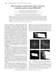
A. Resolution Target<br />
<strong>The</strong> experiment used simulated <strong>image</strong>ry <strong>of</strong> <strong>the</strong> tumbling<br />
E eye chart shown in Fig. 1. Strictly speaking,<br />
NIIRS measures <strong>the</strong> ability to perform specific objectrecognition<br />
tasks listed in <strong>the</strong> NIIRS tables [4,5]. For<br />
<strong>the</strong> purpose <strong>of</strong> this study, however, <strong>image</strong> <strong>quality</strong><br />
equates with <strong>the</strong> ability to recognize <strong>the</strong> orientation<br />
<strong>of</strong> <strong>the</strong> letter E at various scale sizes and contrast ratios.<br />
Thus, study participants did not need to be familiar<br />
with <strong>the</strong> tasks listed in <strong>the</strong> NIIRS tables. This<br />
resolution target has a number <strong>of</strong> o<strong>the</strong>r important features<br />
that enabled <strong>the</strong> use <strong>of</strong> untrained human subjects<br />
in <strong>the</strong> experiment, as opposed to using trained<br />
<strong>image</strong> analysts. <strong>The</strong> object-recognition task being<br />
Table 2.<br />
Statistics <strong>of</strong> General Image-Quality Equation Terms<br />
for Imagery Used to Develop GIQE 4.0 [4]<br />
Term Minimum Mean Maximum<br />
GSD 0:076 m 0:52 m 2:03 m<br />
RER 0.2 0.92 1.3<br />
SNR 2 52.3 130<br />
G 1 10.66 19<br />
H 0.9 1.31 1.9<br />
Fig. 1.<br />
Digital resolution target used for experiment.<br />
10 April 2010 / Vol. 49, No. 11 / APPLIED OPTICS 2133
performed does not vary with <strong>the</strong> scale <strong>of</strong> <strong>the</strong> <strong>image</strong><br />
content. <strong>The</strong> organization <strong>of</strong> <strong>the</strong> eye chart allowed<br />
subjects to locate <strong>the</strong> minimum resolvable features<br />
in an <strong>image</strong> quickly, in comparison to scanning<br />
through a realistic scene for various-sized features.<br />
This allowed us to obtain more data from each subject<br />
in a given amount <strong>of</strong> time. <strong>The</strong> main drawback <strong>of</strong><br />
using <strong>the</strong> eye chart is that it simply is not a natural<br />
scene. As such, <strong>the</strong>re is no reason to believe that results<br />
with <strong>the</strong> eye chart will precisely match results<br />
that would have been obtained had we used actual<br />
aerial <strong>image</strong>ry. We had planned to use realistic syn<strong>the</strong>tic<br />
<strong>image</strong>ry from <strong>the</strong> Digital Imaging and Remote-<br />
Sensing Image Generation (DIRSIG) model [9] in<br />
addition to using <strong>the</strong> eye chart, but did not have<br />
sufficient time to implement this.<br />
<strong>The</strong> eye chart contains a wide range <strong>of</strong> feature sizes.<br />
<strong>The</strong> widths <strong>of</strong> <strong>the</strong> lines making up <strong>the</strong> letter Es in<br />
each row are equivalent to 2.00, 1.59, 1.26, 1.00,<br />
0.794, 0.630, 0.500, 0.397, 0.315, 0.250, 0.198,<br />
0.158, and 0:125 m on <strong>the</strong> ground (in order from <strong>the</strong><br />
top to <strong>the</strong> bottom). This range allowed us to explore<br />
variations in <strong>image</strong> <strong>quality</strong> as large as ΔNIIRS ¼<br />
log 2 ð2=0:125Þ ¼4:0. Adjacent rows <strong>of</strong> <strong>the</strong> eye chart<br />
differ in scale by a factor <strong>of</strong> 1.26, which is equivalent<br />
to ΔNIIRS ¼ log 2 ð1:26Þ ¼0:33. <strong>The</strong> eye chart was<br />
spatially oversampled to minimize artificial sampling<br />
artifacts when simulating <strong>image</strong>s. <strong>The</strong> sampling <strong>of</strong><br />
<strong>the</strong> eye chart was 0:042 m, which is a factor <strong>of</strong> 11 finer<br />
than <strong>the</strong> nominal GSD ¼ 0:46 m <strong>of</strong> <strong>the</strong> simulated<br />
<strong>image</strong>ry.<br />
Note that <strong>the</strong> eye chart exhibits a range <strong>of</strong> contrast<br />
ratios, instead <strong>of</strong> simply having black letters on a<br />
white background [8]. This was done to avoid skewing<br />
<strong>the</strong> results. For example, <strong>the</strong> impact <strong>of</strong> noise<br />
depends on <strong>the</strong> ratio <strong>of</strong> <strong>the</strong> noise amplitude to <strong>the</strong><br />
contrast ratio <strong>of</strong> <strong>the</strong> feature <strong>of</strong> interest. Using only<br />
high-contrast-ratio Es would tend to minimize <strong>the</strong><br />
impact <strong>of</strong> noise on <strong>the</strong> observed ΔNIIRS values,<br />
while using only low-contrast-ratio Es would make<br />
<strong>the</strong> observed ΔNIIRS values overly sensitive to<br />
noise. <strong>The</strong> background reflectance <strong>of</strong> <strong>the</strong> eye chart<br />
was set to 16%. <strong>The</strong> reflectances <strong>of</strong> <strong>the</strong> circular<br />
regions surrounding each E were randomly picked<br />
from a uniform distribution between 6% and 26%.<br />
<strong>The</strong> letter E reflectances were taken from <strong>the</strong> same<br />
random distribution, with <strong>the</strong> constraint that <strong>the</strong> absolute<br />
value <strong>of</strong> <strong>the</strong> reflectance difference between<br />
each E and <strong>the</strong> immediately surrounding circular region<br />
had to be greater than 1%. <strong>The</strong> linear gray scale<br />
for Fig. 1 (and all following simulated <strong>image</strong>s) is such<br />
that black represents 5% reflectance and white<br />
represents 27% reflectance. While an antibunching<br />
method was used to balance <strong>the</strong> effect <strong>of</strong> orientation<br />
[10] in <strong>the</strong> resolution target, <strong>the</strong> effect <strong>of</strong> contrast was<br />
not ideally balanced. Specifically, note that <strong>the</strong><br />
second and third rows <strong>of</strong> <strong>the</strong> eye chart do not contain<br />
very high contrast ratios. In retrospect, an antibunching<br />
method could have been used to yield a<br />
better balance <strong>of</strong> contrast in <strong>the</strong> target.<br />
B. Baseline Imaging Parameters<br />
Figure 2 shows simulated <strong>image</strong>ry <strong>of</strong> <strong>the</strong> resolution<br />
target for a system with <strong>the</strong> baseline imaging parameters<br />
listed in Table 3. <strong>The</strong>se parameters are representative<br />
<strong>of</strong> a current high-resolution panchromatic<br />
remote-sensing system, such as WorldView-1 [11] or<br />
GeoEye-1 [12]. <strong>The</strong> <strong>image</strong> shown in Fig. 2(a) was obtained<br />
by (i) convolving <strong>the</strong> resolution target with a<br />
spectrally averaged optical point spread function<br />
(PSF) for <strong>the</strong> imaging system [13], (ii) convolving<br />
again with <strong>the</strong> pixel impulse response function for<br />
<strong>the</strong> focal plane array, (iii) downsampling this result<br />
based on <strong>the</strong> detector-pixel pitch p, (iv) scaling <strong>the</strong><br />
<strong>image</strong> data to have an average number <strong>of</strong> detected<br />
photoelectrons per pixel N, and (v) adding shot and<br />
detector read noise. For (i), <strong>the</strong> spectrally averaged<br />
optical PSF was computed as a weighted sum <strong>of</strong><br />
monochromatic optical PSFs computed at 81 discrete<br />
wavelengths using <strong>the</strong> gray-world (spatially spectrally<br />
separable) approximation for <strong>the</strong> scene. <strong>The</strong><br />
Fig. 2. Portions <strong>of</strong> <strong>the</strong> simulated baseline <strong>image</strong> (a) before and<br />
(b) after postprocessing. <strong>The</strong> portion shown here corresponds to<br />
rows 4–10 <strong>of</strong> <strong>the</strong> eye chart with corresponding line widths 1.00,<br />
0.794, 0.630, 0.500, 0.397, 0.315, and 0:250 m (from top to bottom).<br />
2134 APPLIED OPTICS / Vol. 49, No. 11 / 10 April 2010
spectrum used for <strong>the</strong> scene was <strong>the</strong> product <strong>of</strong> a solar<br />
irradiance spectrum and a silicon-based detector<br />
responsivity curve. <strong>The</strong> GSD for this <strong>image</strong> is given<br />
by<br />
GSD ¼ pR=f ;<br />
ð2Þ<br />
where R is <strong>the</strong> altitude or target range <strong>of</strong> <strong>the</strong> system<br />
and f is <strong>the</strong> system focal length. <strong>The</strong> RER is calculated<br />
as [4,5]<br />
RER ¼ ERð0:5pÞ − ERð−0:5pÞ;<br />
ð3Þ<br />
where ERðxÞ is <strong>the</strong> normalized edge response for <strong>the</strong><br />
<strong>image</strong>, given by<br />
as <strong>the</strong> signal amplitude for an 8% reflectance target<br />
divided by <strong>the</strong> average noise level (standard deviation)<br />
for an <strong>image</strong> before any postprocessing. Since<br />
<strong>the</strong> average reflectance <strong>of</strong> <strong>the</strong> resolution target is<br />
16%, <strong>the</strong> signal amplitude for an 8% reflectance<br />
target is N=2, where N is <strong>the</strong> average number <strong>of</strong> detected<br />
photoelectrons per pixel. Thus, <strong>the</strong> SNR for an<br />
<strong>image</strong> is computed as<br />
N<br />
SNR ¼ p ffiffiffiffiffiffiffiffiffiffiffiffiffiffiffi ; ð6Þ<br />
2 N þ σ 2<br />
where σ is <strong>the</strong> standard deviation <strong>of</strong> <strong>the</strong> detector read<br />
noise. <strong>The</strong> postprocessed <strong>image</strong> shown in Fig. 2(b)<br />
was obtained by convolving <strong>the</strong> <strong>image</strong> <strong>of</strong> Fig. 2(a)<br />
with a symmetric 3 × 3 sharpening kernel <strong>of</strong> <strong>the</strong> form<br />
ERðxÞ ¼<br />
Z ∞ 0<br />
dx 0 Z ∞ −∞<br />
dysðx − x 0 ; yÞ; ð4Þ<br />
ðx; yÞ are <strong>image</strong> plane coordinates, and sðx; yÞ is <strong>the</strong><br />
net PSF <strong>of</strong> an <strong>image</strong>, which includes effects from<br />
<strong>the</strong> optical PSF, <strong>the</strong> pixel impulse response, and<br />
an <strong>image</strong>-sharpening kernel (if <strong>the</strong> <strong>image</strong> was postprocessed).<br />
ERðxÞ is also used to compute <strong>the</strong> edgeovershoot<br />
factor H as<br />
2<br />
3<br />
−0:2 −0:4 −0:2<br />
w ¼ 4 −0:4 3:4 −0:4 5: ð7Þ<br />
−0:2 −0:4 −0:2<br />
<strong>The</strong> values <strong>of</strong> this kernel were chosen to yield an<br />
RER ¼ 0:925 for <strong>the</strong> postprocessed <strong>image</strong>, which is<br />
about equal to <strong>the</strong> mean value <strong>of</strong> RER ¼ 0:92 for<br />
<strong>the</strong> <strong>image</strong>ry used to develop GIQE 4.0 (see Table 2).<br />
<strong>The</strong> postprocessing noise gain G is given by<br />
<br />
H ¼ ERð1:25pÞ;<br />
maxfERðxÞg on interval x ∈ ½1p; 3pŠ;<br />
when<br />
dERðxÞ<br />
dx<br />
≥ 0 for all x ∈ ½1p; 3pŠ<br />
: ð5Þ<br />
o<strong>the</strong>rwise<br />
<strong>The</strong> H term acts as a penalty for edge-ringing artifacts.<br />
When using small (3 × 3 or 5 × 5), low-fidelity<br />
<strong>image</strong>-sharpening kernels, <strong>the</strong> net system transfer<br />
function is <strong>of</strong>ten boosted above unity at mid-spatial<br />
frequencies to achieve a desired net amount <strong>of</strong> <strong>image</strong><br />
sharpening, resulting in significant edge-overshoot<br />
artifacts. <strong>The</strong> SNR term used in <strong>the</strong> GIQE is defined<br />
Table 3. Simulation Parameters for Baseline Image Shown in Fig. 2<br />
Parameter<br />
Value<br />
Altitude<br />
496 km<br />
Primary mirror diameter, D<br />
0:60 m<br />
Obscuration diameter<br />
0:23 m<br />
Focal length, f<br />
8:8 m<br />
F-number, FN ¼ f =D 14.7<br />
Detector-pixel pitch, p<br />
8:16 μm<br />
Spectral bandwidth<br />
0:4–1:1 μm<br />
Mean wavelength, λ 0<br />
0:644 μm<br />
Detector sampling ratio, Q ¼ λ 0 FN=p 1.19<br />
Average number <strong>of</strong> photoelectrons per pixel, N 162,462<br />
Detector read noise, σ<br />
50 photoelectrons<br />
G ¼<br />
rffiffiffiffiffiffiffiffiffiffiffiffiffiffiffiffiffiffi<br />
P<br />
w 2 m;n<br />
ðm;nÞ<br />
P ; ð8Þ<br />
w m;n<br />
ðm;nÞ<br />
where w m;n represents an individual element <strong>of</strong> w.<br />
Table 4 lists <strong>the</strong> individual <strong>image</strong>-<strong>quality</strong> terms for<br />
<strong>the</strong> baseline <strong>image</strong>ry, along with overall NIIRS-level<br />
predictions from GIQE 3.0 and 4.0.<br />
C. Calibrated Reference Imagery<br />
A set <strong>of</strong> reference <strong>image</strong>s was simulated to have calibrated<br />
ΔNIIRS differences in <strong>image</strong> <strong>quality</strong>. <strong>The</strong><br />
first approach we considered for doing this was to<br />
vary <strong>the</strong> SNR for <strong>the</strong> baseline imaging scenario. This<br />
method, however, was discarded for a number <strong>of</strong> reasons.<br />
<strong>The</strong> SNR coefficient in <strong>the</strong> GIQE is based largely<br />
on <strong>the</strong> psychophysical impact <strong>of</strong> noise on <strong>the</strong><br />
human visual system. <strong>The</strong> <strong>general</strong> appearance <strong>of</strong><br />
low-SNR <strong>image</strong>s is considerably different than that<br />
<strong>of</strong> high-SNR <strong>image</strong>s. <strong>The</strong> SNR coefficients for GIQE<br />
3.0 and 4.0 do not agree. Finally, Ref. [6] found that<br />
10 April 2010 / Vol. 49, No. 11 / APPLIED OPTICS 2135
Table 4.<br />
Image-Quality Parameters for Baseline Image<br />
Parameter Before Processing After Processing<br />
GSD 0:46 m 0:46 m<br />
RER 0.459 0.925<br />
SNR 200 200<br />
G 1 3.42<br />
H 0.876 1.057<br />
GIQE 3.0 NIIRS 5.21 5.93<br />
GIQE 4.0 NIIRS 4.75 5.32<br />
GIQE 4.0 broke down for very low SNR values. <strong>The</strong><br />
next approach for generating <strong>the</strong> reference <strong>image</strong>ry<br />
was to vary <strong>the</strong> primary mirror diameter D, which, in<br />
turn, affects <strong>the</strong> RER <strong>of</strong> <strong>the</strong> simulated <strong>image</strong>ry. Advantages<br />
<strong>of</strong> this approach are that it is deterministic<br />
and <strong>the</strong> resulting changes in <strong>image</strong> <strong>quality</strong> can be<br />
readily understood from <strong>the</strong> notion that each factor<br />
<strong>of</strong> 2 difference in resolution is equivalent to<br />
ΔNIIRS ¼ 1. <strong>The</strong> disadvantage <strong>of</strong> this approach is<br />
that GIQE 4.0 has a breakpoint at RER ¼ 0:9. Thus,<br />
ΔNIIRS predictions from GIQE 3.0 and 4.0 for reference<br />
<strong>image</strong>s generated by this technique would disagree.<br />
Ultimately, <strong>the</strong> method used to generate <strong>the</strong><br />
reference <strong>image</strong>ry was to vary <strong>the</strong> GSD while maintaining<br />
a fixed RER ¼ 0:925 for <strong>the</strong> <strong>image</strong>ry. In terms<br />
<strong>of</strong> fundamental system parameters, this could be accomplished<br />
ei<strong>the</strong>r by changing <strong>the</strong> system altitude or<br />
simultaneously changing <strong>the</strong> detector-pixel pitch and<br />
primary mirror diameter. This approach is also deterministic<br />
and based on <strong>the</strong> notion that ΔNIIRS ¼<br />
1 is equivalent to a 2× change in resolution. Fur<strong>the</strong>rmore,<br />
both GIQE 3.0 and 4.0 (since RER > 0:9) predict<br />
<strong>the</strong> same variation <strong>of</strong> <strong>image</strong> <strong>quality</strong> for this<br />
scenario. This provides confidence in <strong>the</strong> <strong>quality</strong> <strong>of</strong><br />
<strong>the</strong> reference <strong>image</strong>s. In this scenario, <strong>the</strong> <strong>quality</strong><br />
<strong>of</strong> <strong>the</strong> reference <strong>image</strong>ry varies as<br />
ΔNIIRS ¼ log 2 ðGSD 0 =GSDÞ;<br />
ð9Þ<br />
where GSD 0 ¼ 0:46 m is <strong>the</strong> ground sample distance<br />
for <strong>the</strong> baseline <strong>image</strong>. A set <strong>of</strong> 38 reference <strong>image</strong>s<br />
were created, spanning a range <strong>of</strong> ΔNIIRS ¼þ0:21<br />
to −2:38 in increments <strong>of</strong> ΔNIIRS ¼ 0:07. Figure 3<br />
shows <strong>the</strong> ΔNIIRS ¼ −0:98 reference <strong>image</strong> for comparison<br />
with <strong>the</strong> baseline <strong>image</strong> shown in Fig. 2(b).<br />
<strong>The</strong> corresponding 2× difference in spatial resolution<br />
is apparent on comparison <strong>of</strong> <strong>the</strong> different rows <strong>of</strong> <strong>the</strong><br />
eye chart in each <strong>image</strong>.<br />
D. Subjective Image-Quality Evaluation<br />
Observed ΔNIIRS values were obtained for simulated<br />
<strong>image</strong>s from aberrated systems by having human<br />
subjects make visual comparisons with <strong>the</strong><br />
calibrated reference <strong>image</strong> set using a MATLAB<br />
[14] graphical user interface (GUI). A number <strong>of</strong> precautions<br />
were taken to optimize this process. All<br />
<strong>image</strong>s were displayed on a NEC MultiSync<br />
LCD2190UXi monitor calibrated to <strong>the</strong> IDEX-EPD<br />
pr<strong>of</strong>ile using a GretagMacbeth Eye-One Display colorimeter<br />
and <strong>the</strong> NEC SpectraViewII calibration<br />
s<strong>of</strong>tware. Subjects were permitted to adjust <strong>the</strong> display<br />
magnification to optimize <strong>the</strong> viewing eye scale<br />
for each <strong>image</strong> evaluation. 4 × 4 Lagrange interpolation<br />
[15] was used to resample each <strong>image</strong> for display<br />
at <strong>the</strong> monitor screen resolution. This was done to<br />
avoid <strong>image</strong> reinterpolation by ei<strong>the</strong>r MATLAB or<br />
<strong>the</strong> display hardware. <strong>The</strong> GUI was designed to alternately<br />
display <strong>image</strong> pairs (one aberrated <strong>image</strong><br />
and one reference <strong>image</strong>) in a s<strong>of</strong>tcopy flicker sequence.<br />
Image pairs were coregistered and displayed<br />
on <strong>the</strong> same absolute gray scale, and subjects could<br />
adjust <strong>the</strong> flicker rate to suit personal preference.<br />
<strong>The</strong> s<strong>of</strong>tcopy flicker sequence was deemed to be more<br />
comfortable for human subjects and likely to produce<br />
less eye strain than displaying <strong>image</strong>s side by side.<br />
Finally, subjects could change <strong>the</strong> reference <strong>image</strong><br />
being displayed in <strong>the</strong> flicker sequence.<br />
For each aberrated <strong>image</strong>, subjects were instructed<br />
to choose <strong>the</strong> reference <strong>image</strong> that was a best match<br />
in terms <strong>of</strong> <strong>the</strong> ability to recognize <strong>the</strong> orientation <strong>of</strong><br />
<strong>the</strong> Es in <strong>the</strong> eye chart. Because resolution, noise,<br />
and artifacts affect <strong>image</strong> <strong>quality</strong> differently, subjects<br />
were told to base <strong>the</strong>ir choice on <strong>the</strong> average ability<br />
to perform this task using <strong>the</strong> two smallest scale<br />
lines for which Es were recognizable in <strong>the</strong> aberrated<br />
<strong>image</strong>ry. <strong>The</strong> GUI presented <strong>the</strong> aberrated <strong>image</strong>s in<br />
random order to each subject and recorded each subject’s<br />
selections. <strong>The</strong> ΔNIIRS values <strong>of</strong> <strong>the</strong> selected<br />
reference <strong>image</strong>s were <strong>the</strong>n assigned to be <strong>the</strong> observed<br />
ΔNIIRS for <strong>the</strong> corresponding aberrated<br />
<strong>image</strong>s. Six subjects (ages between 20 and 35) participated<br />
in <strong>the</strong> experiment, each <strong>of</strong> whom evaluated<br />
56 <strong>image</strong>s. While some <strong>of</strong> <strong>the</strong> subjects had prescription<br />
glasses, <strong>the</strong>y were not required to wear <strong>the</strong>m<br />
during <strong>the</strong> study.<br />
Fig. 3. Portion <strong>of</strong> simulated reference <strong>image</strong> corresponding to<br />
ΔNIIRS ¼ −0:98 compared to <strong>the</strong> baseline <strong>image</strong> shown in Fig. 2(b).<br />
3. Results<br />
This section describes <strong>the</strong> <strong>image</strong>ry simulated with<br />
aberrations and <strong>the</strong> corresponding human-subject<br />
results.<br />
2136 APPLIED OPTICS / Vol. 49, No. 11 / 10 April 2010
A. Defocus Aberration<br />
Twenty-eight test <strong>image</strong>s were simulated with different<br />
defocus aberration amounts and SNR levels by<br />
modifying <strong>the</strong> parameters <strong>of</strong> <strong>the</strong> baseline imaging<br />
scenario. Defocus was included in <strong>the</strong> simulation<br />
as a rotationally symmetric, quadratic wavefront error<br />
in <strong>the</strong> pupil <strong>of</strong> <strong>the</strong> imaging system. <strong>The</strong> defocus<br />
amount was quantified by <strong>the</strong> peak-to-valley (P–V)<br />
amplitude (in units <strong>of</strong> <strong>the</strong> mean wavelength λ 0 )<strong>of</strong><br />
aberration across <strong>the</strong> area <strong>of</strong> <strong>the</strong> obscured pupil.<br />
Images were simulated with defocus amounts <strong>of</strong><br />
f0; 0:125; 0:25; 0:375; 0:5; 0:627; 0:75g waves P–V <strong>of</strong><br />
defocus. <strong>The</strong> SNR <strong>of</strong> <strong>the</strong> <strong>image</strong>ry was modified by<br />
simply changing N, <strong>the</strong> average number <strong>of</strong> photons<br />
per pixel <strong>of</strong> <strong>the</strong> <strong>image</strong>ry. High, medium, and low<br />
8% reflectivity SNR values <strong>of</strong> 200, 50, and 10 were<br />
obtained using N = 162,462, 12,071, and 1220 photoelectrons<br />
per pixel, respectively. All <strong>of</strong> <strong>the</strong> o<strong>the</strong>r imaging<br />
parameters were <strong>the</strong> same as those listed in<br />
Table 3. Figure 4(a) shows an example <strong>image</strong> with<br />
0.5 wave P–V <strong>of</strong> defocus and SNR ¼ 50 before postprocessing.<br />
Blurring due to defocus can be recognized<br />
by comparing this <strong>image</strong> with Fig. 2(a).<br />
<strong>The</strong> postprocessing for <strong>the</strong> aberrated <strong>image</strong>ry did<br />
not use a simple 3 × 3 sharpening kernel, as was used<br />
for <strong>the</strong> reference <strong>image</strong>ry. Instead, a Fourier-domain<br />
Wiener filter [16–19] with more degrees <strong>of</strong> freedom<br />
was used to enable higher-fidelity aberration compensation.<br />
Also, <strong>the</strong> Wiener filter automatically<br />
yields postprocessed <strong>image</strong>ry that is optimum in<br />
terms <strong>of</strong> <strong>the</strong> expected mean-squared error without<br />
requiring a human observer “in <strong>the</strong> loop.” Specifically,<br />
<strong>the</strong> Wiener filter used here was based on <strong>the</strong><br />
formulation <strong>of</strong> Eq. (19) in [19], using filter parameters<br />
c n ¼ 0:2 and c a ¼ 5. <strong>The</strong> Wiener filter requires<br />
exact knowledge <strong>of</strong> <strong>the</strong> PSF (and thus, <strong>the</strong><br />
aberrations) <strong>of</strong> <strong>the</strong> imaging system, as well as lessstringent<br />
statistical knowledge <strong>of</strong> both <strong>the</strong> object<br />
being <strong>image</strong>d and <strong>the</strong> noise sources. Reference [19]<br />
gives a method <strong>of</strong> estimating from an <strong>image</strong> <strong>the</strong><br />
power spectra for <strong>the</strong> object Φ o and <strong>the</strong> noise Φ n ,<br />
where <strong>the</strong> noise is assumed to be white (Φ n equals<br />
a constant) and Φ o has <strong>the</strong> form<br />
where M represents <strong>the</strong> number <strong>of</strong> pixels along each<br />
dimension <strong>of</strong> an <strong>image</strong> (M ¼ 313 for each <strong>of</strong> <strong>the</strong> aberrated<br />
<strong>image</strong>s in this study), and <strong>the</strong> values A ¼<br />
0:365A 0 and α ¼ 1:57 were predetermined by numerically<br />
fitting <strong>the</strong> power spectrum model <strong>of</strong><br />
Eq. (10) to <strong>the</strong> fast Fourier transform (FFT) <strong>of</strong> <strong>the</strong><br />
resolution target. Note that <strong>the</strong>se expressions assume<br />
that unitary FFTs are used to compute Fourier-domain<br />
quantities and that ρ has units <strong>of</strong><br />
Fourier-domain samples. Using <strong>the</strong>se analytic expressions<br />
and predetermined values resulted in less<br />
postprocessing variability than if <strong>the</strong> power spectra<br />
parameters were estimated from <strong>the</strong> noisy <strong>image</strong>ry.<br />
Figure 4(b) shows <strong>the</strong> Wiener filter result for <strong>the</strong><br />
aberrated <strong>image</strong> with 0.5 wave P–V <strong>of</strong> defocus and<br />
SNR ¼ 50. <strong>The</strong> Wiener filter has improved <strong>the</strong> edge<br />
sharpness, but has also amplified <strong>the</strong> noise in<br />
<strong>the</strong> <strong>image</strong>.<br />
A total <strong>of</strong> 28 <strong>image</strong>s were simulated for this portion<br />
<strong>of</strong> <strong>the</strong> experiment: a set <strong>of</strong> seven <strong>image</strong>s with SNR ¼<br />
200 and each <strong>of</strong> <strong>the</strong> defocus values listed above, a set<br />
<strong>of</strong> seven with SNR ¼ 50, and two sets <strong>of</strong> seven with<br />
SNR ¼ 10. Figure 5 shows <strong>the</strong> variation <strong>of</strong> <strong>the</strong> <strong>image</strong>-<br />
Φ o ðρÞ ¼<br />
<br />
A<br />
2<br />
0<br />
for ρ ¼ 0<br />
A 2 ρ −2α for ρ ≠ 0 : ð10Þ<br />
ρ is a Fourier-domain radial spatial-frequency coordinate<br />
and A 0 , A, and α are power spectrum parameters.<br />
Here, however, <strong>the</strong> noise power spectrum<br />
is computed as<br />
Φ n ¼ N þ σ 2 ;<br />
ð11Þ<br />
where N is <strong>the</strong> average variance <strong>of</strong> <strong>the</strong> <strong>image</strong> shot<br />
noise and σ 2 is <strong>the</strong> variance <strong>of</strong> <strong>the</strong> detector read<br />
noise. For <strong>the</strong> object power spectrum, <strong>the</strong> value <strong>of</strong><br />
A 0 was computed from knowledge <strong>of</strong> <strong>the</strong> average signal<br />
level as<br />
A 0 ¼ MN;<br />
ð12Þ<br />
Fig. 4. Portion <strong>of</strong> simulated <strong>image</strong> with 0.5 waves P–V <strong>of</strong> defocus<br />
and SNR ¼ 50 (a) before and (b) after postprocessing.<br />
10 April 2010 / Vol. 49, No. 11 / APPLIED OPTICS 2137
defocus ¼ 0:75 wave P–V case was discarded as an<br />
outlier, because <strong>the</strong> observed ΔNIIRS value differed<br />
from <strong>the</strong> average by 0.7. Comparison <strong>of</strong> results from<br />
different subjects revealed that noise affected each<br />
subject differently, e.g., <strong>the</strong> observed ΔNIIRS values<br />
from some subjects where consistently ei<strong>the</strong>r higher<br />
or lower than <strong>the</strong> mean for <strong>the</strong> low SNR ¼ 10 <strong>image</strong>ry.<br />
This would explain <strong>the</strong> larger spread, <strong>of</strong> about<br />
0:2, in <strong>the</strong> ΔNIIRS values shown in Fig. 6 for <strong>the</strong><br />
low SNR case. Section 4 contains a detailed analysis<br />
<strong>of</strong> <strong>the</strong> experimental results.<br />
B. Mid-Spatial-Frequency Aberration<br />
Several <strong>image</strong>s were also simulated for a segmentedaperture<br />
system with mid-spatial-frequency figure<br />
errors on each segment. For this case, <strong>the</strong> system primary<br />
mirror was modeled as a ring <strong>of</strong> six hexagonal<br />
segments, each with a flat-to-flat diameter <strong>of</strong> 0:2 m,<br />
such that <strong>the</strong> overall diameter <strong>of</strong> <strong>the</strong> primary mirror<br />
was about equal to <strong>the</strong> value <strong>of</strong> D ¼ 0:6 m used for<br />
<strong>the</strong> baseline imaging scenario (see Table 3). Figure 7<br />
shows <strong>the</strong> spatial distribution <strong>of</strong> <strong>the</strong> mid-spatial-<br />
Fig. 5. Variation <strong>of</strong> log 2 ðRERÞ,G=SNR, and H with <strong>the</strong> amount <strong>of</strong><br />
defocus for SNR ¼ 200, 50, and 10. <strong>The</strong> marked points represent<br />
<strong>the</strong> values for <strong>the</strong> <strong>image</strong>s that were evaluated by human subjects.<br />
<strong>quality</strong> terms log 2 ðRERÞ, G=SNR, and H as a function<br />
<strong>of</strong> <strong>the</strong> amount <strong>of</strong> defocus for each SNR case.<br />
<strong>The</strong> marked points on each curve correspond to<br />
<strong>the</strong> values for <strong>the</strong> simulated <strong>image</strong>ry used in <strong>the</strong> experiment.<br />
Note that <strong>the</strong> vertical scale is equal for<br />
each plot. While <strong>the</strong> plots do not include <strong>the</strong> GIQE<br />
coefficients, it is obvious from <strong>the</strong> figure that RER<br />
is <strong>the</strong> dominant term affecting <strong>image</strong> <strong>quality</strong>.<br />
Figure 6 shows <strong>the</strong> observed ΔNIIRS values obtained<br />
from all six human subjects, along with<br />
ΔNIIRS values calculated from a modified GIQE<br />
(see Section 4). Note that many <strong>of</strong> <strong>the</strong> data markers<br />
in Fig. 6 represent multiple observations, because<br />
<strong>the</strong> ΔNIIRS values <strong>of</strong> <strong>the</strong> reference <strong>image</strong> set were<br />
quantized to increments <strong>of</strong> 0.07, and multiple observers<br />
<strong>of</strong>ten matched <strong>the</strong> same reference <strong>image</strong> to each<br />
aberrated <strong>image</strong>, especially for <strong>the</strong> SNR ¼ 200 and<br />
50 <strong>image</strong> sets. <strong>The</strong> spread in <strong>the</strong> observed ΔNIIRS<br />
values for <strong>the</strong>se <strong>image</strong> sets are <strong>of</strong> <strong>the</strong> order <strong>of</strong><br />
0:1. This seems reasonable, given that differences<br />
<strong>of</strong> ΔNIIRS ¼ 0:1 are <strong>general</strong>ly considered to be<br />
barely noticeable. One datum for <strong>the</strong> SNR ¼ 50,<br />
Fig. 6. Observed ΔNIIRS values obtained from human subjects<br />
for defocused <strong>image</strong>ry with SNR ¼ 200, 50, and 10. <strong>The</strong> dashed<br />
curves represent <strong>the</strong> modified GIQE, ΔNIIRS ¼ 0:291þ<br />
log 2 ðRERÞ − 2:229 G=SNR, obtained from <strong>the</strong> analysis in Section 4.<br />
2138 APPLIED OPTICS / Vol. 49, No. 11 / 10 April 2010
frequency figure errors across <strong>the</strong> segmented primary.<br />
This particular aberration function is based<br />
on measurements (data supplied by NASA Marshall<br />
Space Flight Center) <strong>of</strong> <strong>the</strong> <strong>the</strong>rmal deformation <strong>of</strong><br />
<strong>the</strong> surface <strong>of</strong> a lightweight mirror [20]. <strong>The</strong> dominant<br />
spatial frequency <strong>of</strong> this aberration is 10 cycles<br />
per segment. <strong>The</strong> o<strong>the</strong>r fundamental system parameters<br />
(altitude, focal length, pixel pitch, spectral<br />
bandwidth, mean wavelength, and detector read<br />
noise) had <strong>the</strong> same values listed in Table 3 for<br />
<strong>the</strong> baseline imaging scenario. Figure 8(a) shows<br />
an example <strong>image</strong> simulated with 0.15 waves rootmean-squared<br />
(RMS) <strong>of</strong> mid-spatial-frequency aberration<br />
and SNR ¼ 50 before postprocessing. While<br />
defocus aberration results in blurred <strong>image</strong>ry, <strong>the</strong><br />
primary effect <strong>of</strong> a mid-spatial-frequency aberration<br />
is to reduce <strong>image</strong> contrast. This is a result <strong>of</strong> <strong>the</strong> optical<br />
PSF having a narrow central peak surrounded<br />
by a halo or intensity pedestal. As with <strong>the</strong> defocused<br />
<strong>image</strong>ry, a Wiener filter was used for postprocessing<br />
aberration compensation. Figure 8(b) shows <strong>the</strong> result<br />
<strong>of</strong> <strong>the</strong> Wiener filter for this example.<br />
As for <strong>the</strong> defocus case, 28 <strong>image</strong>s were simulated<br />
with different aberration amounts and SNRs: a set <strong>of</strong><br />
seven <strong>image</strong>s with mid-spatial-frequency aberration<br />
levels <strong>of</strong> f0; 0:125; 0:25; 0:375; 0:5; 0:627; 0:75g waves<br />
RMS and SNR ¼ 200, ano<strong>the</strong>r set <strong>of</strong> seven with<br />
SNR ¼ 50, and two sets <strong>of</strong> seven with SNR ¼ 10.<br />
Figure 9 contains plots <strong>of</strong> log 2 ðRERÞ, G=SNR, and<br />
H versus <strong>the</strong> aberration amount for each SNR case.<br />
Note that <strong>the</strong> RER for <strong>the</strong> segmented-aperture system<br />
is actually <strong>the</strong> geometric mean <strong>of</strong> <strong>the</strong> RER along<br />
<strong>the</strong> vertical and horizontal directions [4]. Likewise<br />
for <strong>the</strong> edge-overshoot term H. <strong>The</strong> observed ΔNIIRS<br />
values for <strong>the</strong>se <strong>image</strong>s along with ΔNIIRS values<br />
computed from a modified GIQE are shown in Fig. 10.<br />
Compared with <strong>the</strong> results for <strong>the</strong> defocused <strong>image</strong>ry<br />
shown in Fig. 6, <strong>the</strong>re appears to be a bit more variability<br />
in <strong>the</strong> observed ΔNIIRS values for this case.<br />
All <strong>of</strong> <strong>the</strong> data for <strong>the</strong> SNR ¼ 10, aberration ¼ 0:3<br />
waves RMS case were discarded, because this <strong>image</strong>ry<br />
was matched to <strong>the</strong> worst reference <strong>image</strong> nine<br />
out <strong>of</strong> twelve times. Several subjects commented that<br />
<strong>the</strong>y did this because <strong>the</strong> reference set did not include<br />
<strong>image</strong>s with sufficiently poor <strong>quality</strong> for comparison<br />
with <strong>the</strong> <strong>image</strong>ry having SNR <strong>of</strong> 10 and<br />
0.3 waves RMS aberration.<br />
Fig. 7. Spatial distribution <strong>of</strong> mid-spatial-frequency aberrations<br />
for a segmented primary mirror.<br />
4. Analysis<br />
<strong>The</strong> results <strong>of</strong> <strong>the</strong> human-subject experiment were<br />
compared to six different <strong>image</strong>-<strong>quality</strong> <strong>equation</strong>s.<br />
Table 5 shows <strong>the</strong> analysis <strong>of</strong> <strong>the</strong> defocused-<strong>image</strong>ry<br />
results. <strong>The</strong> first column lists <strong>the</strong> <strong>image</strong>-<strong>quality</strong><br />
<strong>equation</strong>s that were considered. <strong>The</strong> first <strong>equation</strong><br />
is essentially GIQE 3.0, while <strong>the</strong> second is GIQE<br />
3.0 without <strong>the</strong> edge-overshoot term H. Similarly,<br />
<strong>the</strong> third and fourth <strong>equation</strong>s represent GIQE 4.0<br />
with and without H, respectively. <strong>The</strong> fifth <strong>equation</strong><br />
represents <strong>the</strong> ΔNIIRS variation from log 2 ðRERÞ<br />
alone. <strong>The</strong> sixth <strong>equation</strong> contains a G=SNR term<br />
with a coefficient that provides <strong>the</strong> best agreement<br />
with <strong>the</strong> experimental data. <strong>The</strong> table lists<br />
Fig. 8. Portion <strong>of</strong> simulated <strong>image</strong> with 0.15 waves RMS <strong>of</strong> midspatial-frequency<br />
aberration and SNR ¼ 50 (a) before and (b) after<br />
postprocessing.<br />
10 April 2010 / Vol. 49, No. 11 / APPLIED OPTICS 2139
a ΔNIIRS value predicted by an <strong>image</strong>-<strong>quality</strong> <strong>equation</strong>,<br />
ΔNIIRS obs;avg is <strong>the</strong> average <strong>of</strong> <strong>the</strong> observed<br />
ΔNIIRS obs;k values, <strong>the</strong> subscript k is used to index<br />
various data points, and K is <strong>the</strong> number <strong>of</strong> data<br />
points used in computing each statistic. μ e and σ e<br />
were calculated individually for <strong>the</strong> SNR ¼ 200,<br />
50, and 10 datasets, as well as for <strong>the</strong> whole set <strong>of</strong><br />
defocused <strong>image</strong>ry. <strong>The</strong> constant term for each <strong>image</strong>-<strong>quality</strong><br />
<strong>equation</strong> was adjusted to yield zero mean<br />
error (μ e ¼ 0) for <strong>the</strong> SNR ¼ 200 <strong>image</strong>ry. This was<br />
done to highlight <strong>the</strong> fact that each <strong>equation</strong> fits<br />
<strong>the</strong> SNR ¼ 200 data fairly well, with σ e ≈ 0:1, which<br />
is consistent with <strong>the</strong> 0:1 variation in <strong>the</strong> observed<br />
ΔNIIRS values from different subjects for SNR ¼<br />
200 (see Section 3.A). <strong>The</strong> <strong>equation</strong>s differ, however,<br />
in how well <strong>the</strong>y fit <strong>the</strong> lower SNR data. For example,<br />
<strong>the</strong> GIQE 4.0 <strong>equation</strong> had μ e ¼ 0:291 for <strong>the</strong> SNR ¼<br />
10 data, while <strong>the</strong> <strong>equation</strong> in <strong>the</strong> bottom row <strong>of</strong><br />
Table 5 had μ e ¼ 0:040. A large μ e value (≥0:2) indicates<br />
a fitting error that is visually noticeable (given<br />
that <strong>image</strong>-<strong>quality</strong> differences <strong>of</strong> ΔNIIRS ≥ 0:2 are<br />
Fig. 9. Variation <strong>of</strong> log 2 ðRERÞ,G=SNR, and H with <strong>the</strong> amount <strong>of</strong><br />
mid-spatial-frequency aberration for SNR ¼ 200, 50, and 10. <strong>The</strong><br />
marked points represent <strong>the</strong> values for <strong>the</strong> <strong>image</strong>s that were evaluated<br />
by human subjects.<br />
quantitative statistics that reflect how well each<br />
<strong>equation</strong> agrees with <strong>the</strong> experimental results. <strong>The</strong>se<br />
statistics are <strong>the</strong> mean error μ e , RMS error σ e , and <strong>the</strong><br />
coefficient <strong>of</strong> determination R 2 , computed as<br />
μ e ¼ 1 X<br />
ðΔNIIRS<br />
K<br />
obs;k − ΔNIIRS iqe;k Þ;<br />
k<br />
ð13Þ<br />
σ 2 e ¼ 1 X<br />
ðΔNIIRS<br />
K<br />
obs;k − ΔNIIRS iqe;k Þ 2 ;<br />
k<br />
ð14Þ<br />
P<br />
ðΔNIIRS obs;k − ΔNIIRS iqe;k Þ 2<br />
R 2 k<br />
¼ 1 − P<br />
ðΔNIIRS obs;k − ΔNIIRS obs;avg Þ 2 ;<br />
k<br />
ð15Þ<br />
where ΔNIIRS obs;k represents an observed ΔNIIRS<br />
value from <strong>the</strong> experiment, ΔNIIRS iqe;k represents<br />
Fig. 10. Observed ΔNIIRS values obtained from human subjects<br />
for <strong>image</strong>ry with mid-spatial-frequency aberrations and<br />
SNR ¼ 200, 50, and 10. <strong>The</strong> dashed curves represent <strong>the</strong> modified<br />
GIQE, ΔNIIRS ¼ 0:283 þ log 2 ðRERÞ − 2:574 G=SNR, obtained<br />
from <strong>the</strong> analysis in Section 4.<br />
2140 APPLIED OPTICS / Vol. 49, No. 11 / 10 April 2010
Table 5.<br />
Analysis <strong>of</strong> Results for Imagery with Defocus Aberration<br />
SNR ¼ 200 SNR ¼ 50 SNR ¼ 10 All SNRs<br />
ΔNIIRS Equation μ e σ e μ e σ e μ e σ e μ e σ e R 2 Comment<br />
1:799 þ log 2 ðRERÞ − G=SNR − 1:48 H 0.000 0.108 0.072 0.119 0.244 0.369 0.140 0.274 0.812 GIQE 3.0<br />
0:238 þ log 2 ðRERÞ − G=SNR 0.000 0.110 0.023 0.102 0.154 0.221 0.083 0.173 0.925 GIQE 3.0 w/o H term<br />
0:884 þ 0:951log 2 ðRERÞ<br />
0.000 0.096 0.085 0.127 0.291 0.366 0.168 0.271 0.815 GIQE 4.0<br />
− 0:344 G=SNR − 0:656 H<br />
0:192 þ 0:951log 2 ðRERÞ − 0:344 G=SNR 0.000 0.096 0.064 0.111 0.251 0.304 0.142 0.228 0.870 GIQE 4.0 w/o H term<br />
0:195 þ log 2 ðRERÞ 0.000 0.100 0.070 0.118 0.246 0.291 0.141 0.220 0.878 log 2 ðRERÞ term only<br />
0:291 þ log 2 ðRERÞ − 2:229 G=SNR 0.000 0.130 −0:035 0.122 0.040 0.167 0.011 0.148 0.945 Fitted G=SNR term<br />
considered to be readily perceived). <strong>The</strong> R 2 values in<br />
<strong>the</strong> second to last column indicate overall agreement<br />
with <strong>the</strong> experimental results.<br />
<strong>The</strong> main points we wish to draw from <strong>the</strong> information<br />
in Table 5 are enumerated as follows.<br />
1. Both GIQE 3.0 and 4.0 are inaccurate for <strong>the</strong><br />
SNR ¼ 10 <strong>image</strong>ry. <strong>The</strong> positive μ e values indicate<br />
that <strong>the</strong> predicted ΔNIIRS values are overly optimistic,<br />
i.e., <strong>the</strong> GIQE predictions are consistently better<br />
than <strong>the</strong> observed <strong>image</strong> <strong>quality</strong>. <strong>The</strong> fact that μ e ><br />
0:2 means that this discrepancy is at a visually noticeable<br />
level. This does not imply that ei<strong>the</strong>r GIQE<br />
3.0 or 4.0 is insufficient for predicting <strong>the</strong> <strong>quality</strong> <strong>of</strong><br />
unaberrated <strong>image</strong>ry as <strong>the</strong>y were originally<br />
intended to be used.<br />
2. Dropping <strong>the</strong> H term from GIQE 3.0 and 4.0<br />
yielded better agreement with <strong>the</strong> experimental results.<br />
An argument in support <strong>of</strong> this observation<br />
is that <strong>the</strong> Fourier-domain Wiener filter used here<br />
does not oversharpen <strong>the</strong> <strong>image</strong>ry, in <strong>the</strong> sense that<br />
<strong>the</strong> net modulation transfer function is boosted above<br />
unity. As such, any edge overshoot from <strong>the</strong> Wiener<br />
filter is associated with <strong>the</strong> Gibb’s phenomena. Unlike<br />
<strong>the</strong> <strong>image</strong>ry used in <strong>the</strong> development <strong>of</strong> GIQE<br />
4.0, which exhibited edge-overshoot values in <strong>the</strong><br />
range <strong>of</strong> H ¼ 0:9–1:9, <strong>the</strong> Wiener filter yields a maximum<br />
value <strong>of</strong> H ¼ 1:03 [as explained in Ref. [8] and<br />
reflected by <strong>the</strong> values plotted in Fig. 5]. In this case,<br />
one might expect <strong>the</strong> H term to play a smaller role<br />
determining <strong>the</strong> <strong>image</strong> <strong>quality</strong> <strong>of</strong> Wiener-filtered<br />
<strong>image</strong>ry. For this reason, <strong>the</strong> H term was left out<br />
<strong>of</strong> <strong>the</strong> fitted <strong>image</strong>-<strong>quality</strong> <strong>equation</strong> on <strong>the</strong> bottom<br />
row <strong>of</strong> Table 5. Had a lower-fidelity Wiener filter<br />
been used for aberration compensation, due ei<strong>the</strong>r<br />
to imperfect knowledge <strong>of</strong> <strong>the</strong> system transfer function<br />
or limited degrees <strong>of</strong> freedom, H might have<br />
played a more significant role.<br />
3. <strong>The</strong> log 2 ðRERÞ term alone agrees fairly well<br />
with <strong>the</strong> observed ΔNIIRS values. This is consistent<br />
with <strong>the</strong> notion that a ΔNIIRS ¼ 1 is equivalent to a<br />
factor <strong>of</strong> 2 change in spatial resolution and <strong>the</strong> fact<br />
that RER is a dominant <strong>image</strong>-<strong>quality</strong> factor.<br />
4. <strong>The</strong> <strong>equation</strong> on <strong>the</strong> bottom row <strong>of</strong> Table 5,<br />
with a log 2 ðRERÞ term and a fitted G=SNR term, exhibited<br />
<strong>the</strong> best agreement with <strong>the</strong> experimental<br />
data with R 2 ¼ 0:945. <strong>The</strong> G=SNR coefficient value<br />
<strong>of</strong> −2:229 was obtained by minimizing <strong>the</strong> RMS error<br />
σ e for <strong>the</strong> entire set <strong>of</strong> defocused <strong>image</strong>ry. This value<br />
differs considerably from <strong>the</strong> G=SNR coefficients <strong>of</strong><br />
GIQE 3.0 and 4.0. In <strong>general</strong>, aberrated <strong>image</strong>ry requires<br />
more aggressive postprocessing (more boosting<br />
<strong>of</strong> <strong>the</strong> mid and high spatial frequencies <strong>of</strong> an<br />
<strong>image</strong>) than unaberrated <strong>image</strong>ry. Also, a number<br />
<strong>of</strong> studies have shown that <strong>the</strong> impact <strong>of</strong> a fixed<br />
amount <strong>of</strong> noise on <strong>image</strong> interpretability depends<br />
on <strong>the</strong> spatial power spectrum <strong>of</strong> <strong>the</strong> noise [21–23].<br />
From <strong>the</strong>se two observations, it is not surprising that<br />
<strong>the</strong> G=SNR coefficient obtained here for aberrated<br />
<strong>image</strong>ry differs from <strong>the</strong> GIQE 3.0 and 4.0 coefficients<br />
obtained from a regression analysis <strong>of</strong> unaberrated<br />
<strong>image</strong>ry. Figure 6 shows ΔNIIRS values<br />
computed from this modified GIQE in comparison<br />
with <strong>the</strong> observed ΔNIIRS values. While <strong>the</strong> modified<br />
GIQE had <strong>the</strong> best overall fit, <strong>the</strong>re are residual<br />
trends in <strong>the</strong> data that are not captured by this <strong>equation</strong>.<br />
For example, <strong>the</strong> ΔNIIRS predictions are a bit<br />
optimistic (by about 0.20 ΔNIIRS) for <strong>the</strong> SNR ¼ 10<br />
cases with more than 0.5 waves P–V <strong>of</strong> defocus aberration,<br />
as can be seen in Fig. 6. Perhaps <strong>the</strong> addition<br />
<strong>of</strong> a new GIQE term dependent on aberration<br />
strength would provide a better fit. <strong>The</strong> development<br />
Table 6.<br />
Analysis <strong>of</strong> Results for Imagery with Mid-Spatial-Frequency Aberration<br />
SNR ¼ 200 SNR ¼ 50 SNR ¼ 10 All SNRs<br />
ΔNIIRS Equation μ e σ e μ e σ e μ e σ e μ e σ e R 2 Comment<br />
1:734 þ log 2 ðRERÞ − G=SNR − 1:48 H 0.000 0.115 0.078 0.130 0.198 0.394 0.112 0.283 0.835 GIQE 3.0<br />
0:195 þ log 2 ðRERÞ − G=SNR 0.000 0.094 0.057 0.102 0.156 0.331 0.087 0.236 0.885 GIQE 3.0 w/o H term<br />
0:825 þ 0:951log 2 ðRERÞ<br />
0.000 0.122 0.113 0.163 0.295 0.458 0.167 0.328 0.777 GIQE 4.0<br />
− 0:344 G=SNR − 0:656 H<br />
0:143 þ 0:951log 2 ðRERÞ − 0:344 G=SNR 0.000 0.112 0.104 0.148 0.276 0.428 0.155 0.306 0.806 GIQE 4.0 w/o H term<br />
0:139 þ log 2 ðRERÞ 0.000 0.118 0.111 0.154 0.278 0.427 0.158 0.307 0.805 log 2 ðRERÞ term only<br />
0:283 þ log 2 ðRERÞ − 2:574 G=SNR 0.000 0.075 −0:029 0.089 −0:036 0.250 −0:024 0.180 0.933 Fitted G=SNR term<br />
10 April 2010 / Vol. 49, No. 11 / APPLIED OPTICS 2141
<strong>of</strong> such a new term was beyond <strong>the</strong> scope <strong>of</strong> this paper.<br />
Never<strong>the</strong>less, it seems remarkable that <strong>the</strong> modified<br />
GIQE <strong>equation</strong> with a single fitted coefficient<br />
fits <strong>the</strong> observed ΔNIIRS values as well as it does,<br />
given <strong>the</strong> differences between this study and prior<br />
work on <strong>the</strong> GIQE noted earlier.<br />
<strong>The</strong> corresponding analysis for <strong>the</strong> <strong>image</strong>ry with<br />
mid-spatial-frequency aberrations is summarized<br />
in Table 6. <strong>The</strong>se statistics support <strong>the</strong> points drawn<br />
from <strong>the</strong> experiment with defocused <strong>image</strong>ry. Of particular<br />
interest is <strong>the</strong> similarity between <strong>the</strong> fitted<br />
G=SNR coefficient value <strong>of</strong> −2:574 for <strong>the</strong> midspatial-frequency<br />
aberration case to <strong>the</strong> value <strong>of</strong><br />
−2:229 obtained for <strong>the</strong> defocus aberration case. This<br />
suggests that a G=SNR coefficient <strong>of</strong> approximately<br />
−2:3 might be more <strong>general</strong>ly applicable to a wider<br />
range <strong>of</strong> aberration types. A larger study including<br />
diverse aberrations, however, is needed to substantiate<br />
this. Note that <strong>the</strong> geometric mean <strong>of</strong> <strong>the</strong> RER<br />
and H terms computed along <strong>the</strong> horizontal and vertical<br />
<strong>image</strong> dimensions should be used in <strong>the</strong> GIQE<br />
for asymmetric aberrations like coma and astigmatism<br />
(as was done for <strong>the</strong> <strong>image</strong>ry in Subsection 3.B).<br />
5. Summary<br />
We have described experimental methods for obtaining<br />
quantitative ΔNIIRS <strong>image</strong>-<strong>quality</strong> data from<br />
human subjects. Several details <strong>of</strong> doing this were<br />
addressed in order to avoid systematic errors and<br />
to facilitate <strong>the</strong> use <strong>of</strong> untrained subjects. This approach<br />
was <strong>the</strong>n used to investigate <strong>the</strong> net loss in<br />
<strong>image</strong> <strong>quality</strong> due to aberrations at different SNR levels<br />
for <strong>the</strong> case in which <strong>the</strong> aberrations are known<br />
and compensated for with postprocessing. Both defocus<br />
and mid-spatial-frequency aberrations were considered.<br />
<strong>The</strong> results <strong>of</strong> this experiment were <strong>the</strong>n<br />
compared with ΔNIIRS models based on <strong>the</strong> GIQE,<br />
which was originally developed for use with unaberrated<br />
<strong>image</strong>ry. Both GIQE version 3.0 and 4.0 were<br />
found to be inaccurate in this scenario for low SNRs.<br />
A modified <strong>image</strong>-<strong>quality</strong> <strong>equation</strong> was proposed for<br />
use with aberrated <strong>image</strong>ry as an alternative. This<br />
<strong>equation</strong> has a log 2 ðRERÞ term, chosen based on<br />
<strong>the</strong> design <strong>of</strong> <strong>the</strong> NIIRS scale, and a G=SNR term<br />
with a coefficient fitted to <strong>the</strong> experimental results.<br />
<strong>The</strong> H term was omitted from <strong>the</strong> modified <strong>image</strong><strong>quality</strong><br />
<strong>equation</strong> because <strong>the</strong> Wiener filter we used<br />
for aberration compensation does not result in large<br />
edge-overshoot artifacts. <strong>The</strong> G=SNR coefficients obtained<br />
for defocus and mid-spatial-frequency aberrations<br />
were −2:229 and −2:574, respectively. Overall,<br />
this modified <strong>image</strong>-<strong>quality</strong> <strong>equation</strong> agrees quite<br />
well with <strong>the</strong> experimental results, yielding coefficients<br />
<strong>of</strong> determination R 2 ¼ 0:945 and 0.933 for<br />
<strong>the</strong> defocus and mid-spatial-frequency aberration<br />
cases, respectively.<br />
<strong>The</strong> authors thank Robert Fiete, Jason Calus, and<br />
James Mooney for much advice in developing <strong>the</strong><br />
methods for <strong>the</strong> human-subject experiment. This<br />
work was supported by <strong>the</strong> U. S. Department <strong>of</strong><br />
Defense.<br />
References<br />
1. For a broad, in-depth review <strong>of</strong> various <strong>image</strong>-<strong>quality</strong> metrics<br />
see H. H. Barrett and K. Myers, Foundations <strong>of</strong> Image Science<br />
(Wiley, 2004).<br />
2. R. D. Fiete, H. H. Barret, W. E. Smith, and K. J. Myers, “Hotelling<br />
trace criterion and its correlation with human-observer<br />
performance,” J. Opt. Soc. Am. A 4, 945–953 (1987).<br />
3. P. G. J. Barten, “Evaluation <strong>of</strong> subjective <strong>image</strong> <strong>quality</strong> with<br />
<strong>the</strong> square-root integral method,” J. Opt. Soc. Am. A 7, 2024–<br />
2031 (1990).<br />
4. J. C. Leachtenauer, W. Malila, J. Irvine, L. Colburn, and N.<br />
Salvaggio, “General <strong>image</strong>-<strong>quality</strong> <strong>equation</strong>: GIQE,” Appl.<br />
Opt. 36, 8322–8328 (1997).<br />
5. J. C. Leachtenauer and R. G. Driggers, Surveillance and<br />
Reconnaissance Systems: Modeling and Performance Prediction<br />
(Artech, 2001).<br />
6. R. D. Fiete and T. Tantalo, “Comparison <strong>of</strong> SNR <strong>image</strong> <strong>quality</strong><br />
metrics for remote sensing systems,” Opt. Eng. 40, 574–<br />
585 (2001).<br />
7. R. D. Fiete, T. A. Tantalo, J. R. Calus, and J. A. Mooney, “Image<br />
<strong>quality</strong> <strong>of</strong> sparse-aperture designs for remote sensing,” Opt.<br />
Eng. 41, 1957–1969 (2002).<br />
8. S. T. Thurman and J. R. Fienup, “Analysis <strong>of</strong> <strong>the</strong> <strong>general</strong> <strong>image</strong><br />
<strong>quality</strong> <strong>equation</strong>,” Proc. SPIE 6978, 69780F (2008).<br />
9. J. R. Schott, S. D. Brown, R. V. Raqueno, H. N. Gross, and G.<br />
Robinson, “An advanced syn<strong>the</strong>tic <strong>image</strong> generation model<br />
and its application to multi/hyperspectral algorithm development,”<br />
Can. J. Remote Sens. 25, 99–111 (1999).<br />
10. F. W. Campbell, J. J. Kulikowski, and J. Levinson, “<strong>The</strong> effect<br />
<strong>of</strong> orientation on <strong>the</strong> visual resolution <strong>of</strong> gratings,” J. Physiol.<br />
187, 427–436 (1966).<br />
11. http://www.digitalglobe.com/index.php/86/WorldView‐1.<br />
12. http://www.satimagingcorp.com/satellite‐sensors/<br />
geoeye‐1.html.<br />
13. J. W. Goodman, Introduction to Fourier Optics, 3rd ed.<br />
(Roberts, 2005).<br />
14. http://www.mathworks.com/products/matlab/.<br />
15. A. K. Jain, Fundamentals <strong>of</strong> Digital Image Processing<br />
(Prentice Hall, 1989).<br />
16. N. Wiener, Extrapolation, Interpolation and Smoothing <strong>of</strong><br />
Stationary Time Series (Wiley, 1950).<br />
17. H. W. Bode and C. E. Shannon, “A simplified derivation <strong>of</strong><br />
linear least square smoothing and prediction <strong>the</strong>ory,” Proc.<br />
IRE 38, 417–425 (1950).<br />
18. C. W. Helstrom, “Image reconstruction by <strong>the</strong> method <strong>of</strong> least<br />
squares,” J. Opt. Soc. Am. 57, 297–303 (1967).<br />
19. S. T. Thurman and J. R. Fienup, “Wiener reconstruction <strong>of</strong> undersampled<br />
<strong>image</strong>ry,” J. Opt. Soc. Am. A 26, 283–288 (2009).<br />
20. J. B. Hadaway, P. Stahl, R. Eng, and B. Hogue, “Cryogenic test<br />
results <strong>of</strong> Hextek mirror,” presented at Mirror Technology<br />
Days 2004, Huntsville, Alabama, USA, 17–19 August 2004.<br />
21. P. F. Judy and R. G. Swennsson, “Lesion detection and signalto-noise<br />
ratio in CT <strong>image</strong>s,” Med. Phys. 8, 13–23 (1981).<br />
22. P. A. Guignard, “A comparative method based on ROC analysis<br />
for <strong>the</strong> quantitation <strong>of</strong> observer performance in scintigraphy,”<br />
Phys. Med. Biol. 27, 1163–1176 (1982).<br />
23. K. J. Myers, H. H. Barrett, M. C. Borgstrom, D. D. Patton, and<br />
G. W. Seeley, “Effect <strong>of</strong> noise correlation on detectability <strong>of</strong><br />
disk signals in medical imaging,” J. Opt. Soc. Am. A 2,<br />
1752–1759 (1985).<br />
2142 APPLIED OPTICS / Vol. 49, No. 11 / 10 April 2010
![Phase retrieval algorithms: a personal tour [Invited] - The Institute of ...](https://img.yumpu.com/25023725/1/190x249/phase-retrieval-algorithms-a-personal-tour-invited-the-institute-of-.jpg?quality=85)