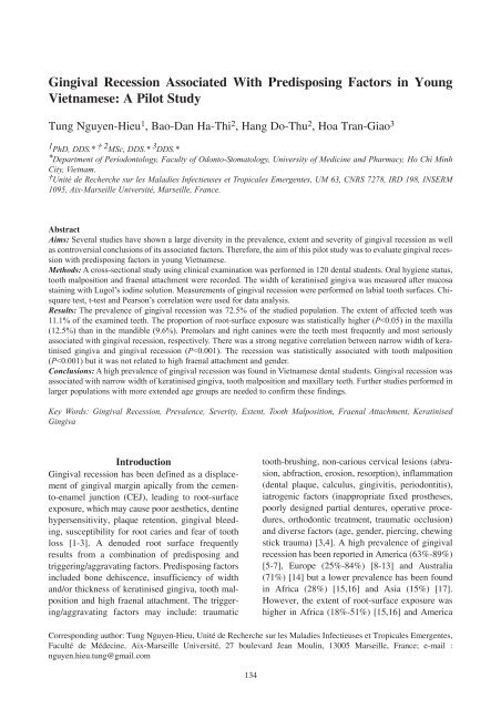
Gingival Recession Associated With Predisposing Factors in Young ...
Gingival Recession Associated With Predisposing Factors in Young ...
Gingival Recession Associated With Predisposing Factors in Young ...

Create successful ePaper yourself
Turn your PDF publications into a flip-book with our unique Google optimized e-Paper software.
<strong>G<strong>in</strong>gival</strong> <strong>Recession</strong> <strong>Associated</strong> <strong>With</strong> <strong>Predispos<strong>in</strong>g</strong> <strong>Factors</strong> <strong>in</strong> <strong>Young</strong><br />
Vietnamese: A Pilot Study<br />
Tung Nguyen-Hieu 1 , Bao-Dan Ha-Thi 2 , Hang Do-Thu 2 , Hoa Tran-Giao 3<br />
1 PhD, DDS.* † 2 MSc, DDS.* 3 DDS.*<br />
*Department of Periodontology, Faculty of Odonto-Stomatology, University of Medic<strong>in</strong>e and Pharmacy, Ho Chi M<strong>in</strong>h<br />
City, Vietnam.<br />
†Unité de Recherche sur les Maladies Infectieuses et Tropicales Emergentes, UM 63, CNRS 7278, IRD 198, INSERM<br />
1095, Aix-Marseille Université, Marseille, France.<br />
Abstract<br />
Aims: Several studies have shown a large diversity <strong>in</strong> the prevalence, extent and severity of g<strong>in</strong>gival recession as well<br />
as controversial conclusions of its associated factors. Therefore, the aim of this pilot study was to evaluate g<strong>in</strong>gival recession<br />
with predispos<strong>in</strong>g factors <strong>in</strong> young Vietnamese.<br />
Methods: A cross-sectional study us<strong>in</strong>g cl<strong>in</strong>ical exam<strong>in</strong>ation was performed <strong>in</strong> 120 dental students. Oral hygiene status,<br />
tooth malposition and fraenal attachment were recorded. The width of kerat<strong>in</strong>ised g<strong>in</strong>giva was measured after mucosa<br />
sta<strong>in</strong><strong>in</strong>g with Lugol’s iod<strong>in</strong>e solution. Measurements of g<strong>in</strong>gival recession were performed on labial tooth surfaces. Chisquare<br />
test, t-test and Pearson’s correlation were used for data analysis.<br />
Results: The prevalence of g<strong>in</strong>gival recession was 72.5% of the studied population. The extent of affected teeth was<br />
11.1% of the exam<strong>in</strong>ed teeth. The proportion of root-surface exposure was statistically higher (P
OHDM - Vol. 11 - No. 3 - September, 2012<br />
(38%-44%) [5-7] than <strong>in</strong> Europe (5%-17%) [8-13],<br />
Australia (4.6%) [14] and Asia (2%) [17]. There<br />
are limited published data concern<strong>in</strong>g the prevalence,<br />
extent, and severity, as well as associated<br />
factors of g<strong>in</strong>gival recession <strong>in</strong> Asia [16,18].<br />
Furthermore, conclusions of the association<br />
between g<strong>in</strong>gival recession and predispos<strong>in</strong>g factors<br />
<strong>in</strong>clud<strong>in</strong>g width of kerat<strong>in</strong>ised g<strong>in</strong>giva [12],<br />
fraenum attachment [13,18] and tooth malposition<br />
[15,18,19] are still controversial [20]. Although the<br />
aetiology of root-surface exposure is multifactorial,<br />
determ<strong>in</strong><strong>in</strong>g the <strong>in</strong>fluence of predispos<strong>in</strong>g factors<br />
has an important role <strong>in</strong> management of g<strong>in</strong>gival<br />
recession such as <strong>in</strong>creas<strong>in</strong>g kerat<strong>in</strong>ised tissue,<br />
fraenectomy or orthodontic treatment.<br />
Aim<br />
The aim of this pilot study was to measure g<strong>in</strong>gival<br />
recession <strong>in</strong> a population of 19-25 year-old dental<br />
students for prelim<strong>in</strong>ary assessment of the prevalence,<br />
extent, severity of g<strong>in</strong>gival recession and its<br />
predispos<strong>in</strong>g factors <strong>in</strong>clud<strong>in</strong>g width of kerat<strong>in</strong>ised<br />
g<strong>in</strong>giva, tooth malposition and fraenal attachment<br />
<strong>in</strong> Vietnamese young adults.<br />
Methods<br />
Target population<br />
A cross-sectional study us<strong>in</strong>g a cl<strong>in</strong>ical exam<strong>in</strong>ation<br />
was performed <strong>in</strong> a population of 120 dental<br />
students of University of Medic<strong>in</strong>e and Pharmacy<br />
of Ho Chi M<strong>in</strong>h City <strong>in</strong> Vietnam. These subjects<br />
were randomly selected from about 600 students of<br />
Faculty of Odonto-Stomatology. Briefly, 80-120<br />
dental students from every class were classified<br />
<strong>in</strong>to ten groups of 8-12 students, from which two<br />
groups were randomly selected. A total of 12<br />
groups <strong>in</strong>clud<strong>in</strong>g 120 students from six classes<br />
agreed to participate <strong>in</strong> this study after read<strong>in</strong>g the<br />
research objectives and sign<strong>in</strong>g a consent form.<br />
Only 19-25 year-old students, non-smok<strong>in</strong>g and<br />
those hav<strong>in</strong>g more than 24 teeth (exclud<strong>in</strong>g third<br />
molars) were <strong>in</strong>cluded. Students with a history of<br />
orthodontic or periodontal treatment were excluded.<br />
The type of toothbrush (soft, medium or hard)<br />
used by the students was recorded.<br />
Cl<strong>in</strong>ical exam<strong>in</strong>ation and measurements<br />
All cl<strong>in</strong>ical exam<strong>in</strong>ations were performed by two<br />
periodontists (B-D H-T and H D-T) who had previously<br />
calibrated by record<strong>in</strong>g all cl<strong>in</strong>ical measurements<br />
to be used <strong>in</strong> the study on ten dental students.<br />
They obta<strong>in</strong>ed a kappa <strong>in</strong>dex of agreement of more<br />
than 0.8. Oral hygiene status was evaluated by<br />
Simplified Oral Hygiene Index (OHI-S) [21].<br />
Labial surfaces of upper first molars and l<strong>in</strong>gual<br />
surfaces of lower first molars (sometimes second<br />
molars because of first molar loss) and the labial<br />
surfaces of upper right and lower left central <strong>in</strong>cisors<br />
were assessed for debris and calculus accumulations.<br />
The OHI-S has two components <strong>in</strong>clud<strong>in</strong>g<br />
the Simplified Debris Index (DI-S) and the<br />
Simplified Calculus Index (CI-S), which are based<br />
on numerical determ<strong>in</strong>ations represent<strong>in</strong>g the<br />
amount of debris and calculus found on the selected<br />
tooth surfaces. Each of these <strong>in</strong>dices has scores<br />
from 0-3 correspond<strong>in</strong>g with an absence of soft<br />
debris/calculus, their amount cover<strong>in</strong>g one-third,<br />
two-thirds, and more than two-thirds of the exam<strong>in</strong>ed<br />
tooth surface, respectively.<br />
All excluded sites <strong>in</strong>clud<strong>in</strong>g absent teeth, tooth<br />
roots and tooth-supported fixed prostheses (crowns<br />
and bridges) were noted. Tooth malpositions<br />
<strong>in</strong>clud<strong>in</strong>g overeruption, rotation and other<br />
malalignment were also recorded. The position of<br />
the CEJ was determ<strong>in</strong>ed by visual observation and<br />
the "slight scratch" of a periodontal probe on the<br />
cervical tooth area <strong>in</strong> a cervico-occlusal direction.<br />
If there were non-carious cervical (wear) lesions or<br />
cervical restorations, the position of the CEJ was<br />
considered as correspond<strong>in</strong>g with that of adjacent<br />
teeth. <strong>G<strong>in</strong>gival</strong> recession was evaluated by us<strong>in</strong>g a<br />
millimetre-graduated periodontal probe to measure<br />
from the free g<strong>in</strong>gival marg<strong>in</strong> to the CEJ at the site<br />
of maximum g<strong>in</strong>gival recession on the labial surface<br />
of each tooth (exclud<strong>in</strong>g third molars). In order<br />
to localise the mucog<strong>in</strong>gival junction (MGJ), both<br />
the g<strong>in</strong>giva and buccal mucosa were r<strong>in</strong>sed with<br />
water, dried and then sta<strong>in</strong>ed with 3% Lugol's<br />
iod<strong>in</strong>e solution (3 g of elemental iod<strong>in</strong>e and 6 g<br />
potassium iod<strong>in</strong>e <strong>in</strong> 300 mL sterile water) by us<strong>in</strong>g<br />
a cotton swab. After two m<strong>in</strong>utes, highly kerat<strong>in</strong>ised<br />
epithelium did not reta<strong>in</strong> the Lugol's iod<strong>in</strong>e<br />
solution while buccal mucosa was sta<strong>in</strong>ed a redbrown<br />
colour. The width of kerat<strong>in</strong>ised g<strong>in</strong>giva of<br />
each tooth (exclud<strong>in</strong>g third molars and previously<br />
excluded sites) was measured by us<strong>in</strong>g a millimetre-graduated<br />
periodontal probe from MGJ to free<br />
g<strong>in</strong>gival marg<strong>in</strong> with 1.0 mm precision. Fraenal<br />
attachments were also assessed accord<strong>in</strong>g to the<br />
Placek et al. (1974) classification [22] <strong>in</strong> which<br />
four levels of labial fraenal attachments are mucosal,<br />
g<strong>in</strong>gival, papillary and papillary penetrat<strong>in</strong>g.<br />
After cl<strong>in</strong>ical exam<strong>in</strong>ation, participants were given<br />
135
OHDM - Vol. 11 - No. 3 - September, 2012<br />
oral hygiene <strong>in</strong>struction and advice on how to control<br />
the progression of g<strong>in</strong>gival recession as well as<br />
suggestions for appropriate treatment.<br />
Data analysis<br />
For assess<strong>in</strong>g the oral hygiene status, OHI-S value<br />
was calculated and classified from 0-1.2 for good<br />
status, 1.3-3.0 for medium status and 3.1-6.0 for<br />
poor status. Values of DI-S and CI-S were comb<strong>in</strong>ed<br />
to obta<strong>in</strong> OHI-S value as follows: DI-S =<br />
(total scores of debris)/(total number of tooth surfaces<br />
with debris), CI-S = (total scores of calculus)/(total<br />
number of tooth surfaces with calculus)<br />
and OHI-S = (DI-S + CI-S) [21]. Descriptive statistics<br />
and the chi-square test were carried out to characterise<br />
the prevalence, extent, severity of g<strong>in</strong>gival<br />
recession, and the predispos<strong>in</strong>g factors. In this<br />
study, the prevalence of g<strong>in</strong>gival recession was the<br />
percentage of patients with at least one g<strong>in</strong>gival<br />
recession <strong>in</strong> the studied population, the extent was<br />
the percentage of teeth associated with root-surface<br />
exposure <strong>in</strong> exam<strong>in</strong>ed teeth, and the severity was<br />
the level of apical migration of marg<strong>in</strong>al g<strong>in</strong>giva<br />
from the CEJ. The Student's t-test was used to compare<br />
kerat<strong>in</strong>ised g<strong>in</strong>giva widths between teeth with<br />
and without root-surface exposure. The correlation<br />
between kerat<strong>in</strong>ised tissue and g<strong>in</strong>gival recession<br />
was assessed by Pearson's correlation coefficient.<br />
These statistical analyses were <strong>in</strong>dependently performed<br />
by a specialist us<strong>in</strong>g statistical software<br />
(Statistical Package for the Social Sciences, version<br />
15.0; SPSS Inc, Chicago, USA). Differences were<br />
considered statistically significant when the P-<br />
value was 1.0 mm) was 87/120<br />
(72.5%). This prevalence was slightly higher <strong>in</strong><br />
females (45/60, 75%) than <strong>in</strong> males (42/60, 70%)<br />
but this difference was not statistically significant.<br />
Among a total of 3269 exam<strong>in</strong>ed teeth (1634<br />
teeth <strong>in</strong> the maxilla and 1635 teeth <strong>in</strong> the<br />
mandible), there were 362 teeth (11.1%) with g<strong>in</strong>gival<br />
recession. The proportion of root denudation<br />
was statistically higher (chi-square test, P molars (74/205, 36.1%) > can<strong>in</strong>es<br />
(34/205, 16.6%) > <strong>in</strong>cisors (13/205, 6.3%). In the<br />
mandible, this was found as follow: premolars<br />
(90/157, 57.3%) > molars (37/157, 23.6%) > <strong>in</strong>cisors<br />
(18/157, 11.5%) > can<strong>in</strong>es (12/157, 7.6%).<br />
Students with g<strong>in</strong>gival recession had mean<br />
measurements of denuded root surface from 1.0-1.9<br />
mm <strong>in</strong> both maxilla and mandible. The most serious<br />
g<strong>in</strong>gival recession was detected at upper right<br />
can<strong>in</strong>es (1.9±1.2 mm) and lower right can<strong>in</strong>es<br />
(1.9±1.3 mm) (Figure 2).<br />
<strong>Predispos<strong>in</strong>g</strong> factors associated with g<strong>in</strong>gival<br />
recession<br />
Overall, the width of kerat<strong>in</strong>ised g<strong>in</strong>giva was more<br />
important <strong>in</strong> the regions of <strong>in</strong>cisors and molars and<br />
less important <strong>in</strong> the regions of can<strong>in</strong>es and premolars.<br />
Teeth with g<strong>in</strong>gival recession had a greater<br />
reduced width of kerat<strong>in</strong>ised g<strong>in</strong>giva than nonaffected<br />
teeth. In particular, these differences were<br />
statistically significant <strong>in</strong> all premolars (t-test,<br />
P
OHDM - Vol. 11 - No. 3 - September, 2012<br />
Figure 1. Extent of g<strong>in</strong>gival<br />
recessions <strong>in</strong> the maxilla<br />
and the mandible.<br />
Figure 2. Severity of g<strong>in</strong>gival<br />
recession <strong>in</strong> the maxilla<br />
and the mandible<br />
Figure 3. Width of kerat<strong>in</strong>ised<br />
g<strong>in</strong>giva between<br />
teeth with and without<br />
g<strong>in</strong>gival recession.(t-test:<br />
**:P
OHDM - Vol. 11 - No. 3 - September, 2012<br />
Table 1. Correlations between g<strong>in</strong>gival recessions and predispos<strong>in</strong>g factors<br />
<strong>Predispos<strong>in</strong>g</strong> factors Number of teeth (%) P*<br />
<strong>With</strong>out recession <strong>With</strong> recession<br />
Normal width of kerat<strong>in</strong>ised g<strong>in</strong>giva (> 2 mm) 2630 (91.3%) 251 (8.7%) 0.05<br />
<strong>G<strong>in</strong>gival</strong>/papillary fraenal attachments 61 (95.3%) 3 (4.7%)<br />
Normal position teeth 2658 (89.9%) 298 (10.1%) 0.05). The proportion of malpositioned<br />
teeth was 313/3269 (9.6%), of which dental rotation<br />
(117/3269, 3.6%) and labial decl<strong>in</strong>ation<br />
(67/3269, 2%) were most frequently found.<br />
Malaligned teeth had a higher prevalence of g<strong>in</strong>gival<br />
recession (64/313, 20.4%) than normally positioned<br />
teeth (298/2956, 10.1%) and this difference<br />
was statistically significant (chi-square test,<br />
P1 mm and an extent of from 2%-18% of teeth. In<br />
the current study, g<strong>in</strong>gival recession was detected<br />
<strong>in</strong> 72.5% of Vietnamese dental students with the<br />
same age range. Indeed, this high prevalence was<br />
similar to that reported <strong>in</strong> France (76%) [12], Italy<br />
(64%) [8], Brazil (64%-77%) [6,7]. The extent of<br />
affected teeth <strong>in</strong> the current study (11.1%) was also<br />
<strong>in</strong> agreement with previous studies <strong>in</strong> France<br />
138
OHDM - Vol. 11 - No. 3 - September, 2012<br />
Table 2. Studies of prevalence extent, severity and associated factors of g<strong>in</strong>gival recession (GR)<br />
(cont<strong>in</strong>ued over)<br />
Authors Year Population Prevalence (% Extent (% teeth <strong>Associated</strong> Non-associated<br />
Study Country [males/females] subjects with GR) with GR) Severity factors factors<br />
Years of age (mean) Exam<strong>in</strong>ed surfaces Most frequent teeth<br />
Tenenbaum 1982 100 dental students 76/100 (76%) 320/2725 (11.7%) - Attached Oral hygiene<br />
Cross-sectional France [68/32] Male = female First premolars g<strong>in</strong>giva status, fraenum<br />
19-26 (21.7) Labial (r = -0.201, attachment<br />
P female brush<strong>in</strong>g<br />
frequency<br />
Ser<strong>in</strong>o et al. 1994 225 subjects (25%) 1373/5168 (26.6%) - Age, loss of <strong>G<strong>in</strong>gival</strong><br />
Cross-sectional Sweden 18-65 18-29 years: (44%) 18-29 years: (7%) approximal <strong>in</strong>flammation<br />
and longitud<strong>in</strong>al Labial Upper first molars attachment<br />
and premolars<br />
(multiple<br />
regression model,<br />
P 3 mm Cigarette smok<strong>in</strong>g, Gender, race,<br />
Cross-sectional Brazil [719/867] 20-29 years: (76.5%) 20-29 years: 18.1% (51.6 %), suprag<strong>in</strong>gival dental visits,<br />
14-103 Male = female Mandibular GR > 3 mm calculus (Wald socio-economic<br />
(37.9 ± 13.3) Labial and l<strong>in</strong>gual <strong>in</strong>cisors (22 %) test, P
OHDM - Vol. 11 - No. 3 - September, 2012<br />
Table 2. Studies of prevalence extent, severity and associated factors of g<strong>in</strong>gival recession (GR) (cont<strong>in</strong>ued)<br />
Authors Year Population Prevalence (% Extent (% teeth <strong>Associated</strong> Non-associated<br />
Study Country [males/females] subjects with GR) with GR) Severity factors factors<br />
Years of age (mean) Exam<strong>in</strong>ed surfaces Most frequent teeth<br />
Thomson et al. 2006 915 subjects 628/882 (71.2%) (4.6%) - - Age<br />
Longitud<strong>in</strong>al New [469/446] Labial and l<strong>in</strong>gual Lower <strong>in</strong>cisors<br />
Zealand 32<br />
Slutzkey& Lev<strong>in</strong> 2007 303 subjects (14.6%) (1.6%) GR = 1-2 mm Past orthodontic Dental plaque,<br />
Cross-sectional Israel [126/177] Male > female (79.5%), treatment and oral g<strong>in</strong>givitis,<br />
18-22 Labial surface GR > 3 mm pierc<strong>in</strong>g (Fisher smok<strong>in</strong>g<br />
(20.5%) exact test and<br />
Pearson chi-square<br />
test, P female Maxilla < Mandible (0.8 %), traumatic tooth-<br />
15-68 (P 6 mm bleed<strong>in</strong>g <strong>in</strong>dex, dental visits<br />
(24.6% subjects) (1.8 %) tobacco<br />
consumption<br />
(multiple<br />
regression model,<br />
P
OHDM - Vol. 11 - No. 3 - September, 2012<br />
gival recession than females. Moreover, the proportion<br />
of affected teeth <strong>in</strong> the current study was statistically<br />
higher <strong>in</strong> the maxilla than <strong>in</strong> the mandible<br />
whereas two previous studies [6,13] reported an<br />
opposite f<strong>in</strong>d<strong>in</strong>g. This contradiction could be<br />
expla<strong>in</strong>ed by the assessment of g<strong>in</strong>gival recession<br />
of both labial and l<strong>in</strong>gual tooth surfaces <strong>in</strong> these<br />
two studies. Indeed, the frequent accumulation of<br />
calculus on the l<strong>in</strong>gual surface of lower <strong>in</strong>cisors<br />
<strong>in</strong>creased the proportion of root-surface exposure<br />
<strong>in</strong> mandible [7,13,16]. Therefore, teeth most frequently<br />
associated with g<strong>in</strong>gival recession also varied<br />
accord<strong>in</strong>g to which tooth surfaces were exam<strong>in</strong>ed.<br />
Studies assess<strong>in</strong>g both labial and l<strong>in</strong>gual surfaces<br />
have usually found that the most frequently<br />
affected teeth were mandibular <strong>in</strong>cisors [6,7,13,<br />
14,16] whereas studies assess<strong>in</strong>g only labial surfaces<br />
have suggested premolars or upper first<br />
molars as most frequently associated with g<strong>in</strong>gival<br />
recession [8,9,11,12,19]. Accord<strong>in</strong>gly, maxillary<br />
and mandibular premolars were most frequently<br />
associated with g<strong>in</strong>gival recession <strong>in</strong> the current<br />
study. The narrowest width of kerat<strong>in</strong>ised g<strong>in</strong>giva<br />
was also found <strong>in</strong> premolar regions. Moreover, the<br />
study of Joshipura et al. (1994) suggested that g<strong>in</strong>gival<br />
recession might be due to tooth-brush<strong>in</strong>g<br />
force <strong>in</strong> premolars or debris/calculus accumulation<br />
<strong>in</strong> molars [28]. A significant negative correlation<br />
has also been found between g<strong>in</strong>gival recession and<br />
dental plaque [29]. However, the role of calculus as<br />
an aetiological factor or consequence of g<strong>in</strong>gival<br />
recession is disputed [6].<br />
Although many studies have confirmed the<br />
<strong>in</strong>fluence of toothbrush type, tooth-brush<strong>in</strong>g<br />
method and tooth-brush<strong>in</strong>g frequency on the <strong>in</strong>cidence<br />
of g<strong>in</strong>gival recession [5,8,9,13,18,19], a systematic<br />
review by Rajapakse et al. (2007) concluded<br />
that data to support or refute this relation were<br />
<strong>in</strong>conclusive [30]. The highest level of evidence<br />
lead<strong>in</strong>g to this conclusion was presented <strong>in</strong> just one<br />
abstract of an <strong>in</strong>dustry-sponsored randomised controlled<br />
cl<strong>in</strong>ical trial and after presentation at a scientific<br />
conference, this full trial had not been published<br />
[31]. Interest<strong>in</strong>gly, <strong>in</strong> the current study it was<br />
observed that the most serious g<strong>in</strong>gival recession<br />
was found <strong>in</strong> right can<strong>in</strong>es of both the maxilla and<br />
the mandible. Accord<strong>in</strong>g to the evaluation of rootsurface<br />
exposure <strong>in</strong> left- and right-handed adults,<br />
Tezel et al. (2001) suggested the relationship<br />
between g<strong>in</strong>gival recession and the hand used <strong>in</strong><br />
tooth-brush<strong>in</strong>g. In right-handed subjects, denuded<br />
root surfaces were frequently detected <strong>in</strong> premolar<br />
and can<strong>in</strong>e regions of upper right and lower right<br />
arches. A similar result was also observed <strong>in</strong> lefthanded<br />
subjects: g<strong>in</strong>gival recession was usually<br />
detected <strong>in</strong> their upper left and lower left arches<br />
[32]. The majority of Vietnamese dental students<br />
were right-handed, used soft/medium toothbrushes<br />
and had a good level of oral hygiene. This suggested<br />
a correlation between g<strong>in</strong>gival recession and<br />
tooth-brush<strong>in</strong>g. Moreover, because of the transitional<br />
site between anterior and posterior teeth, the<br />
specific position of can<strong>in</strong>es <strong>in</strong> dental arch could<br />
make them more susceptible to traumatic toothbrush<strong>in</strong>g.<br />
Although subjects with periodontitis, or<br />
those who had undergone or had periodontal surgery,<br />
orthodontics, fixed prostheses and smok<strong>in</strong>g<br />
were excluded from this study, other trigger<strong>in</strong>g/<br />
aggravat<strong>in</strong>g factors <strong>in</strong>clud<strong>in</strong>g traumatic toothbrush<strong>in</strong>g<br />
and non-carious cervical lesions (abrasion,<br />
erosion) may be risks for bias. Indeed, toothbrush<strong>in</strong>g<br />
and an acidic diet are probably related to<br />
both tooth wear and dent<strong>in</strong>e hypersensitivity and<br />
may be associated with g<strong>in</strong>gival recession that is<br />
termed "healthy recession" [1]. Traumatic toothbrush<strong>in</strong>g<br />
can cause superficial damage of the g<strong>in</strong>giva<br />
usually termed "g<strong>in</strong>gival abrasions". However,<br />
the relevance of this phenomenon to g<strong>in</strong>gival recession<br />
rema<strong>in</strong>s unclear [33]. Nevertheless it is clear<br />
that toothbrush abrasion may cause wear at the<br />
CEJ, result<strong>in</strong>g <strong>in</strong> the destruction of the support<strong>in</strong>g<br />
periodontium and lead<strong>in</strong>g to g<strong>in</strong>gival recession<br />
[34].<br />
Localis<strong>in</strong>g the MGJ was facilitated by mucosa<br />
sta<strong>in</strong><strong>in</strong>g with 3% Lugol's iod<strong>in</strong>e solution. Indeed,<br />
Lugol sta<strong>in</strong><strong>in</strong>g has been widely used to detect<br />
malignant changes <strong>in</strong> the cervix uteri and the<br />
oesophagus [35], and recently has been used to<br />
detect epithelial dysplasia <strong>in</strong> oral mucosa [36]. This<br />
sta<strong>in</strong><strong>in</strong>g produced a red-brown colour based on the<br />
reaction of iod<strong>in</strong>e with glycogen granules of oral<br />
mucosa whereas content glycogen was <strong>in</strong>versely<br />
related to degree of keratosis, lead<strong>in</strong>g to kerat<strong>in</strong>ised<br />
g<strong>in</strong>giva unsta<strong>in</strong>ed with Lugol's iod<strong>in</strong>e solution<br />
[35,36]. This iod<strong>in</strong>e solution has also been used to<br />
measure the width of kerat<strong>in</strong>ised g<strong>in</strong>giva <strong>in</strong> previous<br />
studies [20,37]. Lang and Löe (1972) found<br />
that <strong>in</strong> areas with less than 2 mm of kerat<strong>in</strong>ised g<strong>in</strong>giva,<br />
<strong>in</strong>flammation persisted <strong>in</strong> spite of effective<br />
oral hygiene and they suggested that at least 2 mm<br />
of kerat<strong>in</strong>ised g<strong>in</strong>giva was necessary to ma<strong>in</strong>ta<strong>in</strong><br />
g<strong>in</strong>gival health [37]. Only one longitud<strong>in</strong>al study<br />
has demonstrated that <strong>in</strong> patients ma<strong>in</strong>ta<strong>in</strong><strong>in</strong>g proper<br />
plaque control, lack of an adequate zone of<br />
141
OHDM - Vol. 11 - No. 3 - September, 2012<br />
attached g<strong>in</strong>giva did not result <strong>in</strong> the <strong>in</strong>creased <strong>in</strong>cidence<br />
of g<strong>in</strong>gival recession [20] whereas several<br />
cross-sectional studies have confirmed a correlation<br />
between the decreased width of kerat<strong>in</strong>ised/<br />
attached g<strong>in</strong>giva and soft-tissue recession [12,38].<br />
In the current cross-sectional study, statistical tests<br />
showed a strong negative correlation between the<br />
lack of kerat<strong>in</strong>ised g<strong>in</strong>giva and root-surface exposure<br />
<strong>in</strong> subjects with good oral hygiene. Additionally,<br />
teeth with narrow width of kerat<strong>in</strong>ised g<strong>in</strong>giva had<br />
a statistically higher prevalence of g<strong>in</strong>gival recession<br />
than teeth without. However, it cannot be concluded<br />
from these f<strong>in</strong>d<strong>in</strong>gs that a narrow width of<br />
kerat<strong>in</strong>ised g<strong>in</strong>giva was a cause or a consequence<br />
of g<strong>in</strong>gival recession. Further longitud<strong>in</strong>al studies<br />
are required to <strong>in</strong>vestigate this question.<br />
Malaligned teeth had a statistically higher proportion<br />
of root-surface exposure than those <strong>in</strong> a<br />
normal position. This f<strong>in</strong>d<strong>in</strong>g was <strong>in</strong> accordance<br />
with previous studies [15,18,19]. It was seen that<br />
tooth malposition, <strong>in</strong>clud<strong>in</strong>g rotation and l<strong>in</strong>gual<br />
<strong>in</strong>cl<strong>in</strong>ation, was usually associated with soft debris<br />
retention and calculus because of difficult access<br />
dur<strong>in</strong>g tooth-brush<strong>in</strong>g. Labially <strong>in</strong>cl<strong>in</strong>ed teeth were<br />
often susceptible to traumatic tooth-brush<strong>in</strong>g and<br />
appeared to be more susceptible to non-carious cervical<br />
lesions. Furthermore, if a tooth erupts close to<br />
the MGJ, there is probably very little or no kerat<strong>in</strong>ised<br />
g<strong>in</strong>giva labially and then g<strong>in</strong>gival recession<br />
can occur [3].<br />
In the current study, no correlation was found<br />
between high fraenal attachment and root-surface<br />
exposure. This f<strong>in</strong>d<strong>in</strong>g was similar to that from a<br />
study <strong>in</strong> France which also showed a low proportion<br />
(3%) of affected teeth with high fraenal attachments<br />
[12]. However, high fraenal attachments<br />
have been reported as significantly associated with<br />
g<strong>in</strong>gival recession <strong>in</strong> two previous studies [13,18].<br />
It has been suggested that g<strong>in</strong>gival/papillary fraenal<br />
attachments could impede access for plaque<br />
removal [13] and that fraenal pull due to lack of<br />
attached g<strong>in</strong>gival is also a factor caus<strong>in</strong>g g<strong>in</strong>gival<br />
recession [18]. Although the fraenal attachment has<br />
been reported as <strong>in</strong>fluenc<strong>in</strong>g plaque accumulation<br />
and g<strong>in</strong>givitis [39], <strong>in</strong> well-motivated <strong>in</strong>dividuals<br />
with good oral hygiene (such as the dental students<br />
who took part <strong>in</strong> the current study) this <strong>in</strong>fluenc<strong>in</strong>g<br />
factor for g<strong>in</strong>gival recession can be reduced or<br />
elim<strong>in</strong>ated. A key f<strong>in</strong>d<strong>in</strong>g <strong>in</strong> the current study was<br />
that 60.5% of teeth related to g<strong>in</strong>gival recession did<br />
not appear to be associated with predispos<strong>in</strong>g factors.<br />
Therefore, further studies will be needed to<br />
<strong>in</strong>vestigate the issue further.<br />
Conclusions<br />
This pilot study performed <strong>in</strong> a small population of<br />
dental students with high awareness of dental care<br />
may not be representative of the whole population<br />
of the Vietnamese young adults. However, some<br />
prelim<strong>in</strong>ary conclusions may be drawn:<br />
• A high prevalence of g<strong>in</strong>gival recession<br />
(72.5%) was found <strong>in</strong> 19-25 year-old<br />
Vietnamese dental students with 11.1%<br />
extent of affected teeth.<br />
• The proportion of root-surface exposure<br />
was statistically higher <strong>in</strong> the maxilla<br />
(12.5%) than <strong>in</strong> the mandible (9.6%).<br />
• Premolars were most frequently associated<br />
with g<strong>in</strong>gival recession and right can<strong>in</strong>es<br />
were the most severely affected teeth.<br />
• There was a strong negative correlation<br />
between a narrow width of kerat<strong>in</strong>ised g<strong>in</strong>giva<br />
and soft-tissue recessions.<br />
• Root-surface exposure was statistically<br />
associated with tooth malposition.<br />
• <strong>G<strong>in</strong>gival</strong> recession was not associated with<br />
high fraenal attachment and gender.<br />
Further studies performed <strong>in</strong> a larger population<br />
of non-dental students with more extended age<br />
groups are required to confirm these f<strong>in</strong>d<strong>in</strong>gs.<br />
Acknowledgements<br />
The authors would like to thank Dr. Trong-Hung<br />
Hoang (Department of Community Dentistry,<br />
Faculty of Odonto-Stomatology, University of<br />
Medic<strong>in</strong>e and Pharmacy of Ho Chi M<strong>in</strong>h City,<br />
Vietnam) for the statistical analysis.<br />
Author contributions<br />
• T N-H and B-D H-T conceived and<br />
designed the study.<br />
• B-D H-T and H D-T performed the cl<strong>in</strong>ical<br />
exam<strong>in</strong>ations.<br />
• T N-H and H T-G wrote the paper.<br />
Conflict of <strong>in</strong>terest and source of fund<strong>in</strong>g<br />
The authors declare that they have no conflicts<br />
of <strong>in</strong>terest and no fund<strong>in</strong>g source.<br />
142
OHDM - Vol. 11 - No. 3 - September, 2012<br />
References<br />
1. Addy M. Tooth brush<strong>in</strong>g, tooth wear and dent<strong>in</strong>e<br />
hypersensitivity: are they associated? International Dental<br />
Journal. 2005; 55: 261-267.<br />
2. Kassab MM, Cohen RE. The etiology and prevalence<br />
of g<strong>in</strong>gival recession. Journal of the American Dental<br />
Association. 2003; 134: 220-225.<br />
3. Tugnait A, Clerehugh V. <strong>G<strong>in</strong>gival</strong> recession: its significance<br />
and management. Journal of Dentistry. 2001; 29: 381-<br />
394.<br />
4. Borghetti A, Monnet-Corti V. Récessions g<strong>in</strong>givales<br />
[<strong>G<strong>in</strong>gival</strong> recession]. In: Borghetti A, Monnet-Corti V, editors.<br />
Chirurgie plastique parodontale [Periodontal Plastic<br />
Surgery]. 2nd ed. Rueil-Malmaison, France: Editions CDP;<br />
2008: p. 77-95. French<br />
5. Khocht A, Simon G, Person P, Denepitiya JL. <strong>G<strong>in</strong>gival</strong><br />
recession <strong>in</strong> relation to history of hard toothbrush use. Journal<br />
of Periodontology. 1993; 64: 900-905.<br />
6. Mar<strong>in</strong>i MG, Greghi SL, Passanezi E, Sant’ana AC.<br />
<strong>G<strong>in</strong>gival</strong> recession: prevalence, extension and severity <strong>in</strong><br />
adults. Journal of Applied Oral Science. 2004; 12: 250-255.<br />
7. Sus<strong>in</strong> C, Haas AN, Oppermann RV, Haugejorden O,<br />
Albandar JM. <strong>G<strong>in</strong>gival</strong> recession: epidemiology and risk <strong>in</strong>dicators<br />
<strong>in</strong> a representative urban Brazilian population. Journal<br />
of Periodontology. 2004; 75: 1377-1386.<br />
8. Checchi L, Daprile G, Gatto MR, Pelliccioni GA.<br />
<strong>G<strong>in</strong>gival</strong> recession and toothbrush<strong>in</strong>g <strong>in</strong> an Italian School of<br />
Dentistry: a pilot study. Journal of Cl<strong>in</strong>ical Periodontology.<br />
1999; 26: 276-280.<br />
9. Kozlowska M, Wawrzyn-Sobczak K, Karczewski JK,<br />
Stokowska W. The oral cavity hygiene as the basic element of<br />
the g<strong>in</strong>gival recession prophylaxis. Roczniki Akademii<br />
Medycznej w Bialymstoku. 2005; 50 Suppl 1: 234-237.<br />
10. Sarfati A, Bourgeois D, Katsahian S, Mora F,<br />
Bouchard P. Risk assessment for buccal g<strong>in</strong>gival recession<br />
defects <strong>in</strong> an adult population. Journal of Periodontology.<br />
2010; 81: 1419-1425.<br />
11. Ser<strong>in</strong>o G, Wennstrom JL, L<strong>in</strong>dhe J, Eneroth L. The<br />
prevalence and distribution of g<strong>in</strong>gival recession <strong>in</strong> subjects<br />
with a high standard of oral hygiene. Journal of Cl<strong>in</strong>ical<br />
Periodontology. 1994; 21: 57-63.<br />
12. Tenenbaum H. A cl<strong>in</strong>ical study compar<strong>in</strong>g the width<br />
of attached g<strong>in</strong>giva and the prevalence of g<strong>in</strong>gival recessions.<br />
Journal of Cl<strong>in</strong>ical Periodontology. 1982; 9: 86-92.<br />
13. Toker H, Ozdemir H. <strong>G<strong>in</strong>gival</strong> recession: epidemiology<br />
and risk <strong>in</strong>dicators <strong>in</strong> a university dental hospital <strong>in</strong><br />
Turkey. International Journal of Dental Hygiene. 2009; 7: 115-<br />
120.<br />
14. Thomson WM, Broadbent JM, Poulton R, Beck JD.<br />
Changes <strong>in</strong> periodontal disease experience from 26 to 32 years<br />
of age <strong>in</strong> a birth cohort. Journal of Periodontology. 2006; 77:<br />
947-954.<br />
15. Arowojolu MO. <strong>G<strong>in</strong>gival</strong> recession at the University<br />
College Hospital, Ibadan: prevalence and effect of some aetiological<br />
factors. African Journal of Medic<strong>in</strong>e and Medical<br />
Sciences. 2000; 29: 259-263.<br />
16. van Palenste<strong>in</strong> Helderman WH, Lembariti BS, van<br />
der Weijden GA, van ‘t Hof MA. <strong>G<strong>in</strong>gival</strong> recession and its<br />
association with calculus <strong>in</strong> subjects deprived of prophylactic<br />
dental care. Journal of Cl<strong>in</strong>ical Periodontology. 1998; 25: 106-<br />
111.<br />
17. Slutzkey S, Lev<strong>in</strong> L. <strong>G<strong>in</strong>gival</strong> recession <strong>in</strong> young<br />
adults: occurrence, severity, and relationship to past orthodontic<br />
treatment and oral pierc<strong>in</strong>g. American Journal of<br />
Orthodontic and Dentofacial Orthopedics. 2008; 134: 652-<br />
656.<br />
18. Dodwad V. Aetiology and severity of g<strong>in</strong>gival recession<br />
among young <strong>in</strong>dividuals <strong>in</strong> Belgaum district <strong>in</strong> India.<br />
Annals of the Dental University of Malaya. 2001; 8: 1-6.<br />
19. Chrysanthakopoulos NA. Aetiology and severity of<br />
g<strong>in</strong>gival recession <strong>in</strong> an adult population sample <strong>in</strong> Greece.<br />
Dental Research Journal (Isfahan). 2011; 8: 64-70.<br />
20. Wennstrom JL. Lack of association between width of<br />
attached g<strong>in</strong>giva and development of soft tissue recession. A 5-<br />
year longitud<strong>in</strong>al study. Journal of Cl<strong>in</strong>ical Periodontology.<br />
1987; 14: 181-184.<br />
21. Greene JC, Vermillion JR. The Simplified Oral<br />
Hygiene Index. Journal of the American Dental Associaiton.<br />
1964; 68: 7-13.<br />
22. Placek M, Skach M, Mrklas L. [Problems with the lip<br />
frenulum <strong>in</strong> parodontology. I. Classification and epidemiology<br />
of tendons of the lip frenulum]. Ceskoslovenská Stomatologie.<br />
1974; 74: 385-391.<br />
23. Maynard JG, Wilson RD. Diagnosis and management<br />
of mucog<strong>in</strong>gival problems <strong>in</strong> children. Dental Cl<strong>in</strong>ics of North<br />
America. 1980; 24: 683-703.<br />
24. Closs LQ, Grehs B, Raveli DB, Ros<strong>in</strong>g CK.<br />
Occurrence, extension, and severity of g<strong>in</strong>gival marg<strong>in</strong> alterations<br />
after orthodontic treatment. World Journal of<br />
Orthodontics. 2008; 9: e1-e6.<br />
25. Muller HP, Stadermann S, He<strong>in</strong>ecke A. <strong>G<strong>in</strong>gival</strong><br />
recession <strong>in</strong> smokers and non-smokers with m<strong>in</strong>imal periodontal<br />
disease. Journal of Cl<strong>in</strong>ical Periodontology. 2002; 29: 129-<br />
136.<br />
26. Koke U, Sander C, He<strong>in</strong>ecke A, Muller HP. A possible<br />
<strong>in</strong>fluence of g<strong>in</strong>gival dimensions on attachment loss and<br />
g<strong>in</strong>gival recession follow<strong>in</strong>g placement of artificial crowns.<br />
International Journal of Periodontics and Restorative<br />
Dentistry. 2003; 23: 439-445.<br />
27. Daprile G, Gatto MR, Checchi L. The evolution of<br />
buccal g<strong>in</strong>gival recessions <strong>in</strong> a student population: a 5-year follow-up.<br />
Journal of Periodontology. 2007; 78: 611-614.<br />
28. Joshipura KJ, Kent RL, DePaola PF. <strong>G<strong>in</strong>gival</strong> recession:<br />
<strong>in</strong>tra-oral distribution and associated factors. Journal of<br />
Periodontology. 1994; 65: 864-871.<br />
29. Addy M, Mostafa P, Newcombe RG. Dent<strong>in</strong>e hypersensitivity:<br />
the distribution of recession, sensitivity and plaque.<br />
Journal of Dentistry. 1987; 15: 242-248.<br />
30. Rajapakse PS, McCracken GI, Gwynnett E, Steen<br />
ND, Guentsch A, Heasman PA. Does tooth brush<strong>in</strong>g <strong>in</strong>fluence<br />
the development and progression of non-<strong>in</strong>flammatory g<strong>in</strong>gival<br />
recession? A systematic review. Journal of Cl<strong>in</strong>ical<br />
Periodontology. 2007; 34: 1046-1061.<br />
31. Matthews DC. No good evidence to l<strong>in</strong>k toothbrush<strong>in</strong>g<br />
trauma to g<strong>in</strong>gival recession. Evidence-Based Dentistry.<br />
2008; 9: 49.<br />
32. Tezel A, Canakci V, Cicek Y, Demir T. Evaluation of<br />
g<strong>in</strong>gival recession <strong>in</strong> left- and right-handed adults.<br />
International Journal of Neuroscience. 2001; 110: 135-146.<br />
33. Addy M, Hunter ML. Can tooth brush<strong>in</strong>g damage<br />
your health? Effects on oral and dental tissues. International<br />
Dental Journal. 2003; 53 Suppl 3: 177-186.<br />
34. Litonjua LA, Andreana S, Bush PJ, Cohen RE.<br />
Toothbrush<strong>in</strong>g and g<strong>in</strong>gival recession. International Dental<br />
Journal. 2003; 53: 67-72.<br />
143
OHDM - Vol. 11 - No. 3 - September, 2012<br />
35. Ohta K, Ogawa I, Ono S, Taki M, Mizuta K,<br />
Miyauchi M, et al. Histopathological evaluation <strong>in</strong>clud<strong>in</strong>g<br />
cytokerat<strong>in</strong> 13 and Ki-67 <strong>in</strong> the border between Lugol-sta<strong>in</strong>ed<br />
and -unsta<strong>in</strong>ed areas. Oncology Reports. 2010; 24: 9-14.<br />
36. Umeda M, Shigeta T, Takahashi H, M<strong>in</strong>amikawa T,<br />
Komatsubara H, Oguni A, et al. Cl<strong>in</strong>ical evaluation of Lugol’s<br />
iod<strong>in</strong>e sta<strong>in</strong><strong>in</strong>g <strong>in</strong> the treatment of stage I-II squamous cell carc<strong>in</strong>oma<br />
of the tongue. International Journal of Oral<br />
Maxillofacial Surgery. 2011; 40: 593-596.<br />
37. Lang NP, Loe H. The relationship between the width<br />
of kerat<strong>in</strong>ized g<strong>in</strong>giva and g<strong>in</strong>gival health. Journal of<br />
Periodontology. 1972; 43: 623-627.<br />
38. Goutoudi P, Koidis PT, Konstant<strong>in</strong>idis A. <strong>G<strong>in</strong>gival</strong><br />
recession: a cross-sectional cl<strong>in</strong>ical <strong>in</strong>vestigation. European<br />
Journal of Prosthodontic and Restorative Dentistry. 1997; 5:<br />
57-61.<br />
39. Addy M, Dummer PM, Hunter ML, K<strong>in</strong>gdon A,<br />
Shaw WC. A study of the association of fraenal attachment, lip<br />
coverage, and vestibular depth with plaque and g<strong>in</strong>givitis.<br />
Journal of Periodontology. 1987; 58: 752-757.<br />
144