Gingival Recession Associated With Predisposing Factors in Young ...
Gingival Recession Associated With Predisposing Factors in Young ...
Gingival Recession Associated With Predisposing Factors in Young ...
Create successful ePaper yourself
Turn your PDF publications into a flip-book with our unique Google optimized e-Paper software.
OHDM - Vol. 11 - No. 3 - September, 2012<br />
oral hygiene <strong>in</strong>struction and advice on how to control<br />
the progression of g<strong>in</strong>gival recession as well as<br />
suggestions for appropriate treatment.<br />
Data analysis<br />
For assess<strong>in</strong>g the oral hygiene status, OHI-S value<br />
was calculated and classified from 0-1.2 for good<br />
status, 1.3-3.0 for medium status and 3.1-6.0 for<br />
poor status. Values of DI-S and CI-S were comb<strong>in</strong>ed<br />
to obta<strong>in</strong> OHI-S value as follows: DI-S =<br />
(total scores of debris)/(total number of tooth surfaces<br />
with debris), CI-S = (total scores of calculus)/(total<br />
number of tooth surfaces with calculus)<br />
and OHI-S = (DI-S + CI-S) [21]. Descriptive statistics<br />
and the chi-square test were carried out to characterise<br />
the prevalence, extent, severity of g<strong>in</strong>gival<br />
recession, and the predispos<strong>in</strong>g factors. In this<br />
study, the prevalence of g<strong>in</strong>gival recession was the<br />
percentage of patients with at least one g<strong>in</strong>gival<br />
recession <strong>in</strong> the studied population, the extent was<br />
the percentage of teeth associated with root-surface<br />
exposure <strong>in</strong> exam<strong>in</strong>ed teeth, and the severity was<br />
the level of apical migration of marg<strong>in</strong>al g<strong>in</strong>giva<br />
from the CEJ. The Student's t-test was used to compare<br />
kerat<strong>in</strong>ised g<strong>in</strong>giva widths between teeth with<br />
and without root-surface exposure. The correlation<br />
between kerat<strong>in</strong>ised tissue and g<strong>in</strong>gival recession<br />
was assessed by Pearson's correlation coefficient.<br />
These statistical analyses were <strong>in</strong>dependently performed<br />
by a specialist us<strong>in</strong>g statistical software<br />
(Statistical Package for the Social Sciences, version<br />
15.0; SPSS Inc, Chicago, USA). Differences were<br />
considered statistically significant when the P-<br />
value was 1.0 mm) was 87/120<br />
(72.5%). This prevalence was slightly higher <strong>in</strong><br />
females (45/60, 75%) than <strong>in</strong> males (42/60, 70%)<br />
but this difference was not statistically significant.<br />
Among a total of 3269 exam<strong>in</strong>ed teeth (1634<br />
teeth <strong>in</strong> the maxilla and 1635 teeth <strong>in</strong> the<br />
mandible), there were 362 teeth (11.1%) with g<strong>in</strong>gival<br />
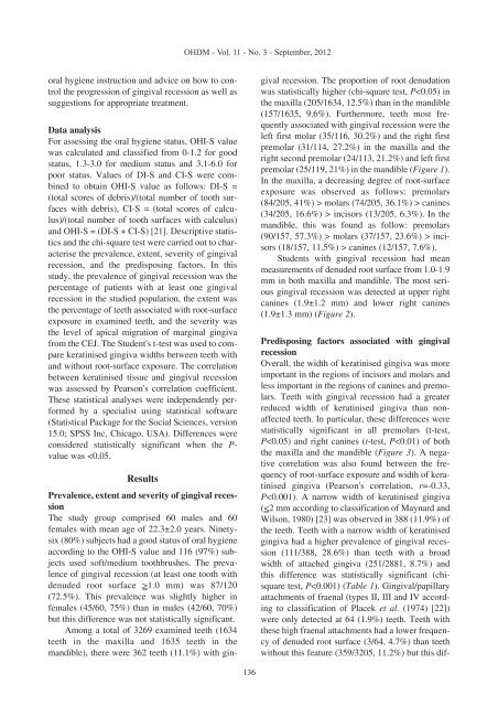
recession. The proportion of root denudation<br />
was statistically higher (chi-square test, P molars (74/205, 36.1%) > can<strong>in</strong>es<br />
(34/205, 16.6%) > <strong>in</strong>cisors (13/205, 6.3%). In the<br />
mandible, this was found as follow: premolars<br />
(90/157, 57.3%) > molars (37/157, 23.6%) > <strong>in</strong>cisors<br />
(18/157, 11.5%) > can<strong>in</strong>es (12/157, 7.6%).<br />
Students with g<strong>in</strong>gival recession had mean<br />
measurements of denuded root surface from 1.0-1.9<br />
mm <strong>in</strong> both maxilla and mandible. The most serious<br />
g<strong>in</strong>gival recession was detected at upper right<br />
can<strong>in</strong>es (1.9±1.2 mm) and lower right can<strong>in</strong>es<br />
(1.9±1.3 mm) (Figure 2).<br />
<strong>Predispos<strong>in</strong>g</strong> factors associated with g<strong>in</strong>gival<br />
recession<br />
Overall, the width of kerat<strong>in</strong>ised g<strong>in</strong>giva was more<br />
important <strong>in</strong> the regions of <strong>in</strong>cisors and molars and<br />
less important <strong>in</strong> the regions of can<strong>in</strong>es and premolars.<br />
Teeth with g<strong>in</strong>gival recession had a greater<br />
reduced width of kerat<strong>in</strong>ised g<strong>in</strong>giva than nonaffected<br />
teeth. In particular, these differences were<br />
statistically significant <strong>in</strong> all premolars (t-test,<br />
P


