
Create successful ePaper yourself
Turn your PDF publications into a flip-book with our unique Google optimized e-Paper software.
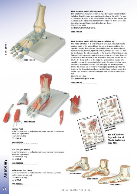
Foot Skeleton Model with Ligaments<br />
This detailed model displays numerous important ligaments and tendons<br />
including the achilles and peroneus longus tendons of the ankle. The model<br />
consists of the bones of the foot and lower portions of the tibia and fibula,<br />
including the introsseous membrane found between them. All the anatomically<br />
important ligaments and tendons are shown.<br />
23x18x30 cm, 0.6 kg<br />
L/D/E/F/I/S/P/J/R/C www.<br />
9985-1000359<br />
Anatomy Skeletons<br />
9985-1000359<br />
9985-1000360<br />
Normal Foot<br />
Superficial structures as well as internal bones, muscles, ligaments and<br />
nerves are represented.<br />
13x24x9 cm; 0.4 kg<br />
L/D/E/F<br />
9985-1000354<br />
Flat Foot (Pes Planus)<br />
Superficial structures as well as internal bones, muscles, ligaments and<br />
nerves are represented.<br />
12x24x10 cm; 0.4 kg<br />
L/D/E/F<br />
9985-1000355<br />
Hollow Foot (Pes Cavus)<br />
Superficial structures as well as internal bones, muscles, ligaments<br />
and nerves are represented.<br />
13x23x10 cm; 0.4 kg<br />
L/D/E/F<br />
Foot Skeleton Model with Ligaments and Muscles<br />
This model is the best of its kind for quality and value. This anatomically<br />
detailed model of the foot and lower leg can be disassembled into 6 removable<br />
parts for detailed study. The model features not only the bones<br />
but also muscles, tendons, ligaments, nerves, arteries, and veins. The frontal<br />
view features the extensor muscles of the lower leg. The tendons can be<br />
followed on their passage under the transverse and crucial crural ligaments<br />
all the way to their insertion points. In addition all tendon sheaths are visible.<br />
On the dorsal portion of the model the gastrocnemius muscle is removable<br />
to reveal deeper anatomical elements. The sole of the foot is represented<br />
in three layers; the first layer displaying the flexor digitorum<br />
brevis. This muscle can be removed revealing the quadratus plantae, the<br />
tendon of the flexor digitorum longus, and the flexor hallucis muscle. This<br />
second layer is in turn removable to display even deeper anatomical details.<br />
23x26x19 cm, 1.1kg<br />
L/D/E/F/I/S/P/J/R/C www.<br />
9985-1000360<br />
9985-1000356<br />
Hand and Wrist<br />
9985-4006659<br />
9985-1001484<br />
9985-1000354<br />
Foot and Joints of Foot<br />
9985-4006662<br />
9985-1001490<br />
You will find our<br />
large selection of<br />
Charts starting on<br />
page 108.<br />
24<br />
9985-1000356<br />
<strong>3B</strong> <strong>Scientific</strong>® <strong>Medical</strong><br />
9985-1000355