
Create successful ePaper yourself
Turn your PDF publications into a flip-book with our unique Google optimized e-Paper software.
9985-1000249<br />
9985-1001249<br />
9985-1001250<br />
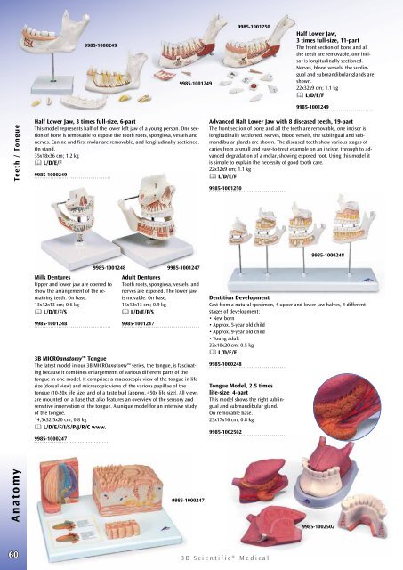
Half Lower Jaw,<br />
3 times full-size, 11-part<br />
The front section of bone and all<br />
the teeth are removable, one incisor<br />
is longitudinally sectioned.<br />
Nerves, blood vessels, the sublingual<br />
and submandibular glands are<br />
shown.<br />
22x32x9 cm; 1.1 kg<br />
L/D/E/F<br />
9985-1001249<br />
Anatomy Teeth / Tongue<br />
Half Lower Jaw, 3 times full-size, 6-part<br />
This model represents half of the lower left jaw of a young person. One section<br />
of bone is removable to expose the tooth roots, spongiosa, vessels and<br />
nerves. Canine and first molar are removable, and longitudinally sectioned.<br />
On stand.<br />
35x18x36 cm; 1.2 kg<br />
L/D/E/F<br />
9985-1000249<br />
Milk Dentures<br />
Upper and lower jaw are opened to<br />
show the arrangement of the remaining<br />
teeth. On base.<br />
13x12x13 cm; 0.6 kg<br />
L/D/E/F/S<br />
9985-1001248<br />
9985-1001248 9985-1001247<br />
Adult Dentures<br />
Tooth roots, spongiosa, vessels, and<br />
nerves are exposed. The lower jaw<br />
is movable. On base.<br />
16x12x13 cm; 0.9 kg<br />
L/D/E/F/S<br />
9985-1001247<br />
<strong>3B</strong> MICROanatomy Tongue<br />
The latest model in our <strong>3B</strong> MICROanatomy series, the tongue, is fascinating<br />
because it combines enlargements of various different parts of the<br />
tongue in one model. It comprises a macroscopic view of the tongue in life<br />
size (dorsal view) and microscopic views of the various papillae of the<br />
tongue (10-20x life size) and of a taste bud (approx. 450x life size). All views<br />
are mounted on a base that also features an overview of the sensory and<br />
sensitive innervation of the tongue. A unique model for an intensive study<br />
of the tongue.<br />
14,5x32,5x20 cm, 0,8 kg<br />
L/D/E/F/I/S/P/J/R/C www.<br />
9985-1000247<br />
9985-1000247<br />
Advanced Half Lower Jaw with 8 diseased teeth, 19‐part<br />
The front section of bone and all the teeth are removable, one incisor is<br />
longitudinally sectioned. Nerves, blood vessels, the sublingual and submandibular<br />
glands are shown. The diseased teeth show various stages of<br />
caries from a small and easy-to-treat example on an incisor, through to advanced<br />
degradation of a molar, showing exposed root. Using this model it<br />
is simple to explain the necessity of good tooth care.<br />
22x32x9 cm; 1.1 kg<br />
L/D/E/F<br />
9985-1001250<br />
Tongue Model, 2.5 times<br />
life-size, 4-part<br />
This model shows the right sublingual<br />
and submandibular gland.<br />
On removable base.<br />
23x17x16 cm; 0.8 kg<br />
9985-1002502<br />
9985-1000248<br />
Dentition Development<br />
Cast from a natural specimen, 4 upper and lower jaw halves, 4 different<br />
stages of development:<br />
• New born<br />
• Approx. 5-year old child<br />
• Approx. 9-year old child<br />
• Young adult<br />
33x10x20 cm; 0.5 kg<br />
L/D/E/F<br />
9985-1000248<br />
9985-1002502<br />
60<br />
<strong>3B</strong> <strong>Scientific</strong>® <strong>Medical</strong>