
You also want an ePaper? Increase the reach of your titles
YUMPU automatically turns print PDFs into web optimized ePapers that Google loves.
Joints<br />
9985-1000175<br />
9985-1000003<br />
9985-1002390<br />
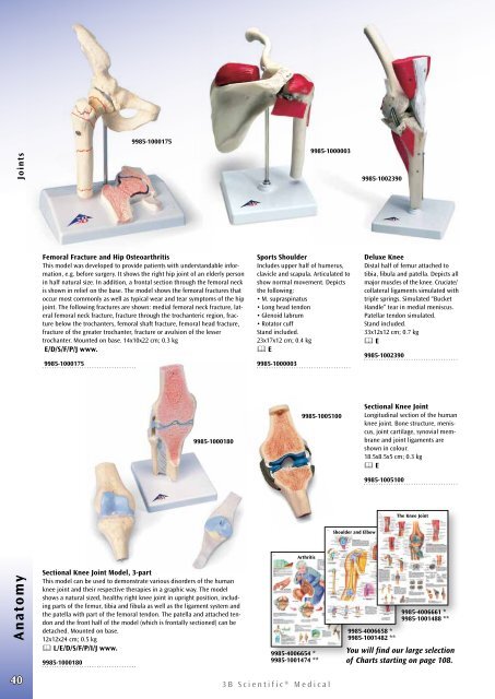
Femoral Fracture and Hip Osteoarthritis<br />
This model was developed to provide patients with un der stand able information,<br />
e.g. before surgery. It shows the right hip joint of an elderly person<br />
in half natural size. In addition, a frontal section through the femoral neck<br />
is shown in relief on the base. The model shows the femoral fractures that<br />
occur most commonly as well as typical wear and tear symptoms of the hip<br />
joint. The following fractures are shown: medial femoral neck fracture, lateral<br />
femoral neck fracture, fracture through the trochanteric region, fracture<br />
below the trochanters, femoral shaft fracture, femoral head fracture,<br />
fracture of the greater trochanter, fracture or avulsion of the lesser<br />
trochanter. Mounted on base. 14x10x22 cm; 0.3 kg<br />
E/D/S/F/P/J www.<br />
9985-1000175<br />
Sports Shoulder<br />
Includes upper half of humerus,<br />
clavicle and scapula. Articulated to<br />
show normal movement. Depicts<br />
the following:<br />
• M. supraspinatus<br />
• Long head tendon<br />
• Glenoid labrum<br />
• Rotator cuff<br />
Stand included.<br />
23x17x12 cm; 0.4 kg<br />
& E<br />
9985-1000003<br />
Deluxe Knee<br />
Distal half of femur attached to<br />
tibia, fibula and patella. Depicts all<br />
major muscles of the knee. Cruciate/<br />
collateral ligaments simulated with<br />
triple springs. Simulated “Bucket<br />
Handle” tear in medial meniscus.<br />
Patellar tendon simulated.<br />
Stand included.<br />
33x12x12 cm; 0.7 kg<br />
& E<br />
9985-1002390<br />
9985-1000180<br />
9985-1005100<br />
Sectional Knee Joint<br />
Longitudinal section of the human<br />
knee joint. Bone structure, meniscus,<br />
joint cartilage, synovial membrane<br />
and joint ligaments are<br />
shown in colour.<br />
18.5x8.5x5 cm; 0.3 kg<br />
& E<br />
9985-1005100<br />
The Knee Joint<br />
Shoulder and Elbow<br />
Anatomy<br />
40<br />
Sectional Knee Joint Model, 3-part<br />
This model can be used to demonstrate various disorders of the human<br />
knee joint and their respective therapies in a graphic way. The model<br />
shows a natural sized, healthy right knee joint in upright position, including<br />
parts of the femur, tibia and fibula as well as the ligament system and<br />
the patella with part of the femoral tendon. The patella and attached tendon<br />
and the front half of the model (which is frontally sectioned) can be<br />
detached. Mounted on base.<br />
12x12x24 cm; 0.5 kg<br />
& L/E/D/S/F/P/I/J www.<br />
9985-1000180<br />
Arthritis<br />
9985-4006654 *<br />
9985-1001474 **<br />
<strong>3B</strong> <strong>Scientific</strong>® <strong>Medical</strong><br />
9985-4006661 *<br />
9985-1001488 **<br />
9985-4006658 *<br />
9985-1001482 **<br />
You will find our large selection<br />
of Charts starting on page 108.