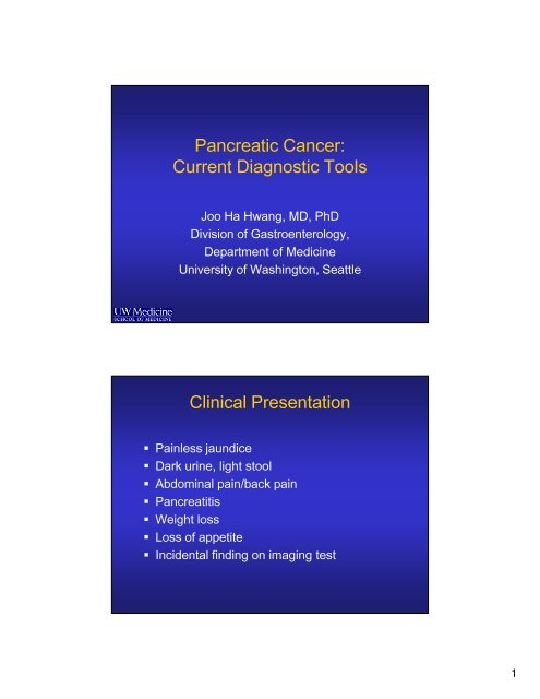
Pancreatic Cancer: Current Diagnostic Tools Clinical Presentation
Pancreatic Cancer: Current Diagnostic Tools Clinical Presentation
Pancreatic Cancer: Current Diagnostic Tools Clinical Presentation

Create successful ePaper yourself
Turn your PDF publications into a flip-book with our unique Google optimized e-Paper software.
<strong>Pancreatic</strong> <strong>Cancer</strong>:<br />
<strong>Current</strong> <strong>Diagnostic</strong> <strong>Tools</strong><br />
Joo Ha Hwang, MD, PhD<br />
Division of Gastroenterology,<br />
Department of Medicine<br />
University of Washington, Seattle<br />
<strong>Clinical</strong> <strong>Presentation</strong><br />
Painless jaundice<br />
Dark urine, light stool<br />
Abdominal pain/back pain<br />
Pancreatitis<br />
Weight loss<br />
Loss of appetite<br />
Incidental finding on imaging test<br />
1
Establishing the Diagnosis<br />
Noninvasive cross-sectional imaging<br />
• CT or MRI<br />
Biopsy<br />
• CT-guided FNA<br />
• US-guided FNA<br />
• EUS-guided FNA<br />
• ERCP brushing/biopsy<br />
GI Anatomy<br />
2
<strong>Pancreatic</strong> Anatomy<br />
<strong>Pancreatic</strong> <strong>Cancer</strong>s - Location<br />
www.radiologyassistant.nl<br />
3
Tumor cannot be assessed T0No evidence of primary tumor TisCarcinoma in situ T1Tumor 2 cm or less T2Tumor limited to pancreas, greater than 2 cm TXPrimary<br />
extends beyond pancreas, no celiac or SMA involvement T4Tumor involves celiac axis or SMA (unresectable primary) Regional Lymph Nodes (N) NX nodes cannot be assessed T3Tumor<br />
No regional lymph nodes N1 Regional lymph metastasis N0<br />
Metastasis (M) MX Presence of distant metastasis cannot be assessed M0 No distant M1 Distant metastasis Distant<br />
Purpose of <strong>Diagnostic</strong> <strong>Tools</strong><br />
Identify the location of the lesion<br />
Diagnosis – tissue sampling<br />
Staging<br />
• Prognosis<br />
• Directs management<br />
Surgery<br />
Chemotherapy<br />
Other interventions<br />
Eligibility for clinical trials<br />
Primary Tumor (T)<br />
TNM Staging<br />
4
I T1-2 IIA T3 N0 IIB T1-3 N1 III T4 M0<br />
IV Any T Any N M1 Stage<br />
AJCC Staging<br />
Stage T N M<br />
Imaging Modalities<br />
Transabdominal Ultrasound<br />
CT scan<br />
MRI/MRCP<br />
Endoscopic Retrograde<br />
Cholangiopancreatogrpahy (ERCP)<br />
Endoscopic Ultrasound (EUS)<br />
PET/PET-CT<br />
5
Transabdominal US<br />
• Non-invasive<br />
• Relatively inexpensive<br />
• Quick to perform<br />
• Usually can identify<br />
tumors that cause biliary<br />
obstruction (pancreatic<br />
head tumors)<br />
• Limited detail<br />
• Operator dependent<br />
• May be sufficient for<br />
staging if metastatic<br />
disease is identified<br />
Computed Tomography (CT)<br />
Primary imaging<br />
modality for evaluation<br />
of pancreatic CA<br />
Multidetector technology<br />
is improving the<br />
resolution<br />
IV contrast is required<br />
• Requires normal kidney<br />
function<br />
6
Computed Tomography (CT)<br />
Typical CT findings:<br />
Hypodense<br />
pancreatic mass<br />
Proximal dilation of<br />
pancreatic duct<br />
Computed Tomography (CT)<br />
Typical CT findings<br />
Biliary ductal dilation<br />
Gallbladder distention<br />
Portal vein narrowing<br />
7
Adenocarcinoma of the Pancreas<br />
MRI/MRCP<br />
Indicated in patients for<br />
patients with contraindication<br />
to CT<br />
Contraindicated in patients<br />
with<br />
Devices and Foreign bodies Pacemakers and implanted electronic devices •Implanted<br />
clips and magnetizable materials<br />
Cochlear implants •Claustrophobia<br />
Aneurysm<br />
www.radiologyassistant.nl<br />
8
Endoscopic Retrograde<br />
Cholangiopancreatography (ERCP)<br />
ERCP<br />
CBD Stricture<br />
Main PD stricture<br />
9
Indications<br />
ERCP<br />
1. Therapeutic drainage of the bile duct when the<br />
patient is jaundiced and…..<br />
In-operable and is planning chemotherapy<br />
Infection of the bile duct<br />
Itching that is medically refractory<br />
Long delay for surgery<br />
2. Obtain tissue for diagnosis<br />
Only if therapeutic intervention is planned<br />
Brushings/Biopsies<br />
Not indicated for diagnosis only<br />
ERCP with Stent Placement<br />
Relieves obstruction<br />
of the bile duct<br />
10
Biliary Stents<br />
Endoscopic Ultrasound (EUS)<br />
Staging to determine resectability<br />
Most sensitive test for detecting small (
EUS of the Pancreas<br />
www.hopkins-gi.org<br />
EUS-FNA<br />
12
Positron Emission Tomography<br />
(PET)<br />
Based on glucose<br />
metabolism<br />
Entire body is imaged<br />
May be useful in<br />
detecting distant<br />
metastasis<br />
Assess response to<br />
therapy<br />
13
<strong>Pancreatic</strong> <strong>Cancer</strong>: PET-CT<br />
correlation<br />
CT with IV contrast<br />
PET-CT fused images<br />
FDG-PET: Primary <strong>Pancreatic</strong><br />
<strong>Cancer</strong> with Liver Metastasis<br />
14
FDG-PET:Liver Metastasis from<br />
<strong>Pancreatic</strong> <strong>Cancer</strong><br />
CT-PET fused images<br />
FDG PET<br />
CA 19-9<br />
Elevated in 70% of patients with pancreatic CA<br />
Can be elevated in other GI malignancies<br />
• Colon CA<br />
• Hepatobiliary CA<br />
• Gastric CA<br />
Can be elevated in benign disease<br />
• Obstructive jaundice<br />
• Cirrhosis<br />
Can be used to monitor therapy<br />
Not a screening test<br />
15
Laparoscopic Staging<br />
Identify metastatic disease not seen on other<br />
imaging modalities<br />
www.radiologyassistant.nl<br />
Therapeutic EUS<br />
Celiac Plexus Block<br />
16
Celiac Plexus Neurolysis<br />
EUS guided Celiac Plexus<br />
Neurolysis<br />
Wiersema MJ et al. Endosonography-guided celiac plexus neurolysis.<br />
Gastrointes Endosc 1996;44: 656-662.<br />
17
MR-HIFU<br />
MRI with integrated HIFU<br />
Control computer for<br />
planning and guidance<br />
3D anatomy and<br />
temperature mapping<br />
thermotherapy<br />
position, timing,<br />
& power control<br />
HIFU Phased Array Transducer<br />
Philips Sonalleve MR-HIFU system<br />
Targeted Drug Delivery<br />
18
Summary<br />
<strong>Current</strong> diagnostic tools<br />
• Imaging – CT, MRI, EUS, PET/CT<br />
• Serology – CA 19-9<br />
Endoscopic therapy<br />
• ERCP stenting of biliary obstruction<br />
• EUS-guided celiac plexus neurolysis<br />
New technologies for treatment are in<br />
development<br />
• EUS-guided injection therapy<br />
• Non-invasive HIFU ablation<br />
19