AlphaScreen⢠MAP Kinase Assay - PerkinElmer
AlphaScreen⢠MAP Kinase Assay - PerkinElmer
AlphaScreen⢠MAP Kinase Assay - PerkinElmer

Create successful ePaper yourself
Turn your PDF publications into a flip-book with our unique Google optimized e-Paper software.
Materials and Methods<br />
The AlphaScreen Protein A <strong>Assay</strong> Kit (catalog number 6760617C) is composed of Donor-<br />
Streptavidin and Acceptor-Protein A beads.<br />
ERK1 cell extract was prepared from insect cells infected with Raf1/MEK1/ERK1, synthetic<br />
biotinylated MBP-derived peptide (FFKNIVTPRTPPPSQGK) was from AnaSpec Inc., and<br />
anti-phospho-MBP antibody was purchased from Upstate Biotechnology Inc. (catalog no. 05-429).<br />
The kinase buffer was composed of 8 mM Hepes (pH 7.4), 4 mM MgCl2, 0.25 mM DTT. The detection<br />
buffer (2.5X concentrated) contained 100 mM Hepes (pH 7.4), 100 mM EDTA and 0.25 % BSA.<br />
The AlphaScreen <strong>MAP</strong> kinase assay involves the following three steps:<br />
1. Mix ERK1 <strong>MAP</strong> kinase, biotinylated MBP-derivedpeptide substrate and ATP in a well of a<br />
384-well plate: (ex.: <strong>PerkinElmer</strong> OptiPlate 384 well plates cat. No. 6007290 and 6007299);<br />
incubate for 30 minutes at room temperature (RT).<br />
2. Quench by adding detection buffer containing EDTA, Donor-Streptavidin and Acceptor-Protein<br />
A beads/anti-phospho-MBP antibody mixture; incubate for 1 hour at RT.<br />
3. Detect AlphaScreen signal using an AlphaQuest HTS Microplate Analyzer or a Fusion-Alpha<br />
Multilabel Reader.<br />
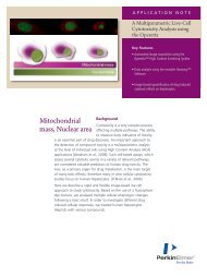
Emission<br />
520-620 nm<br />
Excitation<br />
680 nm<br />
Streptavidin-coated<br />
AlphaScreen Donor Beads<br />
Biotinylated phosphorylated<br />
MBP peptide<br />
Protein A-conjugated<br />
AlphaScreen Acceptor Beads<br />
Anti-phospho MBP antibody<br />
Figure 1. Phosphorylated polypeptide bound by streptavidin-coated Donor beads and by specific anti-phospho-MBP<br />
antibodies bound to Protein A-conjugated Acceptor beads.<br />
3