AlphaScreen⢠MAP Kinase Assay - PerkinElmer
AlphaScreen⢠MAP Kinase Assay - PerkinElmer
AlphaScreen⢠MAP Kinase Assay - PerkinElmer

Create successful ePaper yourself
Turn your PDF publications into a flip-book with our unique Google optimized e-Paper software.
Materials and Methods (continued)<br />
All assays were performed in white, opaque 384-well plates (ex.: <strong>PerkinElmer</strong> OptiPlate 384 well<br />
plates cat. No. 6007290 and 6007299) in a final volume of 25 µL using 5 µL ERK1 extract, 10 µL<br />
biotin-peptide/ATP mix and 10 µL detection mix. For inhibition experiments, ERK1 extract was<br />
pre-incubated with 5 µL staurosporine for 15 minutes prior to adding 5 µL biotin-peptide/ATP<br />
mix. Anti-phospho-MBP antibody was used at 1 nM, and Donor-Streptavidin and Acceptor-Protein<br />
A beads were used at 20 µg/mL each.<br />
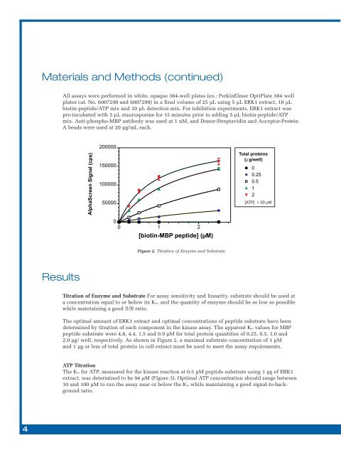
Figure 2. Titration of Enzyme and Substrate.<br />
Results<br />
Titration of Enzyme and Substrate For assay sensitivity and linearity, substrate should be used at<br />
a concentration equal to or below its Km, and the quantity of enzyme should be as low as possible<br />
while maintaining a good S/B ratio.<br />
The optimal amount of ERK1 extract and optimal concentrations of peptide substrate have been<br />
determined by titration of each component in the kinase assay. The apparent Km values for MBP<br />
peptide substrate were 4.8, 4.4, 1.5 and 0.9 µM for total protein quantities of 0.25, 0.5, 1.0 and<br />
2.0 µg/ well, respectively. As shown in Figure 2, a maximal substrate concentration of 1 µM<br />
and 1 µg or less of total protein in cell extract must be used to meet the assay requirements.<br />
ATP Titration<br />
The Km for ATP, measured for the kinase reaction at 0.5 µM peptide substrate using 1 µg of ERK1<br />
extract, was determined to be 94 µM (Figure 3). Optimal ATP concentration should range between<br />
10 and 100 µM to run the assay near or below the Km while maintaining a good signal-to-background<br />
ratio.<br />
4