Phylogeny and taxonomy of obscure genera of microfungi - Persoonia
Phylogeny and taxonomy of obscure genera of microfungi - Persoonia
Phylogeny and taxonomy of obscure genera of microfungi - Persoonia

Create successful ePaper yourself
Turn your PDF publications into a flip-book with our unique Google optimized e-Paper software.
P.W. Crous et al.: Obscure <strong>genera</strong> <strong>of</strong> micr<strong>of</strong>ungi<br />
153<br />
via sympodial proliferation near apex. Conidia 11–16 × 3.5–6<br />
µm, subhyaline, smooth, thin-walled, finely guttulate, fusoidellipsoidal<br />
with obtuse apex <strong>and</strong> tapering from its widest point<br />
in the middle towards a subtruncate base, 1–1.5 µm wide.<br />
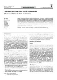
Edenia gomezpompae M.C. González, Anaya, Glenn, Saucedo<br />
& Hanlin, Mycotaxon 101: 254. 2007 — Fig. 10<br />
Leaf spots subcircular, 3–12 mm diam, grey-brown, with a<br />
dark brown, raised border, surrounded by a diffuse, black halo<br />
(absent in smaller spots). Conidiophores in fascicles <strong>of</strong> 5–30,<br />
subcylindrical, medium brown, finely roughened, 3–15-septate,<br />
straight to variously curved or geniculate-sinuous, 50–170 ×<br />
4–6 µm, irregular in width, constricted at some septa, with percurrent<br />
rejuvenation in upper part; fascicles r<strong>and</strong>omly distributed<br />
over lesion, amphigenous, visible as erect, dark brown to black<br />
tufts on lesions, situated on a submerged, brown stroma, up to<br />
60 µm wide <strong>and</strong> 40 µm high, intermingled among leaf trichomes<br />
(fruiting structures <strong>of</strong> a Ramularia sp. <strong>and</strong> ascomata <strong>of</strong> another<br />
fungus also present in some lesions). Conidiogenous cells<br />
15–30 × 3–4 µm, terminal, integrated, becoming paler brown<br />
towards apex, tapering to a subtruncate tip, with several lateral<br />
loci that are somewhat thickened <strong>and</strong> protruding (pimple-like),<br />
up to 1 µm diam, giving rise to conidia via sympodial proliferation<br />
near apex, but some conidiogenous cells also show<br />
signs <strong>of</strong> percurrent proliferation, but this appears to be linked<br />
to rejuvenation, not conidiogenesis. Conidia (11–)13–15(–16)<br />
× (3.5–)4.5–5.5(–6) µm, subhyaline, smooth, thin-walled,<br />
finely guttulate, fusoid-ellipsoidal with obtuse apex <strong>and</strong> tapering<br />
from its widest point in the middle towards a subtruncate<br />
base, 1–1.5 µm wide.<br />
Characteristics in culture — Colonies fluffy, with white hyphal<br />
str<strong>and</strong>s that turn brown with age; surface woolly with abundant<br />
aerial mycelium; margins uneven. On MEA buff to rosy-buff<br />
(surface), brick to dark brick (reverse); on PDA fluffy, cream<br />
to buff (surface), dark brick to buff (reverse); on OA brick with<br />
patches <strong>of</strong> cream to buff. Colonies reaching 25 mm diam after<br />
2 wk at 25 °C, becoming fertile on OA.<br />
Specimens examined. Mexico, Quintana Roo, Isla Mujeres Municipality, El<br />
Eden Ecological reserve, from leaves <strong>of</strong> Callicarpa acuminata (Lamiaceae),<br />
May 2002, A. Saucedo-García & A.L. Anaya, holotype MEXU 25346. – Philippines,<br />
on Senna alata (≡ Cassia alata) (Caesalpiniaceae), Oct. 2008, leg.<br />
C.J.R. Cumagun, isol. P.W. Crous, epitype designated here CBS H-20203,<br />
cultures CPC 15689 = CBS 124106, CPC 15690, 15691.<br />
Notes — The genus Edenia was originally introduced for<br />
a sterile fungus (suspected to be a member <strong>of</strong> the Pleosporaceae),<br />
isolated as an endophyte from leaves <strong>of</strong> Callicarpa<br />
acuminata in Mexico (González et al. 2007). The genus was<br />
characterised by producing numerous sterile, whitish mycelial<br />
str<strong>and</strong>s <strong>and</strong> coils on PDA. The present collection from Cassia<br />
alata in the Philippines has the same colony characteristics,<br />
<strong>and</strong> based on its identical DNA sequence data (GenBank<br />
EF565744.1), we believe that this is the same fungus. What<br />
is interesting, however, is the fact that the latter collection was<br />
made from conidia <strong>of</strong> a dematiaceous hyphomycete sporulating<br />
on leaf spots <strong>of</strong> C. alata. As other fungi were also present on<br />
these spots, its potential role as pathogen remains uncertain.<br />
On host tissue, however, some conidiophores were associated<br />
with a weakly developed layer <strong>of</strong> pale brown stromatic cells. On<br />
OA, cultures became fertile, <strong>and</strong> conidiophores were arranged<br />
around well-developed ostioles <strong>of</strong> submerged pycnidia (with a<br />
similar pale brown stromatic wall to that observed on the host).<br />
It is possible, therefore, that if the field material had been placed<br />
in moist chambers, the pycnidial state would have developed.<br />
The latter state resembles species that are pyronellea-like in<br />
morphology.<br />
Morphologically, the hyphomycete state <strong>of</strong> Edenia resembles<br />
<strong>genera</strong> such as Digitopodium, although species <strong>of</strong> this genus<br />
have rhizoids, <strong>and</strong> 1-septate, pale brown conidia that can also<br />
occur in short chains (Heuchert et al. 2005). It also shares some<br />
similarities with Blastophorum (Matsushima 1971), although<br />
the latter fungus is distinct in having solitary conidiophores with<br />
rhizoids, <strong>and</strong> a hyaline, upper conidiogenous region.<br />
Thedgonia B. Sutton, Trans. Brit. Mycol. Soc. 61: 426. 1973<br />
Type species. Thedgonia ligustrina (Boerema) B. Sutton.<br />
Conidiomata fasciculate, punctiform. Mycelium internal, hyphae<br />
subhyaline, septate, branched, forming substomatal stromata,<br />
hyaline to pale brown. Conidiophores fasciculate, arising from<br />
stromata, simple, rarely branched, subcylindrical, straight to<br />
geniculate-sinuous, continuous to septate, smooth, hyaline to<br />
pale yellowish green. Conidiogenous cells integrated, terminal,<br />
occasionally conidiophores reduced to conidiogenous cells,<br />
sympodial, conidiogenous loci more or less planate, unthickened,<br />
non-pigmented. Conidia in disarticulating chains, rarely<br />
in branched chains, subcylindrical to obclavate, with one to<br />
several transverse eusepta, hyaline or almost so, apex rounded<br />
to truncate, base truncate, hila flat, unthickened, hyaline.<br />
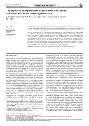
Thedgonia ligustrina (Boerema) B. Sutton, Trans. Brit. Mycol.<br />
Soc. 61: 428. 1973 — Fig. 11<br />
Basionym. Cercospora ligustrina Boerema, Tijdschr. Plantenziekten 68:<br />
117. 1962.<br />
≡ Cercoseptoria ligustrina (Boerema) Arx, Genera <strong>of</strong> Fungi Sporulating<br />
in Pure Culture, ed. 3: 306, Lehre 1981.<br />
Characteristics in culture — On MEA erumpent, slow growing,<br />
5–8 mm after 2 wk, with moderate, white aerial mycelium<br />
<strong>and</strong> smooth, lobate margins; umber in reverse. On OA 5–8 mm<br />
diam after 2 wk, submerged to flattened on surface, sparse aerial<br />
mycelium, <strong>and</strong> smooth, even margins; umber on surface.<br />
Specimens examined. Asia, on Ligustrum sp., H. Evans, CPC 4296 =<br />
W2072, CPC 4297 = W 2073, CPC 4298 = W 1877. – Netherl<strong>and</strong>s, Eefde,<br />
on Ligustrum ovalifolium, 23 Mar. 1959, G.H. Boerema, holotype L, ex-type<br />
culture CBS 148.59; Bilthoven, on L. ovalifolium, 2003, P.W. Crous, CPC<br />
10530 = CBS 124332, CPC 10532, 10533. – South Korea, Namyangju, on<br />
L. ovalifolium, 9 Oct. 2002, leg. H.D. Shin, isol. P.W. Crous, CBS H-20204,<br />
CPC 10019, 10861–10863; Suwon, on L. obtusifolium, 2 Oct. 2007, leg. H.D.<br />
Shin, isol. P.W. Crous, CBS H-20207, CPC 14754–14756.<br />
Notes — Kaiser & Crous (1998) linked ‘Thedgonia’ lupini as<br />
anamorph to Mycosphaerella lupini, <strong>and</strong> thus suggested that<br />
Thedgonia belongs in the Mycosphaerellaceae. Results <strong>of</strong> this<br />
study (Fig. 1), however, show that Thedgonia s.str. belongs to<br />
the Helotiales, <strong>and</strong> is unrelated to the Mycosphaerellaceae.<br />
Furthermore, there is presently no separate anamorph genus<br />
in the Mycosphaerellaceae to accommodate ‘T.’ lupini. Although<br />
‘T.’ lupini resembles species <strong>of</strong> Pseudocercosporella (Braun<br />
1995), it appears to represent a separate phylogenetic lineage.<br />
Trochophora R.T. Moore, Mycologia 47: 90. 1955<br />
Type species. Trochophora fasciculata (Berk. & M.A. Curtis) Goos (syn.<br />
T. simplex (Petch) R.T. Moore).<br />
Colonies hypophyllous, medium to dark brown, consisting <strong>of</strong><br />
numerous synnemata. Stroma absent, but a superficial network<br />
<strong>of</strong> hyphae linking the various synnemata. Conidiophores synnematous,<br />
mostly unbranched <strong>and</strong> straight, or with 1–2 short<br />
branches, straight or curved, cylindrical, individual conidiophores<br />
tightly aggregated, but separating near the apex, pale<br />
to medium brown, smooth. Conidiogenous cells polyblastic,<br />
integrated, terminal, determinate to sympodial, with visible