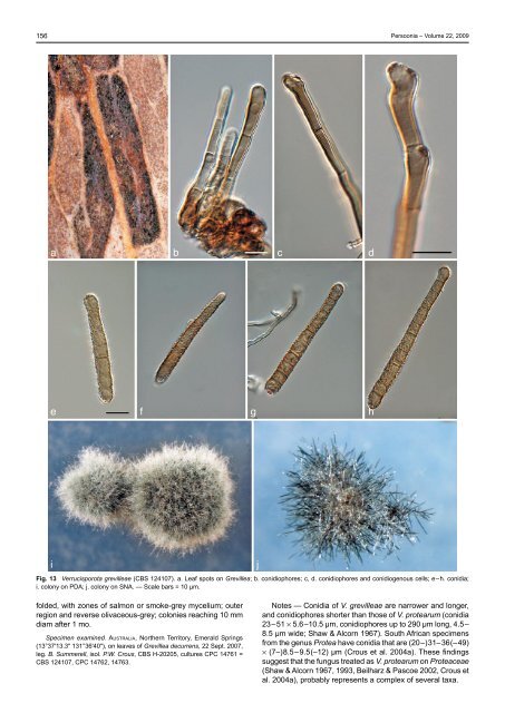
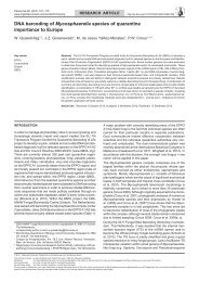
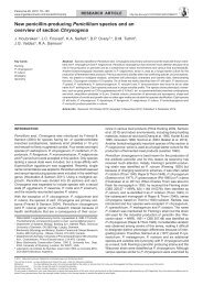
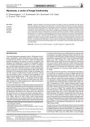
156 <strong>Persoonia</strong> – Volume 22, 2009 a b c d e f g h i Fig. 13 Verrucisporota grevilleae (CBS 124107). a. Leaf spots on Grevillea; b. conidiophores; c, d. conidiophores <strong>and</strong> conidiogenous cells; e–h. conidia; i. colony on PDA; j. colony on SNA. — Scale bars = 10 µm. j folded, with zones <strong>of</strong> salmon or smoke-grey mycelium; outer region <strong>and</strong> reverse olivaceous-grey; colonies reaching 10 mm diam after 1 mo. Specimen examined. Australia, Northern Territory, Emerald Springs (13°37'13.3" 131°36'40"), on leaves <strong>of</strong> Grevillea decurrens, 22 Sept. 2007, leg. B. Summerell, isol. P.W. Crous, CBS H-20205, cultures CPC 14761 = CBS 124107, CPC 14762, 14763. Notes — Conidia <strong>of</strong> V. grevilleae are narrower <strong>and</strong> longer, <strong>and</strong> conidiophores shorter than those <strong>of</strong> V. protearum (conidia 23–51 × 5.6–10.5 µm, conidiophores up to 290 µm long, 4.5– 8.5 µm wide; Shaw & Alcorn 1967). South African specimens from the genus Protea have conidia that are (20–)31–36(–49) × (7–)8.5–9.5(–12) µm (Crous et al. 2004a). These findings suggest that the fungus treated as V. protearum on Proteaceae (Shaw & Alcorn 1967, 1993, Beilharz & Pascoe 2002, Crous et al. 2004a), probably represents a complex <strong>of</strong> several taxa.
P.W. Crous et al.: Obscure <strong>genera</strong> <strong>of</strong> micr<strong>of</strong>ungi 157 Verrucisporota proteacearum (D.E. Shaw & Alcorn) D.E. Shaw & Alcorn, Austral. Syst. Bot. 6: 273. 1993 Basionym. Verrucispora proteacearum D.E. Shaw & Alcorn, Proc. Linn. Soc. New South Wales 92: 171. 1967. Characteristics in culture — On MEA erumpent with sparse aerial mycelium; surface cream to pale olivaceous-grey, folded, with smooth, even margin; reverse brown-vinaceous; reaching 8 mm diam after 2 wk. On PDA erumpent with sparse aerial mycelium <strong>and</strong> smooth to feathery margin; surface cream to pale olivaceous-grey; reverse olivaceous-grey, reaching 8 mm diam after 2 wk. On OA erumpent, with moderate aerial mycelium <strong>and</strong> uneven margin, pale white in middle, pale olivaceous-grey in outer region; reaching 10 mm diam after 2 wk. Specimen examined. Australia, Grevillea sp., V. Beilharz, VPRI31812 = CBS 116003. Notes — Because V. proteacearum was originally described from Finschia (conidia 23–51 × 5.6–10.5 µm; Shaw & Alcorn 1967), there is a strong possibility that the strain listed here from Grevillea (conidia 30–45 × 10–12 µm on OA) may represent a different taxon to the one occurring on Finschia. Although apparently identical based on the LSU phylogeny (see Fig. 1), the ITS sequence <strong>of</strong> this isolate is different to that <strong>of</strong> V. grevilleae (95 % similarity <strong>and</strong> 4 % gaps). Key to species <strong>of</strong> Verrucisporota 1. Conidia wider than 4.5 µm . . . . . . . . . . . . . . . . . . . . . . . . 2 1. Conidia narrower than 4.5 µm . . . . . . . . . . . . . . . . . . . . . 3 2. Conidia up to 56 µm long. . . . . . . . . . . . . . . . . . . . . . . . . 4 2. Conidia longer than 56 µm, 3–7(–12)-septate, (30–)50– 65(–80) × (5–)6–7 µm; on Grevillea . . . . . . . V. grevilleae 3. Conidia mostly up to 30 µm long, (0–)2–3(–7)-septate, 13–30(–70) × 2.75–4 µm; on Capparis . V. kimberleyana 3. Conidia longer, mostly up to 77 µm long, 1–11-septate, (10–)27–77(–108) × 3–4.5 μm; on Struthanthus . . . . . . . . . . . . . . . . . . . . . . . . . . . . . . . . . . . . . . . V. struthanthicola 4. Conidia up to 3-septate, obclavate, 1–3-septate, 32.5–55 × 7–10.5 µm; on Celastrus . . . . . . . . . . . . . . . . . V. indica 4. Conidia more than 3 septa . . . . . . . . . . . . . . . . . . . . . . . 5 5. Conidia up to 32 µm long; (1–)3–4(–5)-septate, 20–32 × 6–10 µm; on Bridelia . . . . . . . . . . . . . . . . . . . . V. brideliae 5. Conidia frequently longer than above . . . . . . . . . . . . . . . 6 6. Conidia 0–6-septate, 18–56 × 4.5–7 µm; on Daviesia (Beilharz & Pascoe 2002) . . . . . . . . . . . . . . . . . . . .V. daviesiae 6. Conidia 3–7-septate, 23–51 × 5.6–10.5 µm; on Finschia . . . . . . . . . . . . . . . . . . . . . . . . . . . . . . . . . V. proteacearum Vonarxia Bat., Publ. Inst. Micol. Univ. Fed. Pernambuco 283: 5. 1960 Type species. Vonarxia anacardii Bat. & J.L. Bezerra. Mycelium immersed <strong>and</strong> superficial, composed <strong>of</strong> branched, septate, pale to medium brown, smooth to finely roughened hyphae. Conidiomata sporodochial; basal stroma composed <strong>of</strong> globose-ellipsoidal, brown, slightly roughened cells. Setae irregularly scattered throughout colony, simple, subulate with a bulbous base, straight to slightly curved, dark brown, smooth to slightly roughened, thick-walled, 5–16-euseptate, septa rather thick, but becoming thinner towards apex. Conidiogenous cells arise from upper cells <strong>of</strong> the stroma, tightly aggregated, doliiform to ellipsoid, pale brown to subhyaline or hyaline, smooth, giving rise to a cluster <strong>of</strong> conidia by means <strong>of</strong> sympodial proliferation, with successive conidia forming at a higher level. Conidia hyaline, smooth-walled, tetraradiate, basal cell subcylindrical to clavate to doliiform, 0–1-septate; upper three arms arise from the apical part <strong>of</strong> the basal cell, 3–10-septate, subcylindrical to cylindrical, apex subobtuse. Vonarxia vagans (Speg.) Aa, <strong>Persoonia</strong> 13: 128. 1986 — Fig. 14 Basionym. Ypsilonia vagans Speg., Revista Mus. La Plata, Secc. Bot. 15: 35. 1908. ≡ Kazulia vagans (Speg.) Nag Raj, Canad. J. Bot. 55: 1621. 1977. On PDA. Mycelium immersed <strong>and</strong> superficial, composed <strong>of</strong> branched, septate, pale to medium brown, smooth to finely roughened, 3–5 µm wide hyphae. Conidiomata sporodochial, flattened to erect <strong>and</strong> globose (especially on WA, not so on MEA or PDA, tending to be more flattened, <strong>and</strong> more hemispherical on OA), up to 300 µm diam; basal stroma up to 70 µm thick, composed <strong>of</strong> globose-ellipsoidal, brown, slightly roughened cells, 5–10 µm diam. Setae irregularly scattered throughout colony, simple, subulate with a bulbous base, straight to slightly curved, dark brown, smooth to slightly roughened, thick-walled (1–1.5 µm diam), (5–)10–12(–16)-septate, septa rather thick, but becoming thinner towards apex, basal cell 10–13 µm wide, with slight taper towards bluntly rounded, obtuse apex, (120–)150–200(–220) µm; width at basal septum (5–)6(–7) µm; width at apical septum, 2–3(–5) µm; apical two cells frequently pale brown; individual cells 10–25 µm long. Conidiogenous cells arise from upper cells <strong>of</strong> the stroma, tightly aggregated, doliiform to ellipsoid, pale brown to subhyaline or hyaline, smooth, 8–10 × 3–5 µm, giving rise to a cluster <strong>of</strong> conidia by means <strong>of</strong> sympodial proliferation, with successive conidia forming at a higher level. Conidia hyaline, smooth-walled, tetraradiate, basal cell subcylindrical to clavate to doliiform, 0–1-septate, 10–15 × (1.5–)2–3 µm (10–18 µm long on OA); upper three arms arise from the apical part <strong>of</strong> the basal cell, 3–5-septate (prominently constricted at septa on WA <strong>and</strong> MEA, up to 10-septate on these media), subcylindrical to cylindrical, apex subobtuse, arms 20–55 µm long (20–90 µm on OA), 1.5–2 µm wide (2–3 µm wide on OA). Characteristics in culture — Colonies on OA spreading, with sparse aerial mycelium, <strong>and</strong> uneven, striate surface, with crenate margin; surface black, with patches <strong>of</strong> mouse-grey, reaching up to 25 mm diam after 1 mo; on PDA spreading, with sparse aerial mycelium <strong>and</strong> crenate margins; surface pale mouse-grey, outer region grey-olivaceous; reverse greyolivaceous, reaching up to 25 mm diam after 1 mo; on MEA spreading, erumpent with sparse aerial mycelium; surface prominently striate, margin crenate; centre black, outer region mouse-grey; reverse black; colonies reaching up to 20 mm diam after 1 mo. Specimens examined. Brazil, São Paulo Horto Botanico, leaves <strong>of</strong> Spiraea cantoniensis, Sept. 1905, leg. Usteri no. 15 bis, holotype LPS 12280; Rio Gr<strong>and</strong>e do Sul, Guaiba, living leaves <strong>of</strong> Stenocalyx uniflorus, 1 Apr. 2008, leg. A.C. Alfenas, isol. P.W. Crous, epitype designated here CBS H-20206, culture ex-type CPC 15151 = CBS 123533, CPC 15152. Notes — The holotype specimen (LPS 12280) was described <strong>and</strong> illustrated in detail by Nag Raj (1977). The species was originally described from leaves <strong>of</strong> Spiraea cantoniensis collected in the São Paulo Botanical Garden, where it occurred on leaves <strong>of</strong> several tree species, suggesting that it is not host specific. The present collection was obtained by incubating Eugenia leaves with leaf spots <strong>of</strong> Phaeophleospora eugeniae in moist chambers, which resulted in a few conidiophores <strong>of</strong> Vonarxia vegans developing. Nag Raj (1977) erected Kazulia for a genus <strong>of</strong> hyphomycetes with dark brown, septate setae, <strong>and</strong> tetraradiate conidia, which he regarded as morphologically distinct, <strong>and</strong> a probable anamorph <strong>of</strong> the Chaetothyriaceae. The fact that he did not compare Kazulia with Vonarxia is not surprising, because