Amebiasis Handout Entamoeba histolytica E. histolytica vs. E. dispar
Amebiasis Handout Entamoeba histolytica E. histolytica vs. E. dispar
Amebiasis Handout Entamoeba histolytica E. histolytica vs. E. dispar

You also want an ePaper? Increase the reach of your titles
YUMPU automatically turns print PDFs into web optimized ePapers that Google loves.
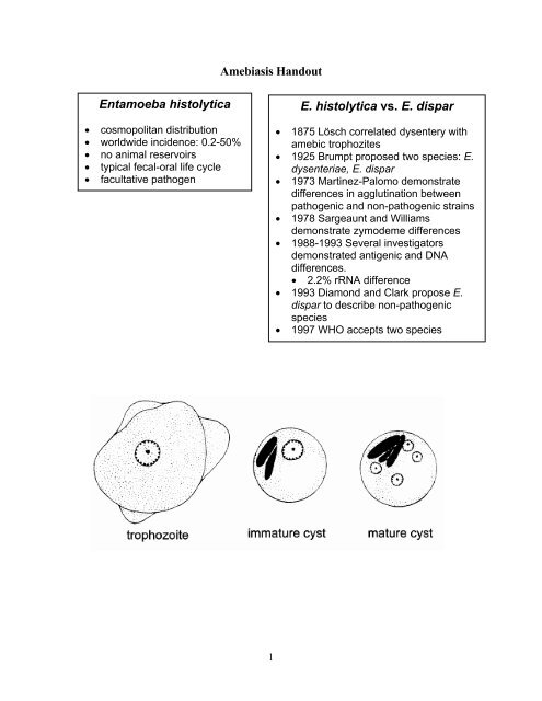
<strong>Amebiasis</strong> <strong>Handout</strong><br />
<strong>Entamoeba</strong> <strong>histolytica</strong><br />
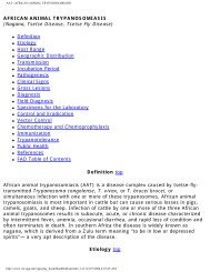
• cosmopolitan distribution<br />
• worldwide incidence: 0.2-50%<br />
• no animal reservoirs<br />
• typical fecal-oral life cycle<br />
• facultative pathogen<br />
E. <strong>histolytica</strong> <strong>vs</strong>. E. <strong>dispar</strong><br />
• 1875 Lösch correlated dysentery with<br />
amebic trophozites<br />
• 1925 Brumpt proposed two species: E.<br />
dysenteriae, E. <strong>dispar</strong><br />
• 1973 Martinez-Palomo demonstrate<br />
differences in agglutination between<br />
pathogenic and non-pathogenic strains<br />
• 1978 Sargeaunt and Williams<br />
demonstrate zymodeme differences<br />
• 1988-1993 Several investigators<br />
demonstrated antigenic and DNA<br />
differences.<br />
• 2.2% rRNA difference<br />
• 1993 Diamond and Clark propose E.<br />
<strong>dispar</strong> to describe non-pathogenic<br />
species<br />
• 1997 WHO accepts two species<br />
1
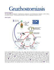
PATHOGENESIS OF AMEBIASIS<br />
non-invasive<br />
• ameba colony on mucosa surface<br />
• asymptomatic cyst passer<br />
• non-dysenteric diarrhea, cramps,<br />
abdominal discomfort<br />
invasive<br />
• necrosis of mucosa → ulcer<br />
• dysentery<br />
• hematophagous trophozoites<br />
• ulcer enlargement → severe<br />
dysentery, colitis, peritonitis,<br />
occasional ameboma<br />
• metastasis → extraintestinal<br />
amebiasis<br />
• dissemination primarily via bloodstream<br />
(eg., portal vein)<br />
• predominantly liver → amebic<br />
abscess<br />
• other sites infrequent (cutaneous,<br />
pulmonary)<br />
• ameba-free stools common<br />
2
1. E. <strong>histolytica</strong> trophozoites colonize the mucosal surface of the large intestine. The<br />
trophozoites adhere to the mucus layer and ingest bacteria and cellular debris from the<br />
lumen. Adherence is mediated by a protein called the eh-lectin, or GIAP, which is expressed<br />
on the surface of trophozoites. This non-invasive infection is usually asymptomatic, or<br />
exhibits symptoms ranging from mild abdominal discomfort to diarrhea and cramps.<br />
2. A breakdown in the mucus barrier can lead to a contact-dependent killing of the epithelial<br />
cells. The Eh-lectin also plays a role in this cytolytic activity. In addition, the breakdown of<br />
the tissue and extracellular matrix (ECM) implies that proteases are also involved in the<br />
pathogenesis. This necrosis of the mucosa will lead to an invasive disease characterized by<br />
dysentery (ie, blood and mucus in the feces).<br />
3. The trophozoites will continue to advance laterally and downward into the submucosa<br />
producing a 'flask-shaped' ulcer. Necrotic material is found in the center of the ulcer and<br />
most of the ameba are at the border between the healthy and damaged tissue. Neutrophils and<br />
other immune effector cells are also killed. The ameba are now ingesting host cells instead of<br />
bacteria and hematophogous trophozoties can be observed. Ulcers can coalesce and lead to<br />
the shedding of patches of mucosa. The severity of the dysentery increases in terms of the<br />
number of stools and the amount of mucus and blood.<br />
4. The trophozoites can also penetrate the muscle and serous layers leading to intestinal<br />
perforations. Perforation of the intestinal wall is a dramatic event that can lead to peritonitis<br />
or leakage into the abdominal cavity. Erosion of blood vessels can lead to massive<br />
hemorrhage. An inflammatory thickening of the intestinal wall, called an ameboma, or<br />
amebic granuloma, can also be formed in response to the ameba. The ameboma presents as a<br />
painful palpable mass that can be mistaken for a tumor.<br />
5. Trophozoites can also gain access to the circulatory system and be disseminated. The liver is<br />
the primary site of extraintestinal amebiasis and hematogenous spread to other organs is rare.<br />
Metastasis to the liver involves the portal vein which carries blood from the colon directly to<br />
the liver. Dissemination to other tissues most often entails the direct extension of hepatic or<br />
colonic lesions. Extraintestinal amebiasis is often characterized by ameba free stools.<br />
3
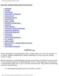
Non-Invasive and Invasive Isolates of <strong>Entamoeba</strong> <strong>histolytica</strong><br />
CRITERIA NON-INVASIVE INVASIVE<br />
In Vitro Culture xenic axenic<br />
ConA Agglutination - +<br />
Complement Resistance - +<br />
Zymodemes (isoenzymes) I & III II<br />
Numerous Antigenic Differences<br />
(eg., GIAP Epitopes) 1-2 1-6<br />
Numerous DNA Sequence Differences<br />
(eg., rRNA)<br />
2.2% sequence diversity<br />
B133<br />
P145<br />
RFLP/DNA Probes<br />
cEH-NP1 cEH-P1<br />
Pathogenic and non-pathogenic isolates of <strong>Entamoeba</strong> <strong>histolytica</strong> exhibit many phenotypic differences. Some<br />
of the first noted differences were the in vitro growth characteristics, agglutination with concanavalin A, and<br />
resistance to complement. Pathogenic strains have the ability to grow in axenic cultures (without bacteria)<br />
whereas the non-pathogenic strains required bacteria for in vitro growth. The ConA agglutination and<br />
complement resistance imply that the outer surfaces of the pathogenic and non-pathogenic strains are<br />
different. Isoenzyme analysis revealed different zymodemes for the pathogenic and non-pathogenic strains.<br />
Similarly, numerous antigenic differences were noted between pathogenic and non-pathogenic isolates. A<br />
well-characterized epitope difference is in a surface protein referred to as galactose-inhibitable adherence<br />
protein (GIAP). GIAP (also called eh-lectin) is also implicated to be involved in the ConA agglutination and<br />
the resistance to complement. Analysis of DNA and sequencing of genes revealed genotypic differences<br />
between the pathogenic and non-pathogenic isolates. A striking variation is the 2.2% difference between the<br />
ribosomal RNA gene sequences of pathogenic and non-pathogenic isolates. Unlike some of the other<br />
differences, the rRNA cannot contribute to the pathogenesis. Furthermore, rRNA sequences of humans and<br />
mice differ by less than 2.2% indicating that the pathogenic and non-pathogenic strains are quite diverged.<br />
These differences led to the formation of a new species call E. <strong>dispar</strong>.<br />
POSSIBLE INVASION FACTORS<br />
• host factors (eg, immune response)?<br />
• parasite factors?<br />
♦ resistance to host response (eg,<br />
complement resistance)<br />
♦ adherence properties (eg, GIAP)<br />
♦ cytolytic properties (adherence +<br />
'amoebapore')<br />
♦ ability to breakdown tissues (eg,<br />
secreted proteases)<br />
4
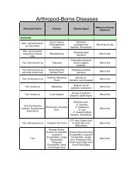
Epidemiologic Risk Factors<br />
Severity<br />
Prevalence<br />
• lower socioeconomic status<br />
• crowding<br />
• lack of indoor plumbing<br />
• endemic area<br />
• institutionalization<br />
• communal living<br />
• promiscuity among male<br />
homosexuals<br />
Modified from Ravdin (1995) Clin. Inf. Dis. 20:1453<br />
• children, esp. neonates<br />
• pregnancy and<br />
postpartum states<br />
• corticosteroid use<br />
• malignancy<br />
• malnutrition<br />
Clinical Syndromes<br />
Associated with <strong>Amebiasis</strong><br />
Intestinal Disease<br />
• asymptomatic cyst passer<br />
• symptomatic nondysenteric<br />
infection<br />
• amebic dysentery (acute)<br />
• fulminant colitis<br />
• + perforation (peritonitis)<br />
• ameboma (amebic granuloma)<br />
• perianal ulceration<br />
Extraintestinal Disease<br />
• liver abscess<br />
• pleuropulmonary amebiasis<br />
• brain and other organs<br />
• cutaneous and genital diseases<br />
Intestinal Symptoms<br />
• range<br />
• mild to intense<br />
• transient to long lasting<br />
• nondysenteric<br />
• diarrhea<br />
• cramps<br />
• flatulence<br />
• nausea<br />
• dysenteric<br />
• blood/mucus in stools<br />
• cramps/pain<br />
• tenesmus<br />
• ameboma<br />
• palpable mass<br />
• obstruction<br />
5
Diagnosis<br />
Intestinal<br />
• stool examination<br />
• cysts and/or trophozoites<br />
• sigmoidoscopy<br />
• lesions, aspirate, biopsy<br />
• antigen detection<br />
• <strong>histolytica</strong>/<strong>dispar</strong><br />
Extraintestinal (hepatic)<br />
• serology<br />
• only E. <strong>histolytica</strong><br />
• current or past?<br />
• imaging<br />
• CT, MRI, ultrasound<br />
• abscess aspiration<br />
• only select cases<br />
• reddish brown liquid<br />
• trophozoites at abscess wall<br />
Disease<br />
Asymptomatic<br />
Nondysenteric, Dysenteric,<br />
or Extraintestinal<br />
<strong>Amebiasis</strong> Treatment<br />
Drug<br />
Iodoquinol, Paromomycin, or<br />
Diloxanide furoate<br />
Metronidazole or Tinidazole +<br />
lumenal agent<br />
6