Causal Agent: Life Cycle:
Causal Agent: Life Cycle:
Causal Agent: Life Cycle:

Create successful ePaper yourself
Turn your PDF publications into a flip-book with our unique Google optimized e-Paper software.
<strong>Causal</strong> <strong>Agent</strong>:<br />
Several protozoan species in the genus Entamoeba infect humans, but not all of them are associated<br />
with disease. Entamoeba histolytica is well recognized as a pathogenic ameba, associated with<br />
intestinal and extraintestinal infections. The other species are important because they may be<br />
confused with E. histolytica in diagnostic investigations.<br />
<strong>Life</strong> <strong>Cycle</strong>:<br />
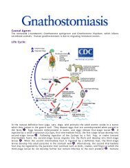
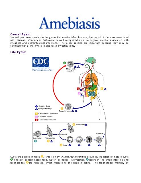
Cysts are passed in feces . Infection by Entamoeba histolytica occurs by ingestion of mature cysts<br />
in fecally contaminated food, water, or hands. Excystation occurs in the small intestine and<br />
trophozoites are released, which migrate to the large intestine. The trophozoites multiply by
inary fission and produce cysts , which are passed in the feces . Because of the protection<br />
conferred by their walls, the cysts can survive days to weeks in the external environment and are<br />
responsible for transmission. (Trophozoites can also be passed in diarrheal stools, but are rapidly<br />
destroyed once outside the body, and if ingested would not survive exposure to the gastric<br />
environment.) In many cases, the trophozoites remain confined to the intestinal lumen ( :<br />
noninvasive infection) of individuals who are asymptomatic carriers, passing cysts in their stool. In<br />
some patients the trophozoites invade the intestinal mucosa ( : intestinal disease), or, through the<br />
bloodstream, extraintestinal sites such as the liver, brain, and lungs ( : extraintestinal disease),<br />
with resultant pathologic manifestations. It has been established that the invasive and noninvasive<br />
forms represent two separate species, respectively E. histolytica and E. dispar, however not all<br />
persons infected with E. histolytica will have invasive disease. These two species are morphologically<br />
indistinguishable. Transmission can also occur through fecal exposure during sexual contact (in<br />
which case not only cysts, but also trophozoites could prove infective).<br />
Geographic Distribution:<br />
Worldwide, with higher incidence of amebiasis in developing countries. In industrialized countries,<br />
risk groups include male homosexuals, travelers and recent immigrants, and institutionalized<br />
populations.<br />
Clinical Features:<br />
A wide spectrum, from asymptomatic infection ("luminal amebiasis"), to invasive intestinal amebiasis<br />
(dysentery, colitis, appendicitis, toxic megacolon, amebomas), to invasive extraintestinal amebiasis<br />
(liver abscess, peritonitis, pleuropulmonary abscess, cutaneous and genital amebic lesions).<br />
Laboratory Diagnosis:<br />
Entamoeba histolytica must be differentiated from other intestinal protozoa including: E. coli, E.<br />
hartmanni, E. gingivalis, Endolimax nana, and Iodamoeba buetschlii (the nonpathogenic amebas);<br />
Dientamoeba fragilis (which is a flagellate not an ameba); and the possibly pathogenic Entamoeba<br />
polecki. Differentiation is possible, but not always easy, based on morphologic characteristics of the<br />
cysts and trophozoites. The nonpathogenic Entamoeba dispar, however, is morphologically identical<br />
to E. histolytica, and differentiation must be based on isoenzymatic or immunologic analysis.<br />
Molecular methods are also useful in distinguishing between E. histolytica and E. dispar and can also<br />
be used to identify E. polecki. Microscopic identification of cysts and trophozoites in the stool is the<br />
common method for diagnosing E. histolytica. This can be accomplished using:<br />
• Fresh stool: wet mounts and permanently stained preparations (e.g., trichrome).<br />
• Concentrates from fresh stool: wet mounts, with or without iodine stain, and permanently<br />
stained preparations (e.g., trichrome). Concentration procedures, however, are not useful for<br />
demonstrating trophozoites.<br />
In addition, E. histolytica trophozoites can also be identified in aspirates or biopsy samples obtained<br />
during colonoscopy or surgery.<br />
Diagnostic findings:<br />
• Microscopy<br />
• Immunodiagnosis<br />
• Molecular methods for discriminating between E. histolytica and E. dispar<br />
• Morphologic comparison with other intestinal parasites<br />
• Bench aid for E. histolytica
Treatment:<br />
For asymptomatic infections, iodoquinol, paromomycin, or diloxanide furoate (not commercially<br />
available in the U.S.) are the drugs of choice. For symptomatic intestinal disease, or extraintestinal,<br />
infections (e.g., hepatic abscess), the drugs of choice are metronidazole or tinidazole, immediately<br />
followed by treatment with iodoquinol, paromomycin, or diloxanide furoate.