Age-Related Changes in Strength, Joint Laxity, and Walking ...
Age-Related Changes in Strength, Joint Laxity, and Walking ...
Age-Related Changes in Strength, Joint Laxity, and Walking ...

Create successful ePaper yourself
Turn your PDF publications into a flip-book with our unique Google optimized e-Paper software.
<strong>Age</strong>-<strong>Related</strong> <strong>Changes</strong> <strong>in</strong> <strong>Strength</strong>, Jo<strong>in</strong>t <strong>Laxity</strong>, <strong>and</strong> Walk<strong>in</strong>g Patterns<br />
Table 1.<br />
Group Characteristics<br />
a P.030.<br />
b P.001.<br />
c P.002.<br />
d P.001.<br />
Young<br />
Control Group<br />
(n15)<br />
Middle-aged<br />
Control Group<br />
(n15)<br />
Older<br />
Control Group<br />
(n14)<br />
Osteoarthritis<br />
Group<br />
(n15)<br />
<strong>Age</strong> (y), X (range) 20.6 (18–25) 49.2 (40–57) 68.8 (60–80) 49.2 (39–57)<br />
Sex (female/male) 8/7 7/8 10/4 7/8<br />
Body mass <strong>in</strong>dex (kg/m 2 ), X (SD) 24.3 (2.8) a,b 28.7 (5.5) a 24.7 (2.5) c 30.7 (4.8) b.c<br />
Alignment (°), X (SD) Not tested 0.1 (1.58) d valgus 1.0 (2.09) d varus 6.33 (2.39) d varus<br />
[ages 40–59 years], <strong>and</strong> 14 older <strong>in</strong>dividuals<br />
[ages 60–80 years])<br />
(Tab. 1). The middle-aged <strong>in</strong>dividuals<br />
were matched by age <strong>and</strong> sex to 15<br />
people with symptomatic, medial<br />
knee OA (Tab. 1). The subjects with<br />
medial knee OA were part of a larger<br />
study of people who were go<strong>in</strong>g to<br />
undergo a high tibial osteotomy.<br />
They had no history of knee ligament<br />
<strong>in</strong>jury; however, those <strong>in</strong>dividuals<br />
with a history of meniscectomy<br />
were <strong>in</strong>cluded. Data on some of<br />
the people with knee OA <strong>and</strong> data<br />
on the middle-aged control group<br />
have been reported previously. 24<br />
Radiographic <strong>in</strong>formation, isometric<br />
quadriceps femoris strength, <strong>and</strong> k<strong>in</strong>ematic,<br />
k<strong>in</strong>etic, <strong>and</strong> electromyographic<br />
(EMG) data dur<strong>in</strong>g walk<strong>in</strong>g<br />
were collected from the more<strong>in</strong>volved<br />
limb of the subjects with<br />
OA <strong>and</strong> a r<strong>and</strong>omly chosen limb of<br />
the control subjects. The test limb of<br />
the control subjects was chosen r<strong>and</strong>omly<br />
to avoid any possible <strong>in</strong>fluence<br />
of limb dom<strong>in</strong>ance.<br />
Procedure<br />
Radiographs. The diagnosis of OA<br />
is based on the presence of knee<br />
pa<strong>in</strong> <strong>in</strong> conjunction with age over 50<br />
years <strong>and</strong> either radiographic evidence<br />
of OA (eg, osteophytes) or<br />
other symptoms such as stiffness or<br />
crepitus. 27 Although none of our<br />
control subjects compla<strong>in</strong>ed of knee<br />
pa<strong>in</strong> or stiffness, st<strong>and</strong><strong>in</strong>g posterioranterior<br />
(approximately 30° of knee<br />
flexion) radiographs of the knees of<br />
the middle-aged <strong>and</strong> older control<br />
groups were obta<strong>in</strong>ed as an added<br />
precaution to rule out the presence<br />
of knee OA. Radiographs were not<br />
taken of the knees of the young subjects<br />
because they had no knee<br />
symptoms <strong>and</strong> no history of knee<br />
<strong>in</strong>jury <strong>and</strong> were unlikely to have undiagnosed<br />
knee OA based on the<br />
above def<strong>in</strong>ition.<br />
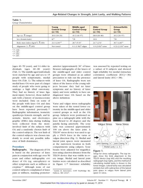
Varus <strong>and</strong> valgus stress radiographs<br />
were taken of the tested lower extremity<br />
<strong>in</strong> the middle-aged <strong>and</strong> older<br />
control groups as well as the OA<br />
group. Subjects were positioned sup<strong>in</strong>e<br />
on a radiograph table with the<br />
knee flexed to 20 degrees <strong>and</strong> the<br />
patella fac<strong>in</strong>g anteriorly. The x-ray<br />
tube was centered approximately<br />
100 cm above the knee jo<strong>in</strong>t. A<br />
TELOS* stress device was used to apply<br />
a 150-N force <strong>in</strong> the varus or<br />
valgus direction (Fig. 1). Medial <strong>and</strong><br />
lateral jo<strong>in</strong>t spaces were measured<br />
at the narrowest location <strong>in</strong> both<br />
compartments us<strong>in</strong>g calipers. X-ray<br />
beams were adjusted for magnification<br />
us<strong>in</strong>g a known distance from the<br />
TELOS device that was visible <strong>in</strong> every<br />
image. Medial <strong>and</strong> lateral jo<strong>in</strong>t<br />
laxities were calculated as described<br />
<strong>in</strong> Figure 1. 28 Interrater reliability<br />
* Aust<strong>in</strong> & Associates, 1109 Sturbridge Rd,<br />
Fallston, MD 21047.<br />
was assessed by repeated test<strong>in</strong>g on<br />
a subset of 8 subjects <strong>and</strong> showed<br />
high reliability for medial (<strong>in</strong>traclass<br />
correlation coefficient [ICC].96)<br />
<strong>and</strong> lateral laxity (ICC.98).<br />
Figure 1.<br />
Setup for stress radiographs. The top images<br />
show the limb alignment <strong>in</strong> the<br />
TELOS device (top left) <strong>and</strong> the result<strong>in</strong>g<br />
radiograph (top right), <strong>and</strong> the method of<br />
calculat<strong>in</strong>g medial laxity is shown <strong>in</strong> the<br />
lower images. Lateral laxity was calculated<br />
similarly but with subtraction of the lateral<br />
jo<strong>in</strong>t space <strong>in</strong> valgus from lateral jo<strong>in</strong>t<br />
space <strong>in</strong> varus.<br />
November 2007 Volume 87 Number 11 Physical Therapy f 3<br />
Downloaded from http://ptjournal.apta.org/ by guest on January 11, 2014