Age-Related Changes in Strength, Joint Laxity, and Walking ...
Age-Related Changes in Strength, Joint Laxity, and Walking ...
Age-Related Changes in Strength, Joint Laxity, and Walking ...

You also want an ePaper? Increase the reach of your titles
YUMPU automatically turns print PDFs into web optimized ePapers that Google loves.
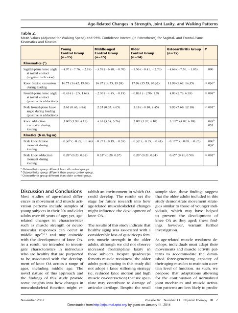
Table 2.<br />
Mean Values (Adjusted for Walk<strong>in</strong>g Speed) <strong>and</strong> 95% Confidence Interval (<strong>in</strong> Parentheses) for Sagittal- <strong>and</strong> Frontal-Plane<br />
K<strong>in</strong>ematics <strong>and</strong> K<strong>in</strong>etics<br />
K<strong>in</strong>ematics (°)<br />
Sagittal-plane knee angle<br />
at <strong>in</strong>itial contact<br />
(negative is flexion)<br />
Knee flexion excursion<br />
dur<strong>in</strong>g load<strong>in</strong>g<br />
Frontal-plane knee angle<br />
at <strong>in</strong>itial contact<br />
(positive is adduction)<br />
Peak frontal-plane knee<br />
angle dur<strong>in</strong>g load<strong>in</strong>g<br />
(positive is adduction)<br />
Knee adduction<br />
excursion dur<strong>in</strong>g<br />
load<strong>in</strong>g<br />
K<strong>in</strong>etics (Nm/kgm)<br />
Peak knee flexion<br />
moment dur<strong>in</strong>g<br />
load<strong>in</strong>g<br />
Peak knee adduction<br />
moment dur<strong>in</strong>g<br />
load<strong>in</strong>g<br />
Young<br />
Control Group<br />
(n15)<br />
a Osteoarthritis group different from all control groups.<br />
b Osteoarthritis group different than young control group.<br />
c Osteoarthritis group different than older control group.<br />
<strong>Age</strong>-<strong>Related</strong> <strong>Changes</strong> <strong>in</strong> <strong>Strength</strong>, Jo<strong>in</strong>t <strong>Laxity</strong>, <strong>and</strong> Walk<strong>in</strong>g Patterns<br />
Middle-aged<br />
Control Group<br />
(n15)<br />
Older<br />
Control Group<br />
(n14)<br />
Osteoarthritis Group<br />
(n15)<br />
4.97 (7.76, 2.18) 3.59 (6.48, 0.70) 5.56 (8.41, 2.70) 4.68 (7.50, 1.85) .800<br />
16.75 (14.42, 19.09) 16.97 (14.55, 19.39) 17.94 (15.55, 20.33) 11.98 (9.62, 14.35) .036 a<br />
0.434 (2.5, 1.64) 2.30 (4.45, 0.15) 0.833 (2.96, 1.9) 4.83 (2.73, 6.93) .004 a<br />
2.62 (0.40, 4.84) 2.35 (0.05, 4.65) 2.18 (0.10, 4.45) 9.93 (7.68, 12.18) .001 a<br />
3.06 b (1.99, 4.12) 4.65 (3.54, 5.76) 3.00 c (1.92, 4.10) 5.10 b,c (4.02, 6.18) .049 b<br />
.055<br />
0.36 b (0.29, 0.44) 0.27 (0.19, 0.35) 0.33 c (0.25, 0.41) 0.17 b,c (0.09, 0.25) .006 b<br />
.039 c<br />
0.28 a (0.23, 0.32) 0.33 a (0.28, 0.37) 0.26 a (0.21, 0.31) 0.45 a (0.41, 0.50) .002 a<br />
P<br />
Discussion <strong>and</strong> Conclusions<br />
Most studies of age-related differences<br />
<strong>in</strong> movement <strong>and</strong> muscle activation<br />
patterns <strong>in</strong>clude samples of<br />
young subjects <strong>in</strong> their 20s <strong>and</strong> older<br />
adults over 60 years of age; yet, agerelated<br />
changes <strong>in</strong> characteristics<br />
such as muscle strength or neuromuscular<br />
responses can occur <strong>in</strong><br />
middle age 7–11 <strong>and</strong> may co<strong>in</strong>cide<br />
with the development of knee OA.<br />
As a result, we <strong>in</strong>tended to <strong>in</strong>vestigate<br />
characteristics <strong>in</strong> <strong>in</strong>dividuals<br />
who are healthy that are purported<br />
to be associated with the development<br />
of knee OA across a range of<br />
ages, <strong>in</strong>clud<strong>in</strong>g middle age. The<br />
novel nature of this approach <strong>and</strong><br />
the f<strong>in</strong>d<strong>in</strong>gs of this study provide<br />
some <strong>in</strong>sights <strong>in</strong>to how changes <strong>in</strong><br />
musculoskeletal function might establish<br />
an environment <strong>in</strong> which OA<br />
could develop. The results set the<br />
stage for future research <strong>in</strong>to how<br />
age-related musculoskeletal changes<br />
might <strong>in</strong>fluence the development of<br />
knee OA.<br />
The results of this study <strong>in</strong>dicate that<br />
healthy ag<strong>in</strong>g was associated with a<br />
considerable loss of quadriceps femoris<br />
muscle strength <strong>in</strong> the older<br />
adults, although we did not observe<br />
<strong>in</strong>creased frontal-plane laxity <strong>in</strong><br />
those subjects. Despite quadriceps<br />
femoris muscle weakness, the older<br />
adults participat<strong>in</strong>g <strong>in</strong> this study did<br />
not adopt a knee stiffen<strong>in</strong>g strategy<br />
(ie, reduced knee motion <strong>and</strong> high<br />
muscle co-contraction) that we speculate<br />
may contribute to damage of<br />
articular cartilage. Despite the small<br />
sample size, these f<strong>in</strong>d<strong>in</strong>gs suggest<br />
that the older adults <strong>in</strong>cluded <strong>in</strong> this<br />
study demonstrate movement strategies<br />
similar to those of younger <strong>in</strong>dividuals,<br />
which may have helped<br />
to prevent the development of<br />
knee OA as they aged; these f<strong>in</strong>d<strong>in</strong>gs,<br />
however, warrant further<br />
<strong>in</strong>vestigation.<br />
As age-related muscle weakness develops,<br />
<strong>in</strong>dividuals must adapt their<br />
movements <strong>and</strong> muscle activity patterns<br />
to accommodate the dim<strong>in</strong>ished<br />
force-generat<strong>in</strong>g capacity of<br />
their ag<strong>in</strong>g muscles to ma<strong>in</strong>ta<strong>in</strong> a certa<strong>in</strong><br />
level of function. As such, we<br />
propose that adaptations allow<strong>in</strong>g<br />
for the cont<strong>in</strong>uation of normalized<br />
jo<strong>in</strong>t mechanics <strong>and</strong> muscle activation<br />
patterns are less likely to predis-<br />
November 2007 Volume 87 Number 11 Physical Therapy f 7<br />
Downloaded from http://ptjournal.apta.org/ by guest on January 11, 2014