Age-Related Changes in Strength, Joint Laxity, and Walking ...
Age-Related Changes in Strength, Joint Laxity, and Walking ...
Age-Related Changes in Strength, Joint Laxity, and Walking ...

You also want an ePaper? Increase the reach of your titles
YUMPU automatically turns print PDFs into web optimized ePapers that Google loves.
<strong>Age</strong>-<strong>Related</strong> <strong>Changes</strong> <strong>in</strong> <strong>Strength</strong>, Jo<strong>in</strong>t <strong>Laxity</strong>, <strong>and</strong> Walk<strong>in</strong>g Patterns<br />
pose the jo<strong>in</strong>t to articular cartilage<br />
damage. A failure to adapt to<br />
strength decl<strong>in</strong>es might contribute<br />
to the development of movement<br />
patterns similar to <strong>in</strong>dividuals with<br />
quadriceps femoris muscle weakness<br />
due to knee jo<strong>in</strong>t pathology.<br />
41–43 Because the older adults <strong>in</strong><br />
this study exhibited similar movement<br />
<strong>and</strong> muscle activity patterns to<br />
those <strong>in</strong> the younger age groups, it<br />
appears that they have discovered a<br />
successful approach to ma<strong>in</strong>ta<strong>in</strong><strong>in</strong>g<br />
normal knee function despite their<br />
quadriceps femoris muscle strength<br />
decl<strong>in</strong>e.<br />
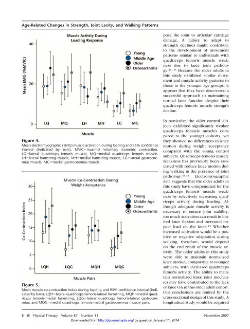
Figure 4.<br />
Mean electromyographic (EMG) muscle activation dur<strong>in</strong>g load<strong>in</strong>g <strong>and</strong> 95% confidence<br />
<strong>in</strong>terval (<strong>in</strong>dicated by bars). MVICmaximal voluntary isometric contraction,<br />
LQlateral quadriceps femoris muscle, MQmedial quadriceps femoris muscle,<br />
LHlateral hamstr<strong>in</strong>g muscle, MHmedial hamstr<strong>in</strong>g muscle, LGlateral gastrocnemius<br />
muscle, MGmedial gastrocnemius muscle.<br />
Figure 5.<br />
Mean muscle co-contraction <strong>in</strong>dex dur<strong>in</strong>g load<strong>in</strong>g <strong>and</strong> 95% confidence <strong>in</strong>terval (<strong>in</strong>dicated<br />
by bars). LQHlateral quadriceps femoris-lateral hamstr<strong>in</strong>g, MQHmedial quadriceps<br />
femoris-medial hamstr<strong>in</strong>g, LQGlateral quadriceps femoris-lateral gastrocnemius,<br />
<strong>and</strong> MQGmedial quadriceps femoris-medial gastrocnemius muscle pairs.<br />
In particular, the older control subjects<br />
exhibited significantly weaker<br />
quadriceps femoris muscles compared<br />
to the younger cohorts, yet<br />
they showed no differences <strong>in</strong> knee<br />
motion dur<strong>in</strong>g weight acceptance<br />
compared with the young control<br />
subjects. Quadriceps femoris muscle<br />
weakness has previously been associated<br />
with reduce knee motion dur<strong>in</strong>g<br />
walk<strong>in</strong>g <strong>in</strong> the presence of jo<strong>in</strong>t<br />
pathology. 33,44 Electromyographic<br />
data suggests that the older adults <strong>in</strong><br />
this study have compensated for the<br />
quadriceps femoris muscle weakness<br />
by selectively <strong>in</strong>creas<strong>in</strong>g quadriceps<br />
activity dur<strong>in</strong>g load<strong>in</strong>g. Although<br />
adequate muscle activity is<br />
necessary to ensure jo<strong>in</strong>t stability,<br />
too much activation can result <strong>in</strong> limited<br />
knee flexion <strong>and</strong> <strong>in</strong>creased impact<br />
load on the knee. 19 Whether<br />
<strong>in</strong>creased activation would be a positive<br />
or negative adaptation dur<strong>in</strong>g<br />
walk<strong>in</strong>g, therefore, would depend<br />
on the end result of the muscle activity.<br />
The older adults <strong>in</strong> this study<br />
were able to ma<strong>in</strong>ta<strong>in</strong> normalized<br />
knee motion, comparable to younger<br />
subjects, with <strong>in</strong>creased quadriceps<br />
femoris activity. The ability to ma<strong>in</strong>ta<strong>in</strong><br />
normalized knee jo<strong>in</strong>t mechanics<br />
may have contributed to the lack<br />
of knee OA <strong>in</strong> this older adult cohort.<br />
Our conclusions are limited by the<br />
cross-sectional design of this study. A<br />
longitud<strong>in</strong>al study would be required<br />
8 f Physical Therapy Volume 87 Number 11 November 2007<br />
Downloaded from http://ptjournal.apta.org/ by guest on January 11, 2014