Maxillary sinus hypoplasia* - ResearchGate
Maxillary sinus hypoplasia* - ResearchGate
Maxillary sinus hypoplasia* - ResearchGate
Create successful ePaper yourself
Turn your PDF publications into a flip-book with our unique Google optimized e-Paper software.
Rhinology, 40, 150-153, 2002<br />
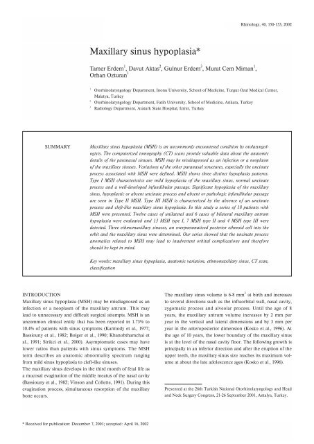
<strong>Maxillary</strong> <strong>sinus</strong> <strong>hypoplasia*</strong><br />
Tamer Erdem 1 , Davut Aktas 2 , Gulnur Erdem 3 , Murat Cem Miman 1 ,<br />
Orhan Ozturan 1<br />
1<br />
2<br />
3<br />
Otorhinolaryngology Department, Inonu University, School of Medicine, Turgut Ozal Medical Center,<br />
Malatya, Turkey<br />
Otorhinolaryngology Department, Fatih University, School of Medicine, Ankara, Turkey<br />
Radiology Department, Ataturk State Hospital, Izmir, Turkey<br />
SUMMARY<br />
<strong>Maxillary</strong> <strong>sinus</strong> hypoplasia (MSH) is an uncommonly encountered condition by otolaryngologists.<br />
The computerized tomography (CT) scans provide valuable data about the anatomic<br />
details of the paranasal <strong>sinus</strong>es. MSH may be misdiagnosed as an infection or a neoplasm<br />
of the maxillary <strong>sinus</strong>es. Variations of the other paranasal structures, especially the uncinate<br />
process associated with MSH were defined. MSH shows three distinct hypoplasia patterns.<br />
Type I MSH characteristics are mild hypoplasia of the maxillary <strong>sinus</strong>, normal uncinate<br />
process and a well-developed infundibular passage. Significant hypoplasia of the maxillary<br />
<strong>sinus</strong>, hypoplastic or absent uncinate process and absent or pathologic infundibular passage<br />
are seen in Type II MSH. Type III MSH is characterized by the absence of an uncinate<br />
process and cleft-like maxillary <strong>sinus</strong> hypoplasia. In this study a series of 18 patients with<br />
MSH were presented. Twelve cases of unilateral and 6 cases of bilateral maxillary antrum<br />
hypoplasia were evaluated and 13 MSH type I, 7 MSH type II and 4 MSH type III were<br />
detected. Three ethmomaxillary <strong>sinus</strong>es, an overpneumatized posterior ethmoid cell into the<br />
orbit and the maxillary <strong>sinus</strong> were determined. Our series showed that the uncinate process<br />
anomalies related to MSH may lead to inadvertent orbital complications and therefore<br />
should be kept in mind.<br />
Key words: maxillary <strong>sinus</strong> hypoplasia, anatomic variation, ethmomaxillary <strong>sinus</strong>, CT scan,<br />
classification<br />
INTRODUCTION<br />
<strong>Maxillary</strong> <strong>sinus</strong> hypoplasia (MSH) may be misdiagnosed as an<br />
infection or a neoplasm of the maxillary antrum. This may<br />
lead to unnecessary and difficult surgical attempts. MSH is an<br />
uncommon clinical entity that has been reported in 1.73% to<br />
10.4% of patients with <strong>sinus</strong> symptoms (Karmody et al., 1977;<br />
Bassiouny et al., 1982; Bolger et al., 1990; Khanobthamchai et<br />
al., 1991; Sirikci et al., 2000). Asymptomatic cases may have<br />
lower ratios than patients with <strong>sinus</strong> symptoms. The MSH<br />
term describes an anatomic abnormality spectrum ranging<br />
from mild <strong>sinus</strong> hypoplasia to cleft-like <strong>sinus</strong>es.<br />
The maxillary <strong>sinus</strong> develops in the third month of fetal life as<br />
a mucosal evagination of the middle meatus of the nasal cavity<br />
(Bassiouny et al., 1982; Vinson and Collette, 1991). During this<br />
evagination process, simultaneous resorption of the maxillary<br />
bone occurs.<br />
The maxillary <strong>sinus</strong> volume is 6-8 mm 3 at birth and increases<br />
to several directions such as the infraorbital wall, nasal cavity,<br />
zygomatic process and alveolar process. Until the age of 8<br />
years, the maxillary antrum volume increases by 2 mm per<br />
year in the vertical and lateral dimensions and by 3 mm per<br />
year in the anteroposterior dimension (Kosko et al., 1996). At<br />
the age of 10 years, the lower boundary of the maxillary <strong>sinus</strong><br />
is at the level of the nasal cavity floor. The following growth is<br />
principally in an inferior direction and after the eruption of the<br />
upper teeth, the maxillary <strong>sinus</strong> size reaches its maximum volume<br />
at about the late adolescence ages (Kosko et al., 1996).<br />
Presented at the 26th Turkish National Otorhinolaryngology and Head<br />
and Neck Surgery Congress, 21-26 September 2001, Antalya, Turkey.<br />
* Received for publication: December 7, 2001; accepted: April 16, 2002
<strong>Maxillary</strong> <strong>sinus</strong> hypoplasia 151<br />
The first attempt to classify the MSH was introduced by<br />
Bassiouny (Bassiouny et al., 1982). Later Bolger et al. (1990)<br />
proposed a new classification, which is currently widely accepted.<br />
According to this classification MSH shows three distinct<br />
hypoplasia patterns. Type I MSH characteristics are mild<br />
hypoplasia of the maxillary <strong>sinus</strong>, normal uncinate process and<br />
well-developed infundibular passage. Significant hypoplasia of<br />
the maxillary <strong>sinus</strong>, hypoplastic or absent uncinate process and<br />
absent or pathologic infundibular passage are seen in Type II<br />
MSH. Type III MSH is characterized by the absence of uncinate<br />
process and cleft-like maxillary <strong>sinus</strong> hypoplasia (Bolger<br />
et al., 1990).<br />
The aim of this study was to present a series of 18 patients<br />
with MSH in the light of the relevant literature and derive clinical<br />
implications.<br />
Figure 1. Bilateral type I MSH well developed infundibular passage<br />
with mild MSH.<br />
MATERIALS AND METHODS<br />
The paranasal <strong>sinus</strong> computerized tomography scans of 280<br />
patients with <strong>sinus</strong> symptoms applying for medical care to the<br />
Malatya Inonu University School of Medicine and Izmir<br />
Ataturk State Hospital were enrolled our study.<br />
Computerized tomographic examinations were performed on<br />
either GE prospeed helical CT or W 950 SR x-ray system CT.<br />
Scans were obtained by 5 mm increments from the glabella to<br />
posterior boundary of the sphenoid <strong>sinus</strong> in coronal planes.<br />
Transverse sections were added to evaluate the posterior ethmoid<br />
cells and sphenoid <strong>sinus</strong>es. All CT scans were evaluated<br />
in both bone and soft tissue windows.<br />
The Bolger’s MSH classification system was used for classification<br />
of our patients with MSH.<br />
Figure 2. Type II MSH, hypoplastic uncinate process and pathologic<br />
infundibular passage with significant MSH.<br />
Figure 3. Type III MSH, slit-like maxillary <strong>sinus</strong>.<br />
RESULTS<br />
MSH was detected in 18 (10 female and 8 male) of 280 patients<br />
(6.4%), with a total of 24 MSH in these 18 patients. MSH was<br />
presenting unilaterally in 12 cases and bilaterally in 6 cases. As<br />
for the severity, 13 MSH type I, 7 MSH type II and 4 MSH<br />
type III were detected (Figures 1 to 3). Among 6 bilateral maxillary<br />
<strong>sinus</strong> hypoplasia, 3 cases with bilateral type I MSH, 2<br />
cases with type I MSH on one side and type III MSH on the<br />
other side, and 1 case with type I + type II MSH were<br />
observed.<br />
MSH was found on the left side in 9 cases and on the right<br />
side in 15 cases. The mean age of the patients was 32 (ranging<br />
from 21 to 56) years. Infundibular pathology and developmental<br />
abnormalities of the uncinate process were observed in all<br />
patients with MSH type II and III. Three overpneumatized<br />
posterior ethmoid cells called ethmomaxillary <strong>sinus</strong> were discovered<br />
(Figure 4).<br />
Any local or systemic reason such as the Treacher-Collins syndrome,<br />
Crouson’s syndrome or Apert’s syndrome, thalassemia,<br />
Paget’s disease or cretinism leading to MSH were not<br />
observed.
152 Erdem et al.<br />
DISCUSSION<br />
Embryogenic developmental abnormalities or acquired reasons<br />
such as infection or trauma leading to arrest of <strong>sinus</strong> pneumatization<br />
may be the cause of MSH (Weed and Cole, 1994). A<br />
developmental abnormality may also be the reason of MSH<br />
during the initial stages of in-utero <strong>sinus</strong> development (Bolger<br />
et al., 1990). Bolger et al. (1990) suggested that developmental<br />
abnormalities of the uncinate process may lead to MSH.<br />
Hypoplastic or absent uncinate processes were observed in all<br />
our patients with MSH type II and type III.<br />
An uncinate process may play an important role for aeration of<br />
the maxillary <strong>sinus</strong> during the expiration phase by altering the<br />
direction of exhaled air into the maxillary <strong>sinus</strong>es (Sirikci et al.,<br />
2000). <strong>Maxillary</strong> <strong>sinus</strong>itis was detected in all of our cases with<br />
absent uncinate process. In our series all patients except those<br />
patients with MSH had a well developed uncinate process ending<br />
at the lamina papyracea, the skull base or the middle concha<br />
and some of them had a free ending.<br />
Milczuk et al. (1993) noticed the relation of MSH and chronic<br />
maxillary <strong>sinus</strong>itis. However, it is not known whether the<br />
<strong>sinus</strong>itis is a primary or secondary condition to the MSH.<br />
Mucosal thickening of the maxillary <strong>sinus</strong> was observed in 4 of<br />
13 MSH type I and in all MSH type II and III cases. In our<br />
series, hypoplastic or an absent uncinate process may be the<br />
reason of the <strong>sinus</strong>itis because of impeding the mucociliary<br />
clearance and lack of altering effect of the well developed uncinate<br />
process on the direction of the expired air (Milczuk et al.,<br />
1993; Sirikci et al., 2000). Weed et al. (1994) observed a clinically<br />
absent natural maxillary ostium associated with a poorly<br />
developed uncinate process and infundibular passage in all 4<br />
patients with MSH. Salib et al. (2001) emphasized the relationship<br />
between MSH and maxillary <strong>sinus</strong>itis and suggested a<br />
posteriorly placed middle meatal antrotomy to avoid the inadvertent<br />
orbital complications.<br />
Paling et al. (1982) suggested that acquired MSH may be secondary<br />
to the <strong>sinus</strong>itis leading to new bone formation and partial<br />
or total obliteration of the maxillary <strong>sinus</strong>. MSH may be<br />
secondary to accidental or surgical trauma during the developmental<br />
period (Kosko et al., 1996). Kosko et al. (1996) reported<br />
five children with MSH secondary to functional endoscopic<br />
<strong>sinus</strong> surgery for chronic refractory <strong>sinus</strong>itis. They proposed<br />
that surgical intervention may lead to MSH, due to activation<br />
of bone formation and removal of pneumatization centers.<br />
The patients did not give any history of accidental or surgical<br />
trauma in our series.<br />
Weed et al. (1994) suggested an analogous mechanism as<br />
hypoventilation between the MSH and chronic otitis media<br />
with effusion since a similar thick mucoid effusion may be<br />
seen in these diseases. Choanal atresia has been accused as the<br />
etiologic agent of MSH. The idea that nasal ventilation may be<br />
important for paranasal <strong>sinus</strong> pneumatization was proposed in<br />
similarity to what middle ear ventilation does for temporal<br />
bone pneumatization. But Behar et al. (2000) found that the<br />
Figure 4. Bilateral ethmomaxillary <strong>sinus</strong> draining into the superior<br />
meatus.<br />
maxillary <strong>sinus</strong> development is independent of posterior nasal<br />
ventilation.<br />
Sirikci et al. (2000) emphasized that there may be an association<br />
between a low-lying roof of the ethmoid and MSH. In<br />
their series, more than one-third of the MSH cases had presented<br />
with a low-lying roof of the ethmoid. This may lead to<br />
inadvertent intracranial complications during functional endoscopic<br />
<strong>sinus</strong> surgery. We found a low-lying ethmoid roof in<br />
two of our cases. One of them had type I and the other has<br />
type III MSH.<br />
An association between maxillary <strong>sinus</strong> hypoplasia and orbital<br />
enlargement has been reported (Bierny and Drayden, 1977;<br />
Modic et al., 1980; Geraghty and Dolan, 1989; Weed and Cole,<br />
1994). Thus, orbit displaces inferiorly.<br />
Khanobthamchai et al. (1991) reported 8 ethmomaxillary<br />
<strong>sinus</strong>es seen above the maxillary <strong>sinus</strong> draining into the superior<br />
meatus. The ethmomaxillary <strong>sinus</strong> is a posterior ethmoidal<br />
air-cell enlarged at the expense of the maxillary <strong>sinus</strong>. An ethmomaxillary<br />
<strong>sinus</strong> should be differentiated from a septated<br />
maxillary <strong>sinus</strong>, with the latter draining into the ethmoid<br />
infundibulum. We found three cases with an ethmomaxillary<br />
<strong>sinus</strong> in our series.<br />
There are several classifications for the MSH. The first attempt<br />
to classify the MSH was achieved by Bassiouny et al (1982) as<br />
isolated, associated with regional abnormalities such as<br />
Treacher-Collins Syndrome, Crouson’s Syndrome and Apert’s<br />
Syndrome, and related to systemic disorders such as thalassemia<br />
and cretinism. Bolger et al. (1990) classified the MSH<br />
based on the degree of maxillary <strong>sinus</strong> pneumatization and lateral<br />
nasal wall abnormalities. Type I MSH characteristics are<br />
composed of mild MSH, a normal uncinate process and a welldeveloped<br />
infundibular passage. Significant MSH, an<br />
hypoplastic or absent uncinate process and an absent or pathologic<br />
infundibular passage are seen in Type II MSH. Type III
<strong>Maxillary</strong> <strong>sinus</strong> hypoplasi 153<br />
Table 1. The rates of MSH according to Bolger’s classification.<br />
Bolger Sirikci Erdem<br />
Type I MSH 14 (66.7%) 7 (33.3%) 13 (54.2%)<br />
Type II MSH 6 (28.6%) 6 (28.6%) 7 (29.2%)<br />
Type III MSH 1 (4.8%) 8 (38.1%) 4 (16.7%)<br />
MSH is characterized by the absence of an uncinate process<br />
and cleft-like MSH. Sirikci et al. (2000) suggested a new classification<br />
of the MSH. While they accepted Bolger’s classification,<br />
they added the orbital enlargement to the type II and type<br />
III MSH and proposed a new definition of the mild and severe<br />
MSH by comparing the maxillary <strong>sinus</strong> size with orbital size.<br />
Table 1 shows the distribution of MSH according to these<br />
types.<br />
CONCLUSION<br />
MSH is a rare, but important condition that may lead to incorrect<br />
diagnosis and inadequate treatments including surgery.<br />
The diagnosis and differential diagnosis of MSH may be<br />
accomplished by paranasal <strong>sinus</strong> CT scans. As uncinate<br />
process anomalies are often presented with MSH, the probability<br />
of inadvertent orbital complications during surgery should<br />
be kept in mind. It is suggested that this anatomical configuration<br />
may result in additional dangers at surgery or need to pay<br />
special attention to the anatomy.<br />
REFERENCES<br />
1. Bassiouny A, Nexlands WC, Hussein A, Yehia Z (1982) <strong>Maxillary</strong><br />
<strong>sinus</strong> hypoplasia and superior orbital fissure asymmetry.<br />
Laryngoscope 92: 441-448.<br />
2. Behar PM, Todd NW (2000) Paranasal <strong>sinus</strong> development and<br />
choanal atresia. Arch Otolaryngol Head Neck Surg 126: 155-157.<br />
3. Bierny J, Dryden R (1977) Orbital enlargement secondary to<br />
paranasal <strong>sinus</strong> hypoplasia. Am J Roentgenol 128: 850-852.<br />
4. Bolger WE, Woodruff WW, Morehead J. Parsons D (1990)<br />
<strong>Maxillary</strong> <strong>sinus</strong> hypoplasia: Classification and description of associated<br />
uncinate process hypoplasia. Otolaryngol Head Neck Surg<br />
103: 759-765.<br />
5. Geraghty JJ, Dolan KD (1989) Computed tomography of the<br />
hypoplastic maxillary <strong>sinus</strong>. Ann Otol Rhinol Laryngol 98: 916-918<br />
6. Karmody CS, Carter B, Vincent ME (1977) Developmental anomalies<br />
of the maxillary <strong>sinus</strong>. Trans Am Acad Ophtalmol<br />
Otolaryngol 84: 723-728.<br />
7. Khanobthamchai K, Shankar L, Hawke M, Bingham B (1991)<br />
Ethmomaxillary <strong>sinus</strong> and hypoplasia of maxillary <strong>sinus</strong>. J<br />
Otolaryngol 20: 425-427.<br />
8. Kosko JR, Hall BE, Tunkel DE (1996) Acquired maxillary <strong>sinus</strong><br />
hypoplasia: A consequence of endoscopic <strong>sinus</strong> surgery?<br />
Laryngoscope 106: 1210-1213.<br />
9. Milczuk HA, Dalley RW, Wessbacher FW, Richardson MA (1993)<br />
Nasal and paranasal <strong>sinus</strong> anomalies in children with chronic<br />
<strong>sinus</strong>itis. Laryngoscope 103: 247-252.<br />
10. Modic MT, Weinstein MA, Berlin J. Duchesneau P (1980)<br />
<strong>Maxillary</strong> <strong>sinus</strong> hypoplasia visualized with computed tomography.<br />
Radiology 135: 383-385.<br />
11. Paling M, Roberts R, Fauci A (1982) Paranasal <strong>sinus</strong> obliteration<br />
in Wegener granulomatosis. Radiology 144: 539-543.<br />
12. Salib RJ, Chaudri SA, Rockley TJ (2001) Sinusitis in the hypoplastic<br />
maxillary antrum: the crucial role of radiology in diagnosis and<br />
management. J Laryngol Otol 115: 676-678.<br />
13. Sirikci A, Bayazit Y, Gümüsburun E, Bayram M, Kanlikama M<br />
(2000) A new approach to the classification of maxillary <strong>sinus</strong><br />
hypoplasia with relevant clinical implications. Surg Radiol Anat<br />
22: 243-247.<br />
14. Vinson RP, Collette RP (1991) <strong>Maxillary</strong> <strong>sinus</strong> hypoplasia masquerading<br />
as chronic <strong>sinus</strong>itis. Postgrad Med 89: 189-190.<br />
15. Weed DT, Cole RR (1994) <strong>Maxillary</strong> <strong>sinus</strong> hypoplasia and vertical<br />
dystopia of the orbit. Laryngoscope 104: 758-762.<br />
Tamer Erdem, MD<br />
Assistant Professor<br />
Inonu University, School of Medicine<br />
Turgut Ozal Medical Center<br />
Otorhinolaryngology Department<br />
Malatya<br />
Turkey<br />
Tel: +90 422 341 06 60 (ext. 4603)<br />
Fax: +90 532 452 00 65<br />
E-mail: terdem71@hotmail.com


