pleural effusion in ICU.pdf - SASSiT
pleural effusion in ICU.pdf - SASSiT
pleural effusion in ICU.pdf - SASSiT
Create successful ePaper yourself
Turn your PDF publications into a flip-book with our unique Google optimized e-Paper software.
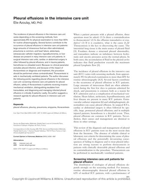
Pleural <strong>effusion</strong>s <strong>in</strong> the <strong>in</strong>tensive care unit<br />
Elie Azoulay, MD, PhD<br />
The <strong>in</strong>cidence of <strong>pleural</strong> <strong>effusion</strong>s <strong>in</strong> the <strong>in</strong>tensive care unit<br />
varies depend<strong>in</strong>g on the screen<strong>in</strong>g methods, from<br />
approximately 8% for physical exam<strong>in</strong>ation to more than 60%<br />
for rout<strong>in</strong>e ultrasonography. Several factors contribute to the<br />
occurrence of <strong>pleural</strong> <strong>effusion</strong>s <strong>in</strong> <strong>in</strong>tensive care unit patients:<br />
large amounts of <strong>in</strong>travenous fluid are often adm<strong>in</strong>istered,<br />
pneumonia is common, and heart failure, atelectasis,<br />
extravascular catheter migration, hypoalbum<strong>in</strong>emia, or liver<br />
disease are present <strong>in</strong> many <strong>in</strong>tensive care unit patients. In<br />
surgical <strong>in</strong>tensive care units, cardiac or abdom<strong>in</strong>al surgery is<br />
often followed by <strong>pleural</strong> <strong>effusion</strong>s, and <strong>in</strong> trauma patients,<br />
hemothorax is a dreaded event. Because no cl<strong>in</strong>ical parameter<br />
excludes <strong>pleural</strong> <strong>in</strong>fection, and because of the impact of<br />
thoracentesis on diagnosis and treatment, this procedure<br />
should be performed unless contra<strong>in</strong>dicated. Thoracentesis is<br />
safe <strong>in</strong> mechanically ventilated patients. The author discusses<br />
the follow<strong>in</strong>g po<strong>in</strong>ts regard<strong>in</strong>g <strong>pleural</strong> <strong>effusion</strong>s <strong>in</strong> the <strong>in</strong>tensive<br />
care unit: screen<strong>in</strong>g <strong>in</strong>tensive care unit patients for <strong>pleural</strong><br />
<strong>effusion</strong>, safety of thoracentesis <strong>in</strong> patients receiv<strong>in</strong>g <strong>in</strong>vasive<br />
mechanical ventilation, dist<strong>in</strong>guish<strong>in</strong>g exudates from<br />
transudates, and diagnos<strong>in</strong>g and manag<strong>in</strong>g <strong>in</strong>fected <strong>pleural</strong><br />
<strong>effusion</strong>s <strong>in</strong> critically ill patients. Lastly, the author suggests a<br />
research agenda for <strong>pleural</strong> <strong>effusion</strong>s <strong>in</strong> <strong>in</strong>tensive care unit<br />
patients.<br />
Keywords<br />
<strong>pleural</strong> <strong>effusion</strong>s, pleurisy, pneumonia, empyema, thoracentesis<br />
Curr Op<strong>in</strong> Pulm Med 2003, 9:291–297 © 2003 Lipp<strong>in</strong>cott Williams & Wilk<strong>in</strong>s.<br />
Service de Réanimation Médicale, Hôpital Sa<strong>in</strong>t Louis et Université Paris,<br />
Assistance Publique–Hôpitaux de Paris, France.<br />
Correspondence to Elie Azoulay, MD, PhD, Service de Réanimation Médicale,<br />
Hôpital Sa<strong>in</strong>t-Louis, 1 Avenue Claude Vellefaux, 75010 Paris, France; e-mail:<br />
elie.azoulay@sls.ap-hop-paris.fr<br />
Current Op<strong>in</strong>ion <strong>in</strong> Pulmonary Medic<strong>in</strong>e 2003, 9:291–297<br />
Abbreviations<br />
<strong>ICU</strong> <strong>in</strong>tensive care unit<br />
LDH lactate dehydrogenase<br />
ISSN 1070–5287 © Lipp<strong>in</strong>cott Williams & Wilk<strong>in</strong>s<br />
When a patient presents with a <strong>pleural</strong> <strong>effusion</strong>, three<br />
questions must be asked: (1) Is there a contra<strong>in</strong>dication<br />
to thoracentesis? (2) Is the <strong>effusion</strong> transudative or exudative?<br />
(3) If it is exudative, what is the cause [1,2]?<br />
Thoracentesis is the key to discover<strong>in</strong>g the cause. The<br />
<strong>in</strong>terstitial lung tissue is the ma<strong>in</strong> source of <strong>pleural</strong> fluid<br />
[3]. Exudates denote a structural <strong>pleural</strong> abnormality,<br />
and transudates <strong>in</strong>dicate an imbalance between hydrostatic<br />
and oncotic pressures across the normal pleura. In<br />
both cases, the accumulation of fluid <strong>in</strong> the <strong>pleural</strong> cavity<br />
<strong>in</strong>dicates that fluid production exceeds the maximum<br />
<strong>pleural</strong> lymphatic flow [3].<br />
The <strong>in</strong>cidence of <strong>pleural</strong> <strong>effusion</strong>s <strong>in</strong> the <strong>in</strong>tensive care<br />
unit (<strong>ICU</strong>) varies with screen<strong>in</strong>g methods, from approximately<br />
8% for physical exam<strong>in</strong>ation to more than 60% for<br />
rout<strong>in</strong>e ultrasonography [4,5]. Several factors contribute<br />
to the occurrence of <strong>pleural</strong> <strong>effusion</strong>s <strong>in</strong> <strong>ICU</strong> patients.<br />
Large amounts of <strong>in</strong>travenous fluid are often adm<strong>in</strong>istered<br />
dur<strong>in</strong>g the first few days to patients admitted for<br />
shock, and pneumonia is common both as a reason for<br />
<strong>ICU</strong> admission and as a complication of mechanical ventilation.<br />
Heart failure, atelectasis, hypoalbum<strong>in</strong>emia, and<br />
liver disease are present <strong>in</strong> many <strong>ICU</strong> patients. Extravascular<br />
catheter migration [6] and subdiaphragmatic abnormalities<br />
can cause <strong>pleural</strong> <strong>effusion</strong>. In surgical <strong>ICU</strong>s,<br />
cardiac or abdom<strong>in</strong>al surgery is often followed by specific,<br />
large, protracted <strong>pleural</strong> <strong>effusion</strong>s; and <strong>in</strong> multiply<br />
<strong>in</strong>jured patients, hemothorax is a dreaded event. Thus,<br />
<strong>pleural</strong> <strong>effusion</strong>s are common <strong>in</strong> <strong>ICU</strong> patients. Nevertheless,<br />
their causes and management are identical to<br />
those <strong>in</strong> other sett<strong>in</strong>gs.<br />
This review of the diagnosis and management of <strong>pleural</strong><br />
<strong>effusion</strong>s <strong>in</strong> <strong>ICU</strong> patients rests on the most recent data<br />
from the literature. The absence of reliable cl<strong>in</strong>ical or<br />
laboratory test criteria for determ<strong>in</strong><strong>in</strong>g the cause of <strong>pleural</strong><br />
<strong>effusion</strong>s and the potentially devastat<strong>in</strong>g consequences<br />
of fail<strong>in</strong>g to diagnose and to treat <strong>pleural</strong> <strong>in</strong>fection<br />
are strong reasons to perform thoracentesis <strong>in</strong><br />
patients with cl<strong>in</strong>ically detectable <strong>pleural</strong> <strong>effusion</strong>s and<br />
no contra<strong>in</strong>dication to the procedure. Thoracentesis can<br />
be performed <strong>in</strong> mechanically ventilated patients.<br />
Screen<strong>in</strong>g <strong>in</strong>tensive care unit patients for<br />
<strong>pleural</strong> <strong>effusion</strong><br />
The distribution of etiologies of <strong>pleural</strong> <strong>effusion</strong>s depends<br />
strongly on the screen<strong>in</strong>g method. For <strong>in</strong>stance,<br />
rout<strong>in</strong>e ultrasonography detected <strong>pleural</strong> <strong>effusion</strong>s <strong>in</strong><br />
62% of medical <strong>ICU</strong> patients, with a predom<strong>in</strong>ance of<br />
291
292 Diseases of the pleura<br />
transudates [5]. In a study conducted by our group <strong>in</strong><br />
medical <strong>ICU</strong> patients, <strong>pleural</strong> <strong>effusion</strong>s detected by<br />
physical exam<strong>in</strong>ation and obscur<strong>in</strong>g at least one third of<br />
the lung field on radiographs were found <strong>in</strong> only 8.4% of<br />
patients [4] (Tables 1 and 2 [7–9]), and were usually<br />
exudates related to <strong>in</strong>fection (empyema or parapneumonic<br />
<strong>effusion</strong>). A satisfactory compromise must be<br />
found between a highly sensitive screen<strong>in</strong>g method that<br />
detects <strong>effusion</strong>s of little cl<strong>in</strong>ical relevance and a less<br />
sensitive method that ma<strong>in</strong>ly detects <strong>effusion</strong>s with a<br />
diagnosis that will affect treatment decisions [4].<br />
In addition to physical exam<strong>in</strong>ation, three screen<strong>in</strong>g<br />
methods are used: pla<strong>in</strong> chest radiographs, <strong>pleural</strong> ultrasonography,<br />
and CT of the chest. Physical exam<strong>in</strong>ation<br />
(auscultation, palpation, and percussion) is very sensitive<br />
for detect<strong>in</strong>g <strong>pleural</strong> <strong>effusion</strong>s. Auscultatory percussion<br />
may detect <strong>effusion</strong>s as small as 50 mL [10]. A pla<strong>in</strong><br />
chest radiograph should be obta<strong>in</strong>ed to confirm the diagnosis.<br />
In non-<strong>ICU</strong> patients, <strong>effusion</strong>s are visible on<br />
lateral views at 50 mL and on conventional posteroanterior<br />
views at 200 mL, whereas obscur<strong>in</strong>g of the hemidiaphragm<br />
occurs only with volumes of more than 500 mL<br />
[11]. Oblique views are recommended as well [12]. In<br />
<strong>ICU</strong> patients receiv<strong>in</strong>g <strong>in</strong>vasive mechanical ventilation, a<br />
daily chest radiograph at the bedside adds to the <strong>in</strong>formation<br />
obta<strong>in</strong>ed by physical exam<strong>in</strong>ation <strong>in</strong> nearly one <strong>in</strong><br />
five patients, although poor image quality may lead to<br />
underestimation or overestimation of <strong>pleural</strong> <strong>in</strong>volvement<br />
[13]. Pleural ultrasonography seems valuable <strong>in</strong><br />
<strong>ICU</strong> patients as a non<strong>in</strong>vasive <strong>in</strong>vestigation that guides,<br />
facilitates, expedites, and improves the safety of thoracentesis<br />
<strong>in</strong> mechanically ventilated patients. Thus,<br />
Lichtenste<strong>in</strong> et al. [14] reported that no complications of<br />
thoracentesis occurred when the <strong>in</strong>ter<strong>pleural</strong> distance<br />
was at least 15 mm and was visible over three <strong>in</strong>tercostal<br />
spaces. CT is extremely sensitive for the diagnosis of<br />
<strong>pleural</strong> <strong>effusion</strong>, and the development of portable CT<br />
devices seems to hold promise.<br />
Thoracentesis is not contra<strong>in</strong>dicated <strong>in</strong><br />
<strong>in</strong>tensive care patients, even those<br />
receiv<strong>in</strong>g <strong>in</strong>vasive mechanical ventilation<br />
Provided basic rules are followed, thoracentesis is safe <strong>in</strong><br />
<strong>ICU</strong> patients [4]. The experience of the operator seems<br />
to <strong>in</strong>fluence the <strong>in</strong>cidence of complications [15]. In our<br />
study of cl<strong>in</strong>ically documented <strong>pleural</strong> <strong>effusion</strong>s, rout<strong>in</strong>e<br />
thoracentesis <strong>in</strong> patients without contra<strong>in</strong>dications to this<br />
procedure (agitation, severe hypoxia, or unstable hemodynamics)<br />
was followed by pneumothorax <strong>in</strong> only 7% of<br />
patients, most of whom were receiv<strong>in</strong>g mechanical ventilation<br />
with positive end-expiratory pressures greater<br />
than 5 mmHg [4]. Chest tube dra<strong>in</strong>age consistently ensured<br />
resolution of the pneumothorax. Thoracentesis altered<br />
the diagnosis <strong>in</strong> 45% of patients and altered the<br />
treatment <strong>in</strong> 33% [4]. Ultrasound guidance reduces the<br />
morbidity of thoracentesis <strong>in</strong> mechanically ventilated patients<br />
[13,14].<br />
The morbidity associated with dra<strong>in</strong>age <strong>in</strong> <strong>ICU</strong> patients<br />
can be reduced by prompt chest tube removal. The optimal<br />
dra<strong>in</strong>age duration for un<strong>in</strong>fected <strong>pleural</strong> <strong>effusion</strong>s<br />
has not been established. A reasonable approach may be<br />
to remove the chest tubes when dra<strong>in</strong>age falls to less<br />
Table 1. Characteristics of medical <strong>in</strong>tensive care unit patients without contra<strong>in</strong>dications to thoracentesis<br />
(n = 82; from Fartoukh et al. [4])<br />
Characteristic<br />
All <strong>effusion</strong>s<br />
(n = 82)<br />
Transudates<br />
(n = 20)<br />
Infectious<br />
exudates (n = 35)<br />
Non<strong>in</strong>fectious<br />
exudates (n = 27)<br />
M<strong>ICU</strong> admission and stay<br />
Admission for pulmonary edema, n (%) 19 (23) 11 (55) † 3 (9)* 5 (19)<br />
SAPS II score at admission, pt (range) 46 (30–56) 51 (38–61) † 48 (28–58)* 40 (26–46)<br />
Length of <strong>ICU</strong> stay, d (range) 11 (6–19) 10 (6–18) 14 (5–23) 11 (7–16)<br />
Mortality, n (%) 29 (35) 9 (45) 11 (32) 9 (34)<br />
Cl<strong>in</strong>ical features at M<strong>ICU</strong> admission<br />
Body temperature, °C (range) 37.7 (37–38.4) 37 (36.6–37.9)* 37.8 (37–38.6) 37.5 (37–38.2)<br />
Bilateral ankle edema, n (%) 40 (49) 17 (85) † 13 (37)* 10 (37)<br />
Unilateral <strong>effusion</strong>, n (%) 40 (49) 6 (30)* 23 (66) 11 (40)<br />
Documented heart failure, n (%) 13 (16) 9 (45) † 1 (3)* 3 (11)<br />
Laboratory data at thoracentesis 11,800 12,450 11,800 12,600<br />
Blood leukocytes/mm 3 (range) (7375–16,350) (8700–16,300) (7675–16,950) (5725–16,250)<br />
Serum fibr<strong>in</strong>ogen, g/L (range) 4.5 (3.4–6) 4.15 (3.1–5.3) 5.7 (4.2–6.9)* 4 (3.3–5.2) ‡<br />
Characteristics of the <strong>effusion</strong><br />
Time from M<strong>ICU</strong> admission to thoracentesis, d (range) 2(0–6) 1 (0–5) 2 (0–10) 2 (0–6)<br />
Clear fluid, n (%) 39 (48) 16 (80)* 10 (29) 13 (48)<br />
Pleural leukocytes/mm 3 , (range) 235 (85–1,180) 131 (42–267)* 1020 (100–2047) † 250 (92–467)<br />
Pleural neutrophils, % (range) 38 (10–80) 25 (11–40)* 80 (63–85) 9 (1–20)<br />
Pleural prote<strong>in</strong>, g/L (range) 26.5 (18–40) 16 (13–21) † 36 (26–50)* 29 (21–40)<br />
Pleural-fluid-to-serum prote<strong>in</strong> ratio (range) 0.47 (0.3–0.65) 0.29 (0.25–0.38) † 0.60 (0.47–0.74)* 0.57 (0.42–0.64)<br />
Pleural fluid-to-serum LDH ratio (range) 1.03 (0.5–2.7) 0.46 (0.33–1.01) † 2.41 (1.06–6.63) ‡ 0.74 (0.45–1.60)<br />
*p < 0.05 between transudates and <strong>in</strong>fectious exudates. † p < 0.05 between transudates and non<strong>in</strong>fectious exudates. ‡ p < 0.05 between <strong>in</strong>fectious<br />
exudates and non<strong>in</strong>fectious exudates. <strong>ICU</strong>, <strong>in</strong>tensive care unit; LDH, lactate dehydrogenase; M<strong>ICU</strong>, medical <strong>in</strong>tensive care unit; SAPS II, Simplified<br />
Acute Physiology Score version II.
Pleural <strong>effusion</strong>s <strong>in</strong> the <strong>ICU</strong> Azoulay 293<br />
Table 2. Causes of <strong>pleural</strong> <strong>effusion</strong>s <strong>in</strong> <strong>in</strong>tensive care unit patients<br />
Causes of <strong>pleural</strong> <strong>effusion</strong>s<br />
Light et al. [7],<br />
n = 150<br />
Colt et al. [8],<br />
n = 205<br />
Heffner et al. [9],<br />
n = 1448<br />
Mattison et al. [5],<br />
n = 62*<br />
Fartoukh et al. [4],<br />
n = 113*<br />
Congestive heart failure 39 (26%) 22 (10.7%) 295 (20.4%) 22 (35.5%) 28 (24.8%)<br />
Hepatic hydrothorax 5 (3.3%) / 43 (3%) 5 (8%) 6 (5.3%)<br />
Nephrotic syndrome 3 (2%) 7 (3.4%) 39 (2.7%) 5 (8%) 1 (0.8%)<br />
Parapneumonic <strong>effusion</strong>s 26 (17.3%) 54 (26.3%) 185 (12.8%) 7 (11.3%) 29 (25.6%)<br />
Empyema / 6 (2.9%) / 1 (1.6%) 12 (10.6%)<br />
Tuberculosis 14 (9.3%) 7 (3.4%) 296 (20.4%) / 2 (1.7%)<br />
Malignancies 43 (28.7%) 109 (53.2%) 438 (30.2%) 2 (3.2%) 11 (9.7%)<br />
Pulmonary embolism 5 (3.3%) / 36 (2.5%) / 5 (4.4%)<br />
Hemothorax 2 (1.3%) / 14 (1%) / 4 (3.4%)<br />
Postsurgical <strong>effusion</strong>s / 9 (4.4%) / / 5 (4.4%)<br />
Atelectasis / 9 (4.4%) / 14 (22.6%) 2 (1.7%)<br />
Pancreatitis 6 (4%) / 5 (0.3%) 1 (1.6%) 2 (1.7%)<br />
Other 7 (4.7%) / 97 (6.7%) / /<br />
Unknown / 25 (12.2%) / 5 (8%) 6 (5.3%)<br />
*Series of patients admitted to <strong>in</strong>tensive care units.<br />
than 200 mL per day [16]. Placement of a small <strong>in</strong>dwell<strong>in</strong>g<br />
<strong>pleural</strong> catheter may reduce morbidity compared<br />
with repeated needle aspiration or chest tube dra<strong>in</strong>age.<br />
In a study of 57 aspirations <strong>in</strong> 23 patients with large<br />
<strong>effusion</strong>s (def<strong>in</strong>ed as opacification of one third of the<br />
hemithorax on the chest radiograph), catheter dra<strong>in</strong>age<br />
seemed effective and was associated with less morbidity<br />
and lower costs than repeated needle aspiration or chest<br />
tube dra<strong>in</strong>age <strong>in</strong> historical control subjects [17].<br />
Causes of <strong>pleural</strong> <strong>effusion</strong>s <strong>in</strong> <strong>in</strong>tensive<br />
care patients<br />
There are no specific causes of <strong>pleural</strong> <strong>effusion</strong>s <strong>in</strong> the<br />
<strong>ICU</strong>. Although complications of <strong>in</strong>vasive procedures [6]<br />
and trauma-related hemothorax [18] are common, all the<br />
causes of <strong>pleural</strong> <strong>effusion</strong>s can be encountered. Transudative<br />
<strong>effusion</strong>s <strong>in</strong> <strong>ICU</strong> patients may be related to congestive<br />
heart failure, hepatic hydrothorax, or nephrotic<br />
syndrome [2]. Exudative <strong>effusion</strong>s may denote an <strong>in</strong>fection<br />
(empyema or parapneumonic <strong>effusion</strong>) or a non<strong>in</strong>fectious<br />
condition (surgery, <strong>in</strong>traabdom<strong>in</strong>al abnormality,<br />
malignancy) [19]. Pleural <strong>effusion</strong>s complicat<strong>in</strong>g heart<br />
surgery have been described recently [2,20]. Their <strong>in</strong>cidence<br />
seems dependent on the surgical technique [21].<br />
We conducted a survey among French <strong>in</strong>tensivists to<br />
evaluate their perceptions of <strong>pleural</strong> <strong>effusion</strong>s <strong>in</strong> the<br />
<strong>ICU</strong>. The respondents thought that nearly one <strong>in</strong> five<br />
<strong>ICU</strong> patients had a <strong>pleural</strong> <strong>effusion</strong> and that congestive<br />
heart failure was the lead<strong>in</strong>g cause (57%), followed by<br />
<strong>in</strong>fection (16%). Intensivists who performed thoracentesis<br />
rout<strong>in</strong>ely thought that this procedure was warranted<br />
to avoid miss<strong>in</strong>g a <strong>pleural</strong> <strong>in</strong>fection [22].<br />
Table 1 reports the characteristics of the patients <strong>in</strong>cluded<br />
<strong>in</strong> our study of <strong>pleural</strong> <strong>effusion</strong>s <strong>in</strong> three medical<br />
<strong>ICU</strong>s over a 1-year period [4]. The <strong>effusion</strong>s were detected<br />
cl<strong>in</strong>ically and confirmed by chest radiographs before<br />
thoracentesis. Of the 82 patients with no contra<strong>in</strong>dications<br />
to thoracentesis, 35 had <strong>in</strong>fected exudative<br />
<strong>effusion</strong>s (21 parapneumonic <strong>effusion</strong>s and 14 empyemas),<br />
27 had un<strong>in</strong>fected exudative <strong>effusion</strong>s, and only<br />
20 had transudates. As shown <strong>in</strong> Table 2, the screen<strong>in</strong>g<br />
method <strong>in</strong>fluences the distribution of causes. Our series<br />
shares a far greater resemblance to data obta<strong>in</strong>ed outside<br />
the <strong>ICU</strong> than to data reported by Mattison et al. [5], who<br />
used ultrasonography and found that most <strong>pleural</strong> <strong>effusion</strong>s<br />
were small transudates. F<strong>in</strong>ally, some <strong>pleural</strong> <strong>effusion</strong>s<br />
rema<strong>in</strong> undiagnosed despite a careful analysis of<br />
cl<strong>in</strong>ical f<strong>in</strong>d<strong>in</strong>gs and laboratory tests <strong>in</strong> blood and <strong>pleural</strong><br />
fluid (repeated thoracentesis) [23]. In <strong>ICU</strong> patients, undiagnosed<br />
venous thromboembolism may expla<strong>in</strong> some<br />
of these <strong>effusion</strong>s [23–25].<br />
Differentiat<strong>in</strong>g exudates from transudates<br />
The criteria of Light et al. [7], which are based on the<br />
ratio of prote<strong>in</strong> or lactate dehydrogenase (LDH) levels <strong>in</strong><br />
the <strong>pleural</strong> fluid and blood, differentiate exudates from<br />
transudates with a negative predictive value of 96% and<br />
a sensitivity of 98%. These criteria were validated <strong>in</strong><br />
pulmonology but rema<strong>in</strong> just as useful <strong>in</strong> <strong>ICU</strong> patients,<br />
despite the common presence of undernutrition, hemodilution,<br />
<strong>in</strong>fection, liver failure, and renal failure [26]. In<br />
patients on diuretic therapy, the album<strong>in</strong> difference between<br />
the serum and the <strong>pleural</strong> fluid is useful for separat<strong>in</strong>g<br />
exudates from transudates [27,28]. Other variables<br />
have been suggested as useful adjuncts to the diagnosis<br />
of exudates, <strong>in</strong>clud<strong>in</strong>g album<strong>in</strong>, cholesterol, amylase,<br />
<strong>pleural</strong> fluid pH, triglycerides, and bilirub<strong>in</strong> [24,29].<br />
Their value has been challenged, however [30,31]. Furthermore,<br />
<strong>in</strong> addition to the fluid-to-serum ratio of LDH,<br />
the absolute LDH level <strong>in</strong> the fluid proved useful <strong>in</strong> a<br />
study of 212 patients studied by Joseph et al. [32]. Table<br />
1 reports the laboratory test f<strong>in</strong>d<strong>in</strong>gs <strong>in</strong> the 82 patients<br />
who underwent thoracentesis <strong>in</strong> our 2002 study <strong>in</strong> three<br />
medical <strong>ICU</strong>s [4]. The data make a conv<strong>in</strong>c<strong>in</strong>g case that<br />
thoracentesis benefits both the diagnosis and the treatment<br />
<strong>in</strong> <strong>ICU</strong> patients, and they confirm the validity of<br />
the criteria of Light et al. [7] <strong>in</strong> the <strong>ICU</strong>. Recently, Romero–Candeira<br />
et al. [33] established that the criteria of
294 Diseases of the pleura<br />
Light et al. [7] were better than cl<strong>in</strong>ical exam<strong>in</strong>ation for<br />
establish<strong>in</strong>g the diagnosis of exudate <strong>in</strong> 249 non-<strong>ICU</strong><br />
patients. In their study [33], the criteria of Light et al. [7]<br />
had a 99.5% sensitivity, and other biochemical parameters<br />
added little to the diagnosis. In difficult cases,<br />
rather than a s<strong>in</strong>gle test or constellation of tests, the<br />
thresholds for biochemical tests that are likely to <strong>in</strong>fluence<br />
the pretest cl<strong>in</strong>ical diagnosis of exudate or transudate<br />
deserve consideration [34].<br />
Other <strong>in</strong>vestigations have been suggested for differentiat<strong>in</strong>g<br />
exudates from transudates. Thicken<strong>in</strong>g of the parietal<br />
pleura can be visualized by CT with contrast agent<br />
<strong>in</strong>jection [35]. On <strong>pleural</strong> sonograms, transudates are<br />
echo free, whereas echoic <strong>effusion</strong>s are likely to be exudates,<br />
particularly when loculation or <strong>pleural</strong> thicken<strong>in</strong>g<br />
is seen, and a homogeneous echoic appearance usually<br />
denotes an empyema or hemothorax [36]. Reagent strips<br />
provide fast and reliable discrim<strong>in</strong>ation between transudates<br />
and exudates (prote<strong>in</strong> level) and, among exudates,<br />
between <strong>in</strong>fected and un<strong>in</strong>fected <strong>effusion</strong>s (leukocyte<br />
esterase level) [26]. Glucose, pH, LDH, and leukocyte<br />
counts <strong>in</strong> the <strong>pleural</strong> fluid are useful for differentiat<strong>in</strong>g<br />
empyema from uncomplicated parapneumonic <strong>effusion</strong>s<br />
[2,37]. Needle or thoracoscopic <strong>pleural</strong> lavage is be<strong>in</strong>g<br />
evaluated, but currently seems useful only for diagnos<strong>in</strong>g<br />
malignant <strong>effusion</strong>s [38,39]. A novel semirigid pleuroscope<br />
is be<strong>in</strong>g evaluated [40]. F<strong>in</strong>ally, cytok<strong>in</strong>e assays <strong>in</strong><br />
<strong>pleural</strong> fluid are useful ma<strong>in</strong>ly for elucidat<strong>in</strong>g the pathophysiology<br />
and genesis of <strong>pleural</strong> <strong>effusion</strong>s. Assays of<br />
adenos<strong>in</strong>e deam<strong>in</strong>ase and <strong>in</strong>terferon- are useful for the<br />
diagnosis of tuberculosis [2].<br />
Infected <strong>pleural</strong> <strong>effusion</strong>s (parapneumonic<br />
<strong>effusion</strong>s and empyema): diagnosis<br />
and management<br />
In response to a <strong>pleural</strong> <strong>in</strong>fectious <strong>in</strong>sult, the mesothelial<br />
cells <strong>in</strong>itiate a massive <strong>in</strong>flammatory reaction that <strong>in</strong>cludes<br />
extravasation of neutrophils and prote<strong>in</strong>s. Cooperation<br />
between neutrophils and mesothelial cells results<br />
<strong>in</strong> the production of pro<strong>in</strong>flammatory cytok<strong>in</strong>es, oxidants,<br />
and prote<strong>in</strong>ases [2,37]. The <strong>in</strong>crease <strong>in</strong> <strong>pleural</strong><br />
permeability results <strong>in</strong> the accumulation of a highly <strong>in</strong>flammatory<br />
exudative <strong>effusion</strong> that may conta<strong>in</strong> the organism<br />
responsible for the pneumonia or empyema.<br />
Pleural dra<strong>in</strong>age and antibiotic therapy may be successful<br />
<strong>in</strong> controll<strong>in</strong>g this acute <strong>in</strong>flammatory response, leav<strong>in</strong>g<br />
m<strong>in</strong>or <strong>pleural</strong> fibrosis as the only residual abnormality.<br />
However, moderate to severe <strong>pleural</strong> fibrosis and<br />
contraction may occur. Early diagnosis and aggressive<br />
treatment are essential to prevent this outcome.<br />
Infection of the <strong>pleural</strong> cavity can result from surgery,<br />
trauma, or lung <strong>in</strong>fection. Although <strong>pleural</strong> <strong>effusion</strong>s develop<br />
<strong>in</strong> more than half the patients with <strong>in</strong>fectious<br />
pneumonia, they usually resolve with antibiotic therapy,<br />
hav<strong>in</strong>g no impact on the management strategy. In <strong>ICU</strong><br />
patients, however, a <strong>pleural</strong> <strong>effusion</strong> may develop as a<br />
complication manifest<strong>in</strong>g as persistent fever or respiratory<br />
status deterioration. Even <strong>in</strong> the absence of major<br />
criteria for dra<strong>in</strong>age, this procedure may improve the patient’s<br />
vital functions. Furthermore, because pneumonia<br />
with parapneumonic <strong>effusion</strong>s and empyema are<br />
common, failure of antibiotic therapy alone with <strong>pleural</strong><br />
cavity loculation and recurrence of sepsis is not <strong>in</strong>frequent<br />
[41].<br />
Recommendations on the medical and surgical treatment<br />
of parapneumonic <strong>pleural</strong> <strong>effusion</strong>s have been developed<br />
recently by the American College of Chest Physicians<br />
based on a pa<strong>in</strong>stak<strong>in</strong>g review of the literature [37].<br />
I will not provide a detailed discussion of the antibiotic<br />
treatment of empyema or parapneumonic <strong>pleural</strong> <strong>effusion</strong>s.<br />
Unless m<strong>in</strong>imal, a parapneumonic <strong>effusion</strong> should<br />
lead to thoracentesis for cytologic and bacteriologic studies,<br />
glucose and LDH assays, and pH determ<strong>in</strong>ation.<br />
Thoracentesis should be performed early [42,43], particularly<br />
<strong>in</strong> immunocompromised patients. Nonloculated<br />
<strong>effusion</strong>s should be dra<strong>in</strong>ed. Recurrent <strong>effusion</strong>s that do<br />
not meet criteria for empyema (positive cultures, high<br />
glucose level, acidic pH, or <strong>pleural</strong> LDH level more than<br />
three times the serum LDH level) can be managed by<br />
close monitor<strong>in</strong>g. However, <strong>in</strong> patients with loculated<br />
<strong>effusion</strong>s or criteria for empyema, a chest tube should be<br />
<strong>in</strong>serted and thrombolytic agents adm<strong>in</strong>istered. When<br />
dra<strong>in</strong>age is <strong>in</strong>complete, thoracoscopy with debridement<br />
of the <strong>pleural</strong> cavity is <strong>in</strong> order. Thoracostomy with decortication<br />
is <strong>in</strong>dicated only when all else fails. These<br />
recommendations leave open the debate on primary surgical<br />
treatment <strong>in</strong> complicated parapneumonic <strong>effusion</strong>s<br />
or empyema [37], particularly because Mandal et al. [44]<br />
found that 42% of patients with primary empyema ultimately<br />
required decortication. Pleural dra<strong>in</strong>age has been<br />
used s<strong>in</strong>ce antiquity [45]. Injection of fibr<strong>in</strong>olytic agents<br />
<strong>in</strong>to the <strong>pleural</strong> cavity was <strong>in</strong>troduced <strong>in</strong> the early 1990s,<br />
and urok<strong>in</strong>ase is now the fibr<strong>in</strong>olytic of choice [46]. Although<br />
dra<strong>in</strong>age with <strong>in</strong>jection of fibr<strong>in</strong>olytics is effective<br />
and safe <strong>in</strong> most patients [47], 10% of patients subsequently<br />
require more <strong>in</strong>vasive procedures such as<br />
video-assisted thoracoscopy. Furthermore, <strong>pleural</strong> decortication<br />
is more likely to be required <strong>in</strong> patients with<br />
anaerobic, staphylococcal, or pneumococcal <strong>in</strong>fections<br />
[44]. Bouros et al. [48] recently reported that videoassisted<br />
thoracoscopic surgery was effective <strong>in</strong> 17 of<br />
20 patients with parapneumonic <strong>effusion</strong> or empyema<br />
refractory to dra<strong>in</strong>age and urok<strong>in</strong>ase <strong>in</strong>stillation. Three<br />
patients required conversion to open thoracotomy for<br />
<strong>pleural</strong> decortication [48]. An important goal is early discrim<strong>in</strong>ation<br />
between patients who will respond to dra<strong>in</strong>age<br />
and fibr<strong>in</strong>olytics, and those who will require surgery.<br />
Huang et al. [49] found that dra<strong>in</strong>age for parapneumonic<br />
<strong>effusion</strong> or empyema failed <strong>in</strong> 47 of 100 patients and
Pleural <strong>effusion</strong>s <strong>in</strong> the <strong>ICU</strong> Azoulay 295<br />
that the variables associated <strong>in</strong>dependently with failure<br />
were loculation and a <strong>pleural</strong> fluid leukocyte count<br />
6,400/mm 3 . In a study of 85 patients with parapneumonic<br />
<strong>effusion</strong> or empyema, dra<strong>in</strong>age and fibr<strong>in</strong>olytics<br />
failed <strong>in</strong> 13 patients, and the only significant predictor<br />
was absence of purulence, which predicted success,<br />
whereas purulence did not predict failure [50]. Survival<br />
was 86% after 4 years [50]. These data seem to identify<br />
patient subsets most likely to benefit from early or primary<br />
surgery. Early surgery lowered mortality <strong>in</strong> a nonrandomized<br />
study [51]. Nevertheless, because dra<strong>in</strong>age<br />
and fibr<strong>in</strong>olytics ensure a favorable outcome <strong>in</strong> most patients,<br />
early surgery may be best reserved for patients at<br />
risk for complications of prolonged dra<strong>in</strong>age (eg, advanced<br />
age, severe comorbidities, or immunodepression)<br />
or with major <strong>pleural</strong> abnormalities by CT. Further studies<br />
are needed to determ<strong>in</strong>e the role for early surgery,<br />
particularly as new and more effective fibr<strong>in</strong>olytic agents<br />
are be<strong>in</strong>g developed [52]. In addition, the use of surgery<br />
is based largely on studies conducted before the <strong>in</strong>tra<strong>pleural</strong><br />
fibr<strong>in</strong>olytic era, and CT data suggest that <strong>pleural</strong><br />
abnormalities may improve after recovery from empyema.<br />
Recently developed experimental models will help<br />
us to optimize current treatment strategies [53].<br />
Similarly, the better overall results obta<strong>in</strong>ed with videoassisted<br />
thoracic surgery than with conventional surgery<br />
[54], particularly at the fibr<strong>in</strong>opurulent phase of empyema<br />
[55,56], and the favorable outcomes achieved recently<br />
with video-assisted thoracoscopic decortication<br />
[57,58] <strong>in</strong>dicate that surgical techniques long considered<br />
to be procedures of last resort are evolv<strong>in</strong>g <strong>in</strong>to less <strong>in</strong>vasive,<br />
safer variants.<br />
Research agenda for <strong>pleural</strong> <strong>effusion</strong>s <strong>in</strong><br />
<strong>in</strong>tensive care patients<br />
Although the data reviewed here <strong>in</strong>dicate that the<br />
diagnosis and treatment of <strong>pleural</strong> <strong>effusion</strong>s should<br />
follow the same rules <strong>in</strong> the <strong>ICU</strong> as they do elsewhere,<br />
several <strong>in</strong>completely resolved issues deserve further<br />
<strong>in</strong>vestigation.<br />
Morbidity ascribable to <strong>pleural</strong> <strong>effusion</strong>s <strong>in</strong> <strong>in</strong>tensive<br />
care patients<br />
Crude mortality rates may be higher <strong>in</strong> <strong>ICU</strong> patients<br />
with <strong>pleural</strong> <strong>effusion</strong>s than <strong>in</strong> those without <strong>pleural</strong> <strong>effusion</strong>s<br />
[4]. I am not aware of any data on the attributable<br />
risk of <strong>pleural</strong> <strong>effusion</strong>s for morbidity and mortality <strong>in</strong><br />
<strong>ICU</strong> patients. El-Ebiary et al. [59] found that <strong>pleural</strong><br />
<strong>effusion</strong> was associated with a poor prognosis <strong>in</strong> patients<br />
with Legionella pneumonia. Others reported that parapneumonic<br />
<strong>effusion</strong>s were more common <strong>in</strong> patients with<br />
greater severity of community-acquired pneumonia, a<br />
longer time to treatment, or positive blood cultures [2]. A<br />
vast epidemiologic study would provide <strong>in</strong>formation on<br />
the impact of <strong>pleural</strong> <strong>effusion</strong>s on <strong>ICU</strong> outcomes such as<br />
time <strong>in</strong> the <strong>ICU</strong>, time on mechanical ventilation, ease of<br />
wean<strong>in</strong>g, nosocomial and iatrogenic adverse events, and<br />
death. These studies will have to take <strong>in</strong>to account and<br />
adjust for the screen<strong>in</strong>g method used, with small, usually<br />
transudative <strong>effusion</strong>s detected by ultrasonography or<br />
CT at one end of the spectrum and cl<strong>in</strong>ically detectable<br />
<strong>effusion</strong>s with radiographic opacification of at least one<br />
third of the hemothorax at the other end.<br />
Optimiz<strong>in</strong>g the management of parapneumonic<br />
<strong>effusion</strong>s and empyema<br />
As shown earlier, three requirements emerge from the<br />
literature on <strong>in</strong>fected <strong>pleural</strong> <strong>effusion</strong>s: thoracentesis<br />
should be performed rout<strong>in</strong>ely and <strong>pleural</strong> fluid biochemistry<br />
analyzed adequately to ensure early diagnosis;<br />
antibiotics, dra<strong>in</strong>age, and fibr<strong>in</strong>olytics should be <strong>in</strong>stituted<br />
as soon as possible; and patients at risk for complications<br />
should be identified (purulence, <strong>in</strong>flammation,<br />
loculation). The role for early video-assisted thoracoscopic<br />
surgery (debridement, <strong>pleural</strong> toilette, optimal<br />
dra<strong>in</strong>age) rema<strong>in</strong>s to be determ<strong>in</strong>ed. This procedure may<br />
prove useful either early (after 48 hours of optimal medical<br />
treatment) or as the primary treatment. The first alternative<br />
would leave time to evaluate novel fibr<strong>in</strong>olytic<br />
agents. This strategy could be reserved for high-risk patients<br />
(immunodepression, advanced age, chronic respiratory<br />
failure) or could be offered to all patients. Evaluation<br />
criteria would have to <strong>in</strong>clude lung function data,<br />
lengths of stay <strong>in</strong> the <strong>ICU</strong> and hospital, mortality, and<br />
cost of the acute condition.<br />
Does <strong>pleural</strong> fluid dra<strong>in</strong>age help to optimize<br />
mechanical ventilation <strong>in</strong> patients with acute<br />
respiratory distress syndrome?<br />
Pleural <strong>effusion</strong>s may develop <strong>in</strong> patients with acute respiratory<br />
distress syndrome and refractory hypoxemia<br />
[60]. Conceivably, dra<strong>in</strong>age may improve oxygenation<br />
and ventilatory mechanics. Pleural <strong>effusion</strong>s of moderate<br />
size had little effect on oxygenation [61]. However, Talmor<br />
et al. [60] found that dra<strong>in</strong>age improved oxygenation<br />
and dynamic compliance <strong>in</strong> 17 of 19 patients with severe<br />
acute respiratory distress syndrome, <strong>in</strong>dependent from<br />
the volume of fluid removed. Gu<strong>in</strong>ard et al. [62] used a<br />
therapeutic optimization strategy <strong>in</strong>clud<strong>in</strong>g dra<strong>in</strong>age <strong>in</strong><br />
36 patients with severe acute respiratory distress syndrome<br />
and found that a response to this strategy was<br />
correlated with survival. In n<strong>in</strong>e patients with <strong>pleural</strong><br />
<strong>effusion</strong>s and no other medical conditions, Agusti et al.<br />
[63] reported that dra<strong>in</strong>age had no effects on ventilationto-perfusion<br />
ratios or oxygenation. Clarify<strong>in</strong>g the impact<br />
of <strong>pleural</strong> dra<strong>in</strong>age <strong>in</strong> patients with acute respiratory distress<br />
syndrome and identify<strong>in</strong>g factors associated with<br />
beneficial effects may be of <strong>in</strong>terest, given the risks associated<br />
with <strong>pleural</strong> dra<strong>in</strong>age <strong>in</strong> these patients. Appropriate<br />
evaluation criteria would <strong>in</strong>clude the PaO 2 -to-<br />
FiO 2 ratio, <strong>pleural</strong> pressure [64,65], esophageal pressure,<br />
lung compliance, and time on mechanical ventilation.
296 Diseases of the pleura<br />
References and recommended read<strong>in</strong>g<br />
1 Light RW: Pleural <strong>effusion</strong>s. Med Cl<strong>in</strong> North Am 1977, 61:1339–1352.<br />
2 Light RW: Cl<strong>in</strong>ical practice. Pleural <strong>effusion</strong>. N Engl J Med 2002, 346:1971–<br />
1977.<br />
3 Miserocchi G: Physiology and pathophysiology of <strong>pleural</strong> fluid turnover. Eur<br />
Respir J 1997, 10:219–225.<br />
4 Fartoukh M, Azoulay E, Galliot R, et al.: Cl<strong>in</strong>ically documented <strong>pleural</strong> <strong>effusion</strong>s<br />
<strong>in</strong> medical <strong>ICU</strong> patients: how useful is rout<strong>in</strong>e thoracentesis? Chest<br />
2002, 121:178–184.<br />
5 Mattison LE, Coppage L, Alderman DF, et al.: Pleural <strong>effusion</strong>s <strong>in</strong> the medical<br />
<strong>ICU</strong>: prevalence, causes, and cl<strong>in</strong>ical implications. Chest 1997, 111:1018–<br />
1023.<br />
6 Strange C: Pleural complications <strong>in</strong> the <strong>in</strong>tensive care unit. Cl<strong>in</strong> Chest Med<br />
1999, 20:317–327.<br />
7 Light RW, Macgregor MI, Luchs<strong>in</strong>ger PC, et al.: Pleural <strong>effusion</strong>s: the diagnostic<br />
separation of transudates and exudates. Ann Intern Med 1972,<br />
77:507–513.<br />
8 Colt HG, Brewer N, Barbur E: Evaluation of patient-related and procedurerelated<br />
factors contribut<strong>in</strong>g to pneumothorax follow<strong>in</strong>g thoracentesis. Chest<br />
1999, 116:134–138.<br />
9 Heffner JE, Brown LK, Barbieri C, et al.: Pleural fluid chemical analysis <strong>in</strong><br />
parapneumonic <strong>effusion</strong>s. A meta-analysis. Am J Respir Crit Care Med 1995,<br />
151:1700–1708.<br />
10 Guar<strong>in</strong>o JR, Guar<strong>in</strong>o JC: Auscultatory percussion: a simple method to detect<br />
<strong>pleural</strong> <strong>effusion</strong>. J Gen Intern Med 1994, 9:71–74.<br />
11 Blackmore CC, Black WC, Dallas RV, et al.: Pleural fluid volume estimation:a<br />
chest radiograph prediction rule. Acad Radiol 1996, 3:103–109.<br />
12 Lawson CC, LeMasters MK, Kawas Lemasters G, et al.: Reliability and validity<br />
of chest radiograph surveillance programs. Chest 2001, 120:64–68.<br />
13 Koh DM, Burke S, Davies N, et al.: Transthoracic US of the chest: cl<strong>in</strong>ical uses<br />
and applications. Radiographics 2002, 22:E1.<br />
14 Lichtenste<strong>in</strong> D, Hulot JS, Rabiller A, et al.: Feasibility and safety of ultrasoundaided<br />
thoracentesis <strong>in</strong> mechanically ventilated patients. Intensive Care Med<br />
1999, 25:955–958.<br />
15 Coll<strong>in</strong>s TR, Sahn SA: Thoracocentesis. Cl<strong>in</strong>ical value, complications, technical<br />
problems, and patient experience. Chest 1987, 91:817–822.<br />
16 Younes RN, Gross JL, Aguiar S, et al.: When to remove a chest tube? A<br />
randomized study with subsequent prospective consecutive validation. J Am<br />
Coll Surg 2002, 195:658–662.<br />
17 Grodz<strong>in</strong> CJ, Balk RA: Indwell<strong>in</strong>g small <strong>pleural</strong> catheter needle thoracentesis <strong>in</strong><br />
the management of large <strong>pleural</strong> <strong>effusion</strong>s. Chest 1997, 111:981–988.<br />
18 Hata N, Tanaka K, Imaizumi T, et al.: Cl<strong>in</strong>ical significance of <strong>pleural</strong> <strong>effusion</strong> <strong>in</strong><br />
acute aortic dissection. Chest 2002, 121:825–830.<br />
19 Yoshii C, Morita S, Tokunaga M, et al.: Bilateral massive <strong>pleural</strong> <strong>effusion</strong>s<br />
caused by uremic pleuritis. Intern Med 2001, 40:646–649.<br />
20 Lee YC, Vaz MA, Ely KA, et al.: Symptomatic persistent post-coronary artery<br />
bypass graft <strong>pleural</strong> <strong>effusion</strong>s requir<strong>in</strong>g operative treatment: cl<strong>in</strong>ical and histologic<br />
features. Chest 2001, 119:795–800.<br />
21 Payne M, Magovern GJ Jr, Benckart DH, et al.: Left <strong>pleural</strong> <strong>effusion</strong> after coronary<br />
artery bypass decreases with a supplemental <strong>pleural</strong> dra<strong>in</strong>. Ann Thorac<br />
Surg 2002, 73:149–152.<br />
22 Azoulay E, Fartoukh M, Similowski T, et al.: Rout<strong>in</strong>e exploratory thoracentesis<br />
<strong>in</strong> <strong>ICU</strong> patients with <strong>pleural</strong> <strong>effusion</strong>s: results of a French questionnaire study.<br />
J Crit Care 2001, 16:98–101.<br />
23 Ansari T, Idell S: Management of undiagnosed persistent <strong>pleural</strong> <strong>effusion</strong>s.<br />
Cl<strong>in</strong> Chest Med 1998, 19:407–417.<br />
24 Light RW: Pleural <strong>effusion</strong> due to pulmonary emboli. Curr Op<strong>in</strong> Pulm Med<br />
2001, 7:198–201.<br />
25 Pastores SM, Halpern NA: Autopsies <strong>in</strong> the <strong>ICU</strong>: we still need them! Crit Care<br />
Med 1999, 27:235–236.<br />
26 Azoulay E, Fartoukh M, Galliot R, et al.: Rapid diagnosis of <strong>in</strong>fectious <strong>pleural</strong><br />
<strong>effusion</strong>s by use of reagent strips. Cl<strong>in</strong> Infect Dis 2000, 31:914–919.<br />
27 Eid AA, Keddissi JI, Samaha M, et al.: Exudative <strong>effusion</strong>s <strong>in</strong> congestive heart<br />
failure. Chest 2002, 122:1518–1523.<br />
28 Romero–Candeira S, Fernandez C, Mart<strong>in</strong> C, et al.: Influence of diuretics on<br />
the concentration of prote<strong>in</strong>s and other components of <strong>pleural</strong> transudates<strong>in</strong><br />
patients with heart failure. Am J Med 2001, 110:681–686.<br />
29 Vaz MA, Marchi E, Vargas FS: Cholesterol <strong>in</strong> the separation of transudates<br />
and exudates. Curr Op<strong>in</strong> Pulm Med 2001, 7:183–186.<br />
30 Branca P, Rodriguez RM, Rogers JT, et al.: Rout<strong>in</strong>e measurement of <strong>pleural</strong><br />
fluid amylase is not <strong>in</strong>dicated. Arch Intern Med 2001, 161:228–232.<br />
31 Vaz MA, Teixeira LR, Vargas FS, et al.: Relationship between <strong>pleural</strong> fluid and<br />
serum cholesterol levels. Chest 2001, 119:204–210.<br />
32 Joseph J, Badr<strong>in</strong>ath P, Basran GS, et al.: Is the <strong>pleural</strong> fluid transudate or<br />
exudate? A revisit of the diagnostic criteria. Thorax 2001, 56:867–870.<br />
33 Romero–Candeira S, Hernandez L, Romero–Brufao S, et al.: Is it mean<strong>in</strong>gful<br />
to use biochemical parameters to discrim<strong>in</strong>ate between transudative and exudative<br />
<strong>pleural</strong> <strong>effusion</strong>s? Chest 2002, 122:1524–1529.<br />
34 Heffner JE, Sahn SA, Brown LK: Multilevel likelihood ratios for identify<strong>in</strong>g<br />
exudative <strong>pleural</strong> <strong>effusion</strong>s. Chest 2002, 121:1916–1920.<br />
35 Aqu<strong>in</strong>o SL, Webb WR, Gushiken BJ: Pleural exudates and transudates: diagnosis<br />
with contrast-enhanced CT. Radiology 1994, 192:803–808.<br />
36 Yang PC, Luh KT, Chang DB, et al.: Value of sonography <strong>in</strong> determ<strong>in</strong><strong>in</strong>g the<br />
nature of <strong>pleural</strong> <strong>effusion</strong>: analysis of 320 cases. AJR Am J Roentgenol 1992,<br />
159:29–33.<br />
37 Colice GL, Curtis A, Deslauriers J, et al.: Medical and surgical treatment of<br />
parapneumonic <strong>effusion</strong>s: an evidence-based guidel<strong>in</strong>e. Chest 2000,<br />
118:1158–1171.<br />
38 Mohamed KH, Mobasher AA, Yousef AI, et al.: Pleural lavage: a novel diagnostic<br />
approach for diagnos<strong>in</strong>g exudative <strong>pleural</strong> <strong>effusion</strong>. Lung 2000,<br />
178:371–379.<br />
39 Noppen M: Normal volume and cellular contents of <strong>pleural</strong> fluid. Curr Op<strong>in</strong><br />
Pulm Med 2001, 7:180–182.<br />
40 Ernst A, Hersh CP, Herth F, et al.: A novel <strong>in</strong>strument for the evaluation of the<br />
<strong>pleural</strong> space: an experience <strong>in</strong> 34 patients. Chest 2002, 122:1530–1534.<br />
41 Strange C, Sahn SA: The def<strong>in</strong>itions and epidemiology of <strong>pleural</strong> space <strong>in</strong>fection.<br />
Sem<strong>in</strong> Respir Infect 1999, 14:3–8.<br />
42 Heffner JE, McDonald J, Barbieri C, et al.: Management of parapneumonic<br />
<strong>effusion</strong>s. An analysis of physician practice patterns. Arch Surg 1995,<br />
130:433–438.<br />
43 Sasse S, Nguyen TK, Mulligan M, et al.: The effects of early chest tube placement<br />
on empyema resolution. Chest 1997, 111:1679–1683.<br />
44 Mandal AK, Thadepalli H, Chettipally U: Outcome of primary empyema thoracis:<br />
therapeutic and microbiologic aspects. Ann Thorac Surg 1998,<br />
66:1782–1786.<br />
45 Aboud FC, Verghese AC: Evarts Ambrose Graham, empyema, and the dawn<br />
of cl<strong>in</strong>ical understand<strong>in</strong>g of negative <strong>in</strong>tra<strong>pleural</strong> pressure. Cl<strong>in</strong> Infect Dis<br />
2002, 34:198–203.<br />
46 Bouros D, Schiza S, Tzanakis N, et al.: Intra<strong>pleural</strong> urok<strong>in</strong>ase versus normal<br />
sal<strong>in</strong>e <strong>in</strong> the treatment of complicated parapneumonic <strong>effusion</strong>s and empyema.<br />
A randomized, double-bl<strong>in</strong>d study. Am J Respir Crit Care Med 1999,<br />
159:37–42.<br />
47 Sahn SA: Use of fibr<strong>in</strong>olytic agents <strong>in</strong> the management of complicated parapneumonic<br />
<strong>effusion</strong>s and empyemas. Thorax 1998, 53:S65–S72.<br />
48 Bouros D, Antoniou KM, Chalkiadakis G, et al.: The role of video-assisted<br />
thoracoscopic surgery <strong>in</strong> the treatment of parapneumonic empyema after the<br />
failure of fibr<strong>in</strong>olytics. Surg Endosc 2002, 16:151–154.<br />
49 Huang HC, Chang HY, Chen CW, et al.: Predict<strong>in</strong>g factors for outcome of<br />
tube thoracostomy <strong>in</strong> complicated parapneumonic <strong>effusion</strong> for empyema.<br />
Chest 1999, 115:751–756.<br />
50 Davies CW, Kearney SE, Gleeson FV, et al.: Predictors of outcome and longterm<br />
survival <strong>in</strong> patients with <strong>pleural</strong> <strong>in</strong>fection. Am J Respir Crit Care Med<br />
1999, 160:1682–1687.<br />
51 Lim TK, Ch<strong>in</strong> NK: Empirical treatment with fibr<strong>in</strong>olysis and early surgery reduces<br />
the duration of hospitalization <strong>in</strong> <strong>pleural</strong> sepsis. Eur Respir J 1999,<br />
13:514–518.<br />
52 Light RW, Nguyen T, Mulligan ME, et al.: The <strong>in</strong> vitro efficacy of Varidase<br />
versus streptok<strong>in</strong>ase or urok<strong>in</strong>ase for liquefy<strong>in</strong>g thick purulent exudative material<br />
from loculated empyema. Lung 2000, 178:13–18.<br />
53 Sasse SA, Caus<strong>in</strong>g LA, Mulligan ME, et al.: Serial <strong>pleural</strong> fluid analysis <strong>in</strong> a<br />
new experimental model of empyema. Chest 1996, 109:1043–1048.<br />
54 Johna S, Alkoraishi A, Taylor E, et al.: Video-assisted thoracic surgery: applications<br />
and outcome. JSLS 1997, 1:41–44.<br />
55 Cass<strong>in</strong>a PC, Hauser M, Hillejan L, et al.: Video-assisted thoracoscopy <strong>in</strong> the<br />
treatment of <strong>pleural</strong> empyema: stage-based management and outcome.<br />
J Thorac Cardiovasc Surg 1999, 117:234–238.
Pleural <strong>effusion</strong>s <strong>in</strong> the <strong>ICU</strong> Azoulay 297<br />
56 Striffeler H, Gugger M, Im Hof V, et al.: Video-assisted thoracoscopic surgery<br />
for fibr<strong>in</strong>opurulent <strong>pleural</strong> empyema <strong>in</strong> 67 patients. Ann Thorac Surg 1998,<br />
65:319–323.<br />
57 Cheng YJ, Wu HH, Chou SH, et al.: Video-assisted thoracoscopic surgery <strong>in</strong><br />
the treatment of chronic empyema thoracis. Surg Today 2002, 32:19–25.<br />
58 Waller DA, Rengarajan A: Thoracoscopic decortication: a role for videoassisted<br />
surgery <strong>in</strong> chronic postpneumonic <strong>pleural</strong> empyema. Ann Thorac<br />
Surg 2001, 71:1813–1816.<br />
59 El-Ebiary M, Sarmiento X, Torres A, et al.: Prognostic factors of severe Legionella<br />
pneumonia requir<strong>in</strong>g admission to <strong>ICU</strong>. Am J Respir Crit Care Med<br />
1997, 156:1467–1472.<br />
60 Talmor M, Hydo L, Gershenwald JG, et al.: Beneficial effects of chest tube<br />
dra<strong>in</strong>age of <strong>pleural</strong> <strong>effusion</strong> <strong>in</strong> acute respiratory failure refractory to positive<br />
end-expiratory pressure ventilation. Surgery 1998, 123:137–143.<br />
61 Gillespie DJ, Rehder K: Effect of positional change on ventilation–perfusion<br />
distribution <strong>in</strong> unilateral <strong>pleural</strong> <strong>effusion</strong>. Intensive Care Med 1989, 15:266–<br />
268.<br />
62 Gu<strong>in</strong>ard N, Beloucif S, Gatecel C, et al.: Interest of a therapeutic optimization<br />
strategy <strong>in</strong> severe ARDS. Chest 1997, 111:1000–1007.<br />
63 Agusti AG, Cardus J, Roca J, et al.: Ventilation–perfusion mismatch <strong>in</strong> patients<br />
with <strong>pleural</strong> <strong>effusion</strong>: effects of thoracentesis. Am J Respir Crit Care<br />
Med 1997, 156:1205–1209.<br />
64 Light RW, Stansbury DW, Brown SE: The relationship between <strong>pleural</strong> pressures<br />
and changes <strong>in</strong> pulmonary function after therapeutic thoracentesis.Am<br />
Rev Respir Dis 1986, 133:658–661.<br />
65 Villena V, Lopez–Encuentra A, Pozo F, et al.: Measurement of <strong>pleural</strong> pressure<br />
dur<strong>in</strong>g therapeutic thoracentesis. Am J Respir Crit Care Med 2000,<br />
162:1534–1538.


