Bio 122L (Laboratory for Bio 322) Exercise The Hill reaction: Effect ...
Bio 122L (Laboratory for Bio 322) Exercise The Hill reaction: Effect ...
Bio 122L (Laboratory for Bio 322) Exercise The Hill reaction: Effect ...

Create successful ePaper yourself
Turn your PDF publications into a flip-book with our unique Google optimized e-Paper software.
Fall 2003<br />
David Herrin, Instructor<br />
Introduction<br />
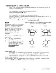
<strong>Bio</strong> <strong>122L</strong> (<strong>Laboratory</strong> <strong>for</strong> <strong>Bio</strong> <strong>322</strong>) <strong>Exercise</strong><br />
<strong>The</strong> <strong>Hill</strong> <strong>reaction</strong>: <strong>Effect</strong> of a Herbicide and Pronase<br />
R. <strong>Hill</strong> first demonstrated that a thylakoid membrane preparation could evolve O 2<br />
without CO 2 being present. <strong>The</strong> <strong>reaction</strong> required light and a suitable hydrogen (i.e.,<br />
electron) acceptor. <strong>The</strong> success of this experiment showed that oxygen is evolved by<br />
the light <strong>reaction</strong>s, and also indicated that water was the electron donor. Today, the <strong>Hill</strong><br />
<strong>reaction</strong> is often referred to as photolysis (i.e., light-dependent splitting) of water with the<br />
subsequent evolution of oxygen. However, it is frequently assayed using an electron<br />
acceptor such as 2,6-dichlorophenol indolephenol (DCIP) that changes color when it is<br />
reduced (see below) rather than assaying the release of oxygen, which is more difficult<br />
to quantify. <strong>The</strong> <strong>reaction</strong> we will use to measure the <strong>Hill</strong> <strong>reaction</strong> is the following:<br />
DCIP (blue) + H 2 O -----> DCIP-H 2 (colorless) + 1/2 O 2<br />
Since half an oxygen molecule would not be released, a more physiological version is:<br />
2DCIP (blue) + 2H 2 0 -----> 2DCIP-H 2 (colorless) + O 2<br />
Thus, with this assay, the extent of the <strong>Hill</strong> <strong>reaction</strong> is a function of the loss of blue<br />
color, which is measured as the loss of absorbance at 620 nm. <strong>The</strong> <strong>Hill</strong> <strong>reaction</strong><br />
assayed in this way is mostly a measure of the light-dependent activity of Photosystem<br />
II, since DCIP accepts electrons from the electron transport chain be<strong>for</strong>e Photosystem I,<br />
probably from the cytochrome f/b 6 complex.<br />
In this lab you will see how this <strong>reaction</strong>, still commonly used to measure<br />
Photosystem II, is assayed, and you will look at the effect of a herbicide (Diuron) and a<br />
protease (pronase) on the electron flow from PSII. You will use isolated membranes<br />
from spinach that consist mainly of thylakoid membrane vesicles enriched in PSII.<br />
<strong>The</strong>se vesicles are large fragments of stacked membranes that close up as vesicles<br />
when the membranes are fragmented during homogenization. <strong>The</strong>se vesicles (from the<br />
stacked regions) are very large, and sediment rapidly in a centrifuge. We will take<br />
advantage of this and isolate the membranes by differential centrifugation. It should be<br />
noted that thylakoid membranes isolated in this way are still right-side out, i.e., the<br />
stromal face of the membrane bilayer faces out as it does in vivo.<br />
Diuron, also known as DCMU (3-(3,4-dichlorophenyl)-1,1-dimethylurea) is one of a<br />
family of compounds that have been used extensively as herbicides. However, they<br />
have fallen out of favor lately, because they are very stable and persist in the<br />
environment. <strong>The</strong>se compounds are purported to be potent inhibitors of photosynthesis,<br />
probably acting at the Quinone b site.<br />
Pronase is a non-specific protease that is isolated from a bacterium. It degrades<br />
proteins at a number of different peptide bond combinations (e.g. aspartic-glutamate,<br />
1
tryptophan-proline, etc.). Proteases can be used to probe the structure and function of<br />
membrane-associated proteins; since most proteases (like pronase) are soluble<br />
enzymes, they can only degrade those portions of membrane proteins that stick out of<br />
the membrane. <strong>The</strong> membrane-embedded portions, and those portions of the proteins<br />
that stick out on the opposite side of the vesicle membrane from the protease (i.e., the<br />
thylakoid lumen, in this case) are protected from degradation. Thus, we will use pronase<br />
to find out if there are any important regions of the proteins that mediate the <strong>Hill</strong> <strong>reaction</strong><br />
(i.e., mainly Photosystem II proteins) that project away from the membrane on the<br />
stromal side.<br />
Protocol<br />
A. Preparation of thylakoid membranes<br />
1. Be<strong>for</strong>e beginning, place a mortar and pestle, a 15 ml centrifuge tube, and, if possible,<br />
a filtration flask on ice. You want to keep the plant material, and enriched<br />
membrane fraction cold to minimize proteolysis.<br />
2. Weigh out ~3 g of fresh spinach (which was kept cold until use) on the top-loading<br />
electronic balance.<br />
3. Shred the leaves with scissors into small pieces, and then homogenize the leaf<br />
pieces with the mortar and pestle, after adding 5 ml of buffer (Tricine, 0.05 M pH<br />
6.5). After grinding <strong>for</strong> ~1 minute, add another 5 ml of buffer and grind <strong>for</strong> another<br />
minute.<br />
4. Line a buchner funnel with 2 Kimwipes, and then filter the green homogenate through<br />
it into a filter flask, using gentle vacuum if necessary. Pour the filtrate into a 15 ml<br />
centrifuge tube, and discard the residue/Kimwipes.<br />
5. Centrifuge the green filtrate <strong>for</strong> ~5 min in a tabletop centrifuge at high speed; a green<br />
pellet should be apparent. Carefully pour off the supernatant (you want the pellet)<br />
and discard it.<br />
6. To the pellet, add ~3 ml (accuracy is not critical here) of cold Tricine buffer and<br />
resuspend this membrane fraction by vortexing (keep tube on ice AMAP).<br />
7. Centrifuge the crude membrane fraction again <strong>for</strong> ~5 min, pour off the supernatant,<br />
and then resuspend the pellet in ~3 ml of cold Tricine buffer with vortexing. This<br />
fraction contains mostly stacked thylakoid membranes.<br />
B. <strong>The</strong> <strong>Hill</strong> Reaction<br />
1. Place a 400 ml beaker filled with tap water (room temperature) adjacent to a light<br />
source (one or two microscope lamps should do).<br />
2
2. Label three 10-15 ml, clear, glass test tubes (labels of 1,2,3 will do).<br />
3. Under subdued light (if possible), pipet 1.25 ml of DCIP (1.1 x 10 -4 M) into each tube.<br />
4. <strong>The</strong>n pipet 2.5 ml of Tricine buffer into each tube.<br />
6. Double-wrap one of the tubes with aluminum foil so that light cannot enter. <strong>The</strong>n<br />
place all three tubes in the water bath (beaker) in the path of the light source <strong>for</strong> two<br />
minutes (this is to pre-warm the <strong>reaction</strong> mixtures).<br />
7. Right be<strong>for</strong>e pipetting the membranes, vortex your thylakoid preparation (to get it as<br />
homogeneous as possible), and then add 0.25 ml of the membrane suspension to<br />
each pre-incubated tube. Mix by quick vortexing, and place the tubes back into the<br />
water bath close to the light source.<br />
8. After 5 min, measure the absorbance (at 620 nm) of the mixture that had been<br />
wrapped in aluminum foil, and from one of the mixtures that had been exposed to<br />
light. You can use water as a blank to zero the machine (to 100% transmittance or 0<br />
absorbance).<br />
Note: To read the absorbance of the mixtures, quickly pour the contents into a cuvette,<br />
insert the cuvette into the machine, and then close the lid. Read the absorbance. <strong>The</strong>n<br />
pour the mixture back into the original tube (in case you need it again). After reading the<br />
dark-incubated tube, add a few grains (a pinch) of ascorbic acid to the tube. Notice what<br />
happens to the solution.<br />
6. After another 5 min (~10 min total) have elapsed, take an absorbance reading (at 620<br />
nm) of the other (#3) light-incubated tube.<br />
7. <strong>The</strong> difference between the absorbance values of the dark and light-incubated tubes<br />
will give you an indication of the extent of the <strong>Hill</strong> <strong>reaction</strong> during the incubation.<br />
C. <strong>Effect</strong> of Diuron and Pronase on the <strong>Hill</strong> Reaction<br />
1. Label four more tubes 1- 4.<br />
2. <strong>The</strong> additions to make to each tube are described in the Table below. One tube will<br />
be the dark-incubated control, and the other 3 will be put in the light. Diuron will be<br />
added to one of the light-incubated tubes, and pronase to another one. After 10<br />
minutes, read the absorbance at 620 nm <strong>for</strong> each tube, to determine the effects of<br />
the treatments on the <strong>Hill</strong> <strong>reaction</strong>.<br />
Note: Be sure to vortex the membrane suspension right be<strong>for</strong>e pipetting it, because the<br />
membranes tend to settle out.<br />
3
TABLE 1 - EFFECT OF DIURON AND PRONASE ON THE HILL REACTION<br />
All the volumes below are in ml. Add the components in the order listed.<br />
Tube Number<br />
Components 1 2 3 4<br />
Tricine Buffer 2.50 2.50 2.40 2.40<br />
Thylakoids 0.25 0.25 0.25 0.25<br />
Diuron (2 mM) 0.00 0.00 0.10 0.00<br />
Pronase (4 mg/ml) 0.00 0.00 0.00 0.10<br />
3. Vortex the tubes to mix the <strong>reaction</strong>s, and then incubate <strong>for</strong> 5 min at room<br />
temperature (to give the pronase a chance to work).<br />
4. Wrap tube #1 in aluminum foil.<br />
5. <strong>The</strong>n add the DCIP to the tubes as follows:<br />
DCIP 1.25 1.25 1.25 1.25<br />
Total Vol. 4.00 4.00 4.00 4.00<br />
Light (minutes) none 10 10 10<br />
Abs. @ 620nm<br />
4
<strong>The</strong> Report<br />
A. Objectives and general protocol<br />
Briefly describe the objectives of the experiment and how it was per<strong>for</strong>med. This part<br />
should be no more than 250 words.<br />
B. Results<br />
<strong>The</strong>n report the data as follows:<br />
1. Construct a table of your absorbance readings <strong>for</strong> the different treatments. Calculate<br />
the ∆A (i.e., change in absorbance compared to the dark control) <strong>for</strong> each<br />
experimental sample, and include the values in the table.<br />
2. What do the changes in absorbance of the <strong>reaction</strong> mixtures reflect? Is it lightdependent?<br />
What is being measured here?<br />
3. Construct a bar graph of the <strong>Hill</strong> <strong>reaction</strong> activity (i.e., ∆A) <strong>for</strong> each of the treatments<br />
in part C.<br />
C. Conclusions and Discussion (or questions to be addressed)<br />
1. What was the effect of Diuron on the <strong>Hill</strong> <strong>reaction</strong> in spinach? Could this result<br />
explain the herbicidal properties of Diuron?<br />
2. What was the effect of pronase on the <strong>Hill</strong> <strong>reaction</strong>? What does this indicate about<br />
photosystem II (or some component of PSII)?<br />
3. Abscorbic acid is often used as an antioxidant in food, and in photosynthesis<br />
research as an electron donor. What was the effect of adding abscorbic acid to the<br />
<strong>reaction</strong> tube in part B? What must it have done to the DCIP? What do you think the<br />
purpose was of doing this test?<br />
4. Indicate why you think you are measuring thylakoid PSII activity in these assays as<br />
opposed to a contaminating redox system from the endoplasmic reticulum or<br />
mitochondria.<br />
5. <strong>The</strong> PSII protein that binds the terminal electron acceptor of PSII, the Qb quinone, is<br />
known to be particularly sensitive to proteases. Could the result you obtained with<br />
the pronase treatment be explained by assuming that pronase damaged this<br />
protein? Can you suggest a way to determine which PSII protein(s) were damaged<br />
by pronase?<br />
6. Do you think Diuron would make a good herbicide? Keep in mind that with any<br />
herbicide, or pesticide, etc., you want the drug to be as specific as possible.<br />
5