Transcription and Translation RNA
Transcription and Translation RNA
Transcription and Translation RNA
Create successful ePaper yourself
Turn your PDF publications into a flip-book with our unique Google optimized e-Paper software.
<strong>Transcription</strong> <strong>and</strong> <strong>Translation</strong><br />
These terms describe the two steps used to transform the information carried in<br />
genes into useful products.<br />
The final product of some genes are <strong>RNA</strong> molecules.<br />
DNA <strong>Transcription</strong> <strong>RNA</strong><br />
The final product for other genes are protein molecules.<br />
First the DNA gene is transcribed into a m<strong>RNA</strong> molecule <strong>and</strong> then the m<strong>RNA</strong> is<br />
translated into a protein.<br />
DNA <strong>Transcription</strong> m<strong>RNA</strong> <strong>Translation</strong> protein<br />
decoding the genetic code<br />
<strong>RNA</strong><br />
Ribonucleic acid (<strong>RNA</strong>) is very much<br />
like DNA. It has a phosphodiester<br />
linked sugar backbone <strong>and</strong> uses<br />
primarily 4 different nitrogenous<br />
bases. The bases are A, G, C <strong>and</strong> U.<br />
U st<strong>and</strong>s for uracil. It is like a<br />
different dialect of the same<br />
language.<br />
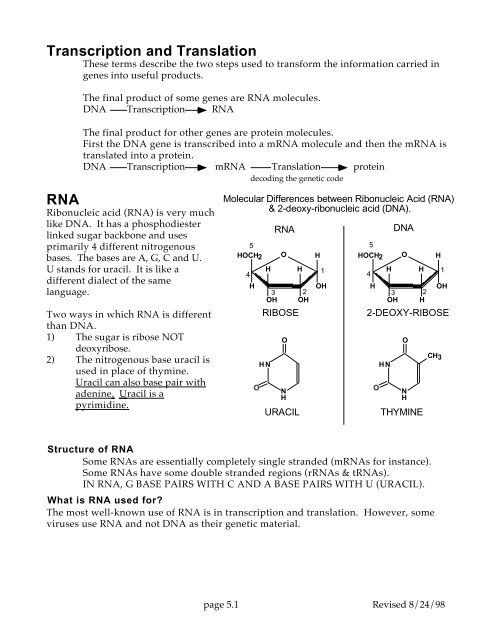
Two ways in which <strong>RNA</strong> is different<br />
than DNA.<br />
1) The sugar is ribose NOT<br />
deoxyribose.<br />
2) The nitrogenous base uracil is<br />
used in place of thymine.<br />
Uracil can also base pair with<br />
adenine. Uracil is a<br />
pyrimidine.<br />
Molecular Differences between Ribonucleic Acid (<strong>RNA</strong>)<br />
& 2-deoxy-ribonucleic acid (DNA).<br />
5<br />
HOCH2<br />
4<br />
H<br />
H<br />
3<br />
OH<br />
O<br />
RIBOSE<br />
H N<br />
<strong>RNA</strong><br />
O<br />
N<br />
H<br />
URACIL<br />
H<br />
2<br />
OH<br />
H<br />
1<br />
OH<br />
HOCH2<br />
4<br />
5<br />
H<br />
H<br />
3<br />
OH<br />
O<br />
H<br />
2<br />
H<br />
H<br />
1<br />
OH<br />
2-DEOXY-RIBOSE<br />
H N<br />
DNA<br />
O<br />
N<br />
H<br />
THYMINE<br />
CH3<br />
Structure of <strong>RNA</strong><br />
Some <strong>RNA</strong>s are essentially completely single str<strong>and</strong>ed (m<strong>RNA</strong>s for instance).<br />
Some <strong>RNA</strong>s have some double str<strong>and</strong>ed regions (r<strong>RNA</strong>s & t<strong>RNA</strong>s).<br />
IN <strong>RNA</strong>, G BASE PAIRS WITH C AND A BASE PAIRS WITH U (URACIL).<br />
What is <strong>RNA</strong> used for?<br />
The most well-known use of <strong>RNA</strong> is in transcription <strong>and</strong> translation. However, some<br />
viruses use <strong>RNA</strong> <strong>and</strong> not DNA as their genetic material.<br />
page 5.1 Revised 8/24/98
Before we continue some terminology<br />
Nucleotide Name Table<br />
Purines<br />
Pyrimidines<br />
Adenine (A) Guanine (G) Cytosine (C) Thymine (T) Uracil (U)<br />
Nucleotides in DNA deoxyadenylate deoxyguanylate deoxycytidylate deoxythymidylate<br />
or thymidylate<br />
Nucleotides in <strong>RNA</strong> adenylate guanylate cytidylate uridylate<br />
Abbreviations<br />
Nucleoside<br />
AMP GMP CMP TMP UMP<br />
monophosphates<br />
Nucleoside<br />
ADP GDP CDP TDP UDP<br />
diphosphates<br />
Nucleoside<br />
ATP GTP CTP TTP UTP<br />
triphosphates<br />
For deoxynucleotides add 'd' in front of the above three.<br />
<strong>Transcription</strong><br />
<strong>Transcription</strong>: DNA is transcribed into <strong>RNA</strong><br />
5'<br />
3'<br />
A<br />
T<br />
C<br />
G<br />
A<br />
T<br />
T<br />
A<br />
G<br />
C<br />
A<br />
T<br />
A<br />
T<br />
G<br />
C<br />
A<br />
T<br />
C<br />
G<br />
3'<br />
5'<br />
Double str<strong>and</strong>ed DNA molecule<br />
first<br />
nucleotide to<br />
be read<br />
5' A<br />
C<br />
A<br />
U<br />
G<br />
moving this way<br />
C<br />
G<br />
A<br />
U<br />
Bases are being read<br />
from left to right<br />
Free pool of<br />
ribonucleotides<br />
for use in making<br />
<strong>RNA</strong><br />
Transcripion<br />
The transcription machinery<br />
reads the nucleotides found in<br />
one of the DNA str<strong>and</strong>s <strong>and</strong><br />
builds a complementary <strong>RNA</strong><br />
molecule. If it reads G in the<br />
DNA then it will put a C on the<br />
end of the <strong>RNA</strong> molecule. If it<br />
reads A in the DNA then it will<br />
add a U on the end of the <strong>RNA</strong><br />
molecule. That is: C is<br />
complementary to G <strong>and</strong> U is<br />
complementary to A.<br />
Single str<strong>and</strong>ed <strong>RNA</strong> molecule that is being built one nucleotide at a time.<br />
Notice:<br />
1. In the diagram above it is the bottom DNA str<strong>and</strong> that is actually being read.<br />
2. The <strong>RNA</strong> that is being synthesized is complementary <strong>and</strong> antiparallel to this DNA<br />
str<strong>and</strong>.<br />
Learn these two terms NOW!!!<br />
• Coding str<strong>and</strong> refers to the DNA str<strong>and</strong> that has the same sequence as the transcribed<br />
<strong>RNA</strong>.<br />
• Template str<strong>and</strong> refers the other str<strong>and</strong>. It is the template that was actually used to<br />
synthesize the <strong>RNA</strong>. It is antiparallel <strong>and</strong> complementary to the <strong>RNA</strong>.<br />
Antisense <strong>and</strong> sense str<strong>and</strong>s are also antiparallel <strong>and</strong> complementary to one another.<br />
page 5.2 Revised 8/24/98
A bit closer look at transcription<br />
Double str<strong>and</strong>ed DNA helix<br />
5' TTTACCATGTTTAAAGGGCCCGACTACTTAACT<br />
|||||||||||||||||||||||||||||||||<br />
3' AAATGGTACAAATTTCCCGGGCAGATGAATTGA<br />
m<strong>RNA</strong><br />
5' UUUACCAUGUUUAAAGGGCCCGUCUACUUAACU<br />
T A A G-G-G-C-C-C-G-A-C<br />
A<br />
U<br />
A<br />
A<br />
T<br />
T G-G-G-C-C-C-G-A-C 3'<br />
| | | | | | | | |<br />
C-C-C-G-G-G-C-T-G<br />
page 5.3 Revised 8/24/98<br />
T<br />
A<br />
C<br />
G<br />
GACTTAGCGTGCATAGCA<br />
||||||||||||||||||<br />
CTGAATCGCACGTATCGA<br />
This diagram is full of very important information. It shows a m<strong>RNA</strong> molecule being<br />
transcribed from a double str<strong>and</strong>ed DNA molecule. The transcription machinery is not<br />
shown.<br />
Notice that:<br />
1. The DNA template str<strong>and</strong> is being read in the 3' to 5' direction.<br />
2. The <strong>RNA</strong> molecule is being synthesized in the 5' to 3' direction.<br />
3. The m<strong>RNA</strong> is antiparallel <strong>and</strong> complementary to the DNA str<strong>and</strong> that is being read.<br />
4. About 12 nucleotides of the m<strong>RNA</strong> are H-bonded to the DNA str<strong>and</strong>.<br />
Actually, it is just such H-bonding that enables the transcription machinery to choose<br />
the correct ribonucleotides when synthesizing an <strong>RNA</strong> molecule. Ribonucleotides<br />
are chosen based on their ability to H-bond or base pair to the DNA template.<br />
<strong>Translation</strong><br />
<strong>Translation</strong>: m<strong>RNA</strong> is translated into a protein.<br />
What are proteins?<br />
A protein is made of a specific sequence of amino acids. If the amino acid sequence of a<br />
protein is wrong then it won't function properly. Amino acids are substantially different<br />
than nucleotides <strong>and</strong> yet the sequence of amino acids in a protein is encoded in the DNA.<br />
The amino acids typically found in proteins are:<br />
phenylalanine F serine S tyrosine Y cysteine C<br />
leucine L tryptophan W proline P histidine H<br />
arginine R glutamine Q methionine M isoleucine I<br />
threonine T asparagine N valine V alanine A<br />
glutamic acid E glycine G aspartic acid D lysine K<br />
Next to the name of each amino acid its single-letter abbreviation.<br />
How can the 4 nucleotides found in DNA encode the 20 different amino acids used to<br />
make a particular protein?<br />
The sugar phosphate backbone is the same throughout the DNA molecule <strong>and</strong> so<br />
information cannot be carried here. The genetic information is carried in the order of the<br />
nitrogenous bases.<br />
3'<br />
5'
Genetic Code<br />
Nucleic acids carry the amino acid sequence of proteins in the form of a genetic code.<br />
This code is read in groups of three nucleotides. Each group of three is called a codon (also<br />
referred to as a triplet codon). The genetic code is a language in which all words are 3 letters<br />
long, <strong>and</strong> in which there is no punctuation, or spaces between the words. The codons are<br />
non-overlapping.<br />
Genetic Code Table<br />
S E C O N D P O S I T I O N<br />
U C A G<br />
F S Y C U<br />
F U F S Y C C T<br />
I L S Stop Stop A H<br />
R L S Stop W G I<br />
S<br />
R<br />
T L P H R U D<br />
C L P H R C<br />
P L P Q R A P<br />
O L P Q R G O<br />
S<br />
S<br />
I I T N S U I<br />
T A I T N S C T<br />
I I T K R A I<br />
O M T K R G O<br />
N<br />
V A D G U<br />
G V A D G C<br />
V A E G A<br />
V A E G G<br />
UCAG on the left, top <strong>and</strong> right sides represent nitrogenous bases in the codon.<br />
Examples of how to use this table:<br />
The codon UUU <strong>and</strong> UUC both encode the amino acid F (phenylalanine).<br />
The codons GCU, GCC, GCA <strong>and</strong> GCG all encode the amino acid A (alanine).<br />
The amino acid M (methionine) is encoded only by the codon AUG.<br />
Notice that we have written the genetic code in ribonucleotides. You can tell because we<br />
have used U in place of T. You will also see it written in terms of deoxynucleotides.<br />
N<br />
page 5.4 Revised 8/24/98
The genetic code is degenerate.<br />
In this context, degeneracy means that some amino acids are encoded by more than one<br />
codon. That is, some codons have synonyms.<br />
What the heck are stop codons?<br />
<strong>Translation</strong> is performed by the ribosomes. When a ribosome encounters a stop codon it<br />
dissociates from the m<strong>RNA</strong> <strong>and</strong> releases the protein. Stop codons are found at the end of<br />
the protein coding region. The codons UAA, UGA <strong>and</strong> UAG are stop codons.<br />
The genetic code is universal<br />
The genetic code shown above is used, essentially unchanged, in all organisms. There are<br />
minor exceptions to this truth, but you should think of these as different dialects of the same<br />
language.<br />
Be completely sure that you know how to read the genetic code table <strong>and</strong> could use it to<br />
translate a m<strong>RNA</strong>.<br />
The Process of <strong>Translation</strong><br />
<strong>Translation</strong> is the process of reading the information carried in a m<strong>RNA</strong> molecule <strong>and</strong><br />
using that information to direct the synthesis of a protein molecule. It is performed by the<br />
ribosomes using both m<strong>RNA</strong>, t<strong>RNA</strong> <strong>and</strong> amino acids.<br />
m<strong>RNA</strong> (made by the process of transcription)<br />
5' A C A U G A A G A C C U G A C C U A A A A A 3'<br />
moving this way<br />
First codon to be read<br />
Amino terminal end<br />
of the nascent polypeptide.<br />
Trans<br />
l ation<br />
F<br />
W<br />
P<br />
G<br />
A M<br />
C<br />
H V<br />
D Q<br />
I<br />
E L<br />
K Y<br />
R N<br />
T S<br />
Amino acid pool.<br />
20 different amino<br />
acids are used to<br />
synthesize proteins.<br />
Each letter represents<br />
a different amino acid.<br />
M<br />
K<br />
T<br />
A protein being synthesized.<br />
Proteins are polymers of amino<br />
acids. The letters<br />
FKSYCWLPHNRIMTKVADEG<br />
represent different amino acids.<br />
The m<strong>RNA</strong> carries the information necessary to build a specific protein. Specifically, it<br />
describes the order of amino acids that the protein should contain. The m<strong>RNA</strong> is like a<br />
sentence. Each 'word' is 3 nucleotides long <strong>and</strong> is called a codon. There are no spaces <strong>and</strong><br />
no punctuation. Each codon specifies a single amino acid. The translation machinery reads<br />
the m<strong>RNA</strong> a codon at time <strong>and</strong> joins together the amino acids specified by the m<strong>RNA</strong>.<br />
Proteins are synthesized in the amino terminus to carboxy terminus direction.<br />
In the example shown above:<br />
page 5.5 Revised 8/24/98
The first amino acid M is methionine it is encoded by the codon AUG. The second<br />
amino acid K is lysine it is encoded by the codon AAG. The third amino acid T is threonine<br />
it is encoded by the codon ACC.<br />
Not all <strong>RNA</strong>s are translated. In fact, only messenger <strong>RNA</strong> (m<strong>RNA</strong>) is translated. Both<br />
transfer <strong>RNA</strong> (t<strong>RNA</strong>) <strong>and</strong> the ribosomal <strong>RNA</strong>s (r<strong>RNA</strong>) are actually part of the translation<br />
machinery.<br />
What are protein molecules composed of?<br />
Proteins are polymers of amino acids.<br />
What are amino acids?<br />
These molecule are called amino acids because (with one exception) they each contain<br />
an amino group (NH 2 ) <strong>and</strong> an acidic carboxylic group (COOH).<br />
General structure of amino acids----------><br />
The alpha carbon is next to the COOH group.<br />
Attached to the α carbon is a hydrogen <strong>and</strong> a nitrogen.<br />
The nitrogen is part of an amino group.<br />
The side chain marked R is different in each amino<br />
acid <strong>and</strong> gives them their characteristic properties.<br />
H<br />
H 2<br />
N C<br />
α<br />
R<br />
COOH<br />
An exception to the above general structure.<br />
In the amino acid, proline, the nitrogen is not part of an amino<br />
groups but is part of an imino ring that involves the α carbon.<br />
The ring in proline causes it to be very rigid, <strong>and</strong> can produce a<br />
bend in a protein. None of the other R groups have rings that<br />
involve the α carbon.<br />
HN<br />
C<br />
α<br />
COOH<br />
H<br />
C<br />
C<br />
imino ring<br />
C<br />
Polypeptides or proteins are polymers of amino acids.<br />
The term polypeptide is usually reserved for very small proteins.<br />
In proteins, amino acids are<br />
joined to form polymers by the<br />
ribosomes.<br />
Condensation reaction:<br />
The amino acids are joined in a<br />
condensation reaction. This<br />
means that the reaction<br />
liberates a water molecule.<br />
The newly formed bond<br />
between the amino acids is<br />
called a peptide bond.<br />
+<br />
H<br />
H N C 3 α<br />
R1<br />
+<br />
H<br />
3<br />
N<br />
O<br />
C<br />
H<br />
C<br />
α<br />
O<br />
O<br />
C<br />
+<br />
N<br />
H<br />
+<br />
H N<br />
3<br />
H<br />
C<br />
α<br />
R 1<br />
R2<br />
Peptide Bond<br />
H<br />
C<br />
α<br />
R<br />
2<br />
H 2 O<br />
O<br />
C<br />
O<br />
C<br />
O<br />
O<br />
page 5.6 Revised 8/24/98
Actually we should draw the peptide bond as ----------><br />
Both O <strong>and</strong> N of the peptide bond have partial double bond<br />
characteristics.<br />
+<br />
C N<br />
H<br />
This double bond character causes the peptide bond to be flat (planar) <strong>and</strong> restricts the<br />
rotation about the peptide bond. Flexibility in proteins is primarily limited to the rotation<br />
around the α carbon's 2 single bonds. This is not true for proline. With proline the<br />
involvement of the α carbon in the imino ring further restriction the flexibility of the<br />
protein.<br />
You remember that nucleic<br />
acids have an inherent<br />
polarity <strong>and</strong> that they are<br />
synthesized in a particular<br />
direction. ------------------><br />
Right?<br />
PPP<br />
N1<br />
5'<br />
P<br />
N2<br />
P<br />
PPP<br />
N3<br />
P<br />
N1<br />
5'<br />
N4<br />
P<br />
3'<br />
OH<br />
N2<br />
P<br />
N3<br />
P<br />
PPP<br />
N4<br />
P<br />
O -<br />
N5<br />
5'<br />
N5<br />
3'<br />
OH<br />
3'<br />
OH<br />
Well, proteins also have an<br />
inherent polarity <strong>and</strong> are<br />
also synthesized in a<br />
particular direction.<br />
One end is called the amino<br />
terminus <strong>and</strong> the other the<br />
carboxy terminus.<br />
+<br />
H N 3<br />
H<br />
C<br />
α<br />
amino<br />
terminus<br />
O<br />
C<br />
N<br />
H<br />
H<br />
C<br />
α<br />
R<br />
R<br />
1 2<br />
O<br />
C<br />
N<br />
H<br />
H<br />
C<br />
α<br />
R 3<br />
O<br />
- C O<br />
carboxy<br />
terminus<br />
Proteins are<br />
synthesized in<br />
the amino to<br />
carboxy<br />
direction.<br />
+<br />
H<br />
3<br />
N<br />
H<br />
C<br />
α<br />
O<br />
C<br />
N<br />
H<br />
H<br />
C<br />
α<br />
R 1<br />
R2<br />
O<br />
C<br />
O -<br />
NH 3<br />
H2O<br />
H<br />
C<br />
α<br />
R<br />
3<br />
O<br />
C<br />
O -<br />
+<br />
H<br />
3<br />
N<br />
H<br />
C<br />
α<br />
O<br />
C<br />
N<br />
H<br />
H<br />
C<br />
α<br />
O<br />
C<br />
N<br />
H<br />
H<br />
C<br />
α<br />
O<br />
C<br />
O -<br />
R<br />
R<br />
1 2<br />
R3<br />
page 5.7 Revised 8/24/98
How do the ribosomes know which amino acids to join together to<br />
form a protein?<br />
Well, of course, this information is carried in a gene. A m<strong>RNA</strong> copy of the gene is<br />
made via transcription <strong>and</strong> then this copy is translated into a protein. That is;<br />
DNA ------------------> m<strong>RNA</strong> ------------------> protein<br />
The translation machinery 'knows' which amino acids to incorporate into the growing<br />
polypeptide because it reads this information from a m<strong>RNA</strong> molecule.<br />
Let's look at this in more detail.<br />
5'<br />
3'<br />
TTTACCATGTTTAAAGGGCCCGACTACTTAACT<br />
|||||||||||||||||||||||||||||||||<br />
AAATGGTACAAATTTCCCGGGCAGATGAATTGA<br />
M<br />
M<br />
proteins being<br />
synthesized<br />
F K D P<br />
F<br />
K<br />
G<br />
m<strong>RNA</strong><br />
5' UUUACCAUGUUUAAAGGGCCCGUCUACUUAACU<br />
T A A G-G-G-C-C-C-G-A-C<br />
A<br />
U<br />
A<br />
A<br />
T<br />
T G-G-G-C-C-C-G-A-C 3'<br />
| | | | | | | | |<br />
C-C-C-G-G-G-C-T-G<br />
ribosomes<br />
T<br />
A<br />
C<br />
G<br />
GACTTAGCGTGCATAGCA<br />
||||||||||||||||||<br />
CTGAATCGCACGTATCGA<br />
This diagram show concomitant translation <strong>and</strong> transcription. Before transcription is<br />
finished, translation has begun. It is the kind of translation that occurs only in prokaryotic<br />
cells. <strong>Translation</strong> nearly always begins with the codon AUG. It is called the start codon.<br />
Each trinucleotide codon is read in succession until a stop codon is reached. Stop codons are<br />
UAA, UAG <strong>and</strong> UGA. The amino terminus is the first to be synthesized <strong>and</strong> the carboxy<br />
terminus of the protein is the last part to be translated.<br />
Many copies of an <strong>RNA</strong> <strong>and</strong> protein molecule can be made from a single<br />
gene.<br />
5' 3'<br />
Three types of <strong>RNA</strong> molecules participate in translation.<br />
1. m<strong>RNA</strong> carries the information from the DNA in a form that can be read by the<br />
ribosomes.<br />
2. r<strong>RNA</strong> is a major component of the ribosomes.<br />
3. t<strong>RNA</strong> carries the amino acid to the ribosomes <strong>and</strong> is actually the agent which<br />
deciphers the genetic code.<br />
<strong>Translation</strong> Cycle<br />
Free Pool<br />
50S<br />
70S<br />
30S<br />
N<br />
N<br />
3'<br />
UAG<br />
N<br />
5'<br />
AUG<br />
Termination<br />
Initiation<br />
amino acid<br />
Elongation<br />
m<strong>RNA</strong> is<br />
eventually<br />
degraded<br />
C<br />
The Start codon<br />
Ribosomes assemble on the m<strong>RNA</strong> around the start codon. Start codons are nearly<br />
always AUG. AUG encodes the amino acid methionine. Therefore, the first amino acid to<br />
be translated in a protein is methionine.<br />
Stop codon<br />
The ribosomes translate the m<strong>RNA</strong> one codon at a time until they hit a stop codon.<br />
Once the stop codon is encountered the ribosomes stop translating <strong>and</strong> dissociate into a large<br />
<strong>and</strong> small subunit <strong>and</strong> release the protein <strong>and</strong> the m<strong>RNA</strong>. The ribosomes re-enter the free<br />
ribosome pool <strong>and</strong> can be used again <strong>and</strong> again. Other ribosomes can continue to translate<br />
the m<strong>RNA</strong> or the m<strong>RNA</strong> may be degraded.<br />
There are 3 stop codons. They are UAA, UAG <strong>and</strong> UGA. Stop codons are also called<br />
nonsense codons.<br />
page 5.9 Revised 8/24/98
Composition of Ribosomes<br />
Prokaryotic ribosomes<br />
Eukaryotic ribosomes<br />
5 S r<strong>RNA</strong><br />
120 bases<br />
+<br />
23 S r<strong>RNA</strong><br />
2900 bases<br />
+<br />
31 ribosomal proteins<br />
16S r<strong>RNA</strong><br />
1540 bases<br />
+<br />
21 ribosomal proteins<br />
50 S subunit<br />
(large)<br />
+<br />
30 S subunit<br />
(small)<br />
60 S subunit<br />
(large)<br />
+<br />
40 S subunit<br />
(small)<br />
5 S r<strong>RNA</strong><br />
120 bases<br />
+<br />
5.8 S r<strong>RNA</strong><br />
160 bases<br />
+<br />
28 S r<strong>RNA</strong><br />
4800 bases<br />
+<br />
50 ribosomal proteins<br />
18 S r<strong>RNA</strong><br />
1900 bases<br />
+<br />
33 ribosomal proteins<br />
5S<br />
23 S<br />
16 S<br />
5.8 S<br />
5S<br />
28 S<br />
18 S<br />
70 S subunit<br />
80 S subunit<br />
The ribosomal subunits are named according to their Svedberg coefficients (S). This is<br />
a unit of measure that describes the sedimentation rate of a particle in a centrifuge. It is<br />
particularly useful with very large macromolecular complexes. The greater the mass the<br />
larger is the Svedberg coefficient. Notice that the units are not additive.<br />
What do ribosomes contain?<br />
Ribosomes are composed to two different types of molecules: 1) <strong>RNA</strong> called r<strong>RNA</strong> or<br />
ribosomal <strong>RNA</strong> <strong>and</strong> 2) proteins, generically referred to as ribosomal proteins. In<br />
prokaryotes, the large ribosomal subunit contains two r<strong>RNA</strong>s (5S <strong>and</strong> 23S) <strong>and</strong> 31 different<br />
ribosomal proteins <strong>and</strong> the small subunit contains a single 16S r<strong>RNA</strong> <strong>and</strong> 21 different<br />
ribosomal proteins. Eukaryotic ribosomes are similar in composition to prokaryotic<br />
ribosomes. The exact size of the r<strong>RNA</strong>s <strong>and</strong> number of unique proteins differ between the<br />
two. One of the biggest differences is the presence of a third r<strong>RNA</strong>, the 5.8S r<strong>RNA</strong>, in the<br />
eukaryotic large subunit. Examine the diagram above very closely. Learn the sizes of the<br />
subunits <strong>and</strong> the r<strong>RNA</strong>s.<br />
page 5.10 Revised 8/24/98
<strong>RNA</strong> has extensive secondary structure<br />
This diagram shows the<br />
secondary structure of<br />
the 16S r<strong>RNA</strong> from the<br />
bacteria, Escherichia<br />
coli. All r<strong>RNA</strong>s have<br />
extensive secondary <strong>and</strong><br />
tertiary structure. The<br />
5'<br />
stems <strong>and</strong> loops are<br />
produced by the<br />
intramolecular base<br />
pairing between<br />
3'<br />
different parts of the<br />
molecule. This is a twodimensional<br />
representation of the<br />
molecule. Actually, the molecule is folded in three dimensions to form a much more<br />
complex shape. This secondary structure is very similar, but not identical, in all organisms.<br />
This reflects the fact that the mechanisms of translation are evolutionarily strongly<br />
conserved.<br />
Function of the ribosomal components<br />
<strong>Translation</strong> requires both subunits. However, some of the labor is divided between the<br />
subunits. The small subunit recognizes the start codon <strong>and</strong> the large subunit synthesizes<br />
the peptide bond that joins the amino acids in a protein.<br />
Notice that the ribosomes are composed of both <strong>RNA</strong> <strong>and</strong> protein. Originally, it was<br />
thought that the r<strong>RNA</strong> served only a structural role, that it formed a scaffold used solely for<br />
the purpose of organizing the various ribosomal proteins. Furthermore, it was thought that<br />
it was the ribosomal proteins that did all of the catalytic work of protein synthesis. This is<br />
not entirely correct.<br />
It is true that the r<strong>RNA</strong> forms a scaffold, however, this is not its sole function. In the<br />
small subunit it is actually the 16S r<strong>RNA</strong> that recognizes the start codon of a m<strong>RNA</strong>. In the<br />
large subunit, it is the 23S r<strong>RNA</strong> that catalyzes peptide bond formation between amino<br />
acids. The various ribosomal proteins enhance both the specificity <strong>and</strong> efficiency of these<br />
processes.<br />
t<strong>RNA</strong><br />
2-dimensional model<br />
3-dimensional model<br />
page 5.11 Revised 8/24/98
Amino<br />
acid<br />
3' amino acid<br />
acceptor site<br />
TΨC loop<br />
Acceptor stem<br />
Acceptor<br />
Stem<br />
D-loop<br />
D-Stem<br />
5' end<br />
CCA<br />
end<br />
TΨC loop<br />
Anticodon<br />
stem<br />
D loop<br />
Variable<br />
loop<br />
Anticodon<br />
stem<br />
D stem<br />
Anticodon<br />
3' acceptor<br />
end<br />
Anticodon loop<br />
A t<strong>RNA</strong> is a single <strong>RNA</strong> chain that is folded into a two dimensional cloverleaf. This then<br />
folds in three dimensions to an L-like structure. t<strong>RNA</strong>s are small <strong>RNA</strong> molecules (usually<br />
in the range of 73 - 93 nucleotides) that participate in the translation of m<strong>RNA</strong>s. They are<br />
not part of the ribosome. But like r<strong>RNA</strong> they have extensive secondary structure produced<br />
by intramolecular base pairing between nucleotides. t<strong>RNA</strong>s are unique in that they contain<br />
many highly modified <strong>and</strong> unusual nucleotides. Immediately following transcription, the<br />
t<strong>RNA</strong> contains only the st<strong>and</strong>ard set of four nucleotides. The unusual nucleotides are<br />
produced by post-transcriptional enzymatic modifications. Some of the modifications help<br />
the cell to unambiguously recognize the t<strong>RNA</strong>. However, the function of most of the<br />
modifications is unknown.<br />
Take careful notice of the features common to all t<strong>RNA</strong>s. They are:<br />
XCCA, D loop, TΨC loop <strong>and</strong> Anticodon loop<br />
Notice that the t<strong>RNA</strong>s have 4 stems <strong>and</strong> 3 loops <strong>and</strong> that each of them has a name.<br />
Furthermore some t<strong>RNA</strong>s have a fourth loop called the Variable Loop. It is located<br />
between the TψC stem <strong>and</strong> the anticodon stem. The D loop is named for a modified<br />
nitrogenous base found within it; dihydrouracil. Similarly, the TψC loop, is named for four<br />
invariant nucleotides found within it. They are thymidylate, pseudouridylate, cytidylate,<br />
guanylate. The XCCA terminus is very important. XCCA refers to the nucleotides found at<br />
this position. X st<strong>and</strong> for any nucleotide, C for cytidylate <strong>and</strong> A for adenylate. The XCCA<br />
stem is the amino acid acceptor site.<br />
It is the t<strong>RNA</strong>s that actually decode the genetic code<br />
This is done via the anticodon loop.<br />
page 5.12 Revised 8/24/98
Threonine<br />
This t<strong>RNA</strong> decodes one of the codons<br />
that specify threonine. This t<strong>RNA</strong> is<br />
referred to as: t<strong>RNA</strong> thr .<br />
t<strong>RNA</strong> which recognizes<br />
a codon for threonine.<br />
Anticodon loop<br />
5'<br />
AGU<br />
The codons for threonine are ACU,<br />
ACC, ACA <strong>and</strong> ACG. This t<strong>RNA</strong><br />
decodes the ACU codon. Notice that<br />
three of the nucleotides in the anticodon<br />
loop are complementary <strong>and</strong><br />
antiparallel to the codon ACU. Notice<br />
that in the diagram that the t<strong>RNA</strong> is<br />
covalently attached to the amino acid<br />
threonine.<br />
t<strong>RNA</strong>s decode codons by base pairing with them.<br />
Threonine<br />
5'<br />
START CODON<br />
STOP CODON<br />
AGU<br />
m<strong>RNA</strong><br />
3' AAAAAAUAUACAAAUGACUUAAGGGCUAUAUCACAUAUAUCAGGAGUAUAUUAUAAA 5'<br />
The anticodon of t<strong>RNA</strong> thr base pairs with the threonine codon. Now, keep this<br />
straight. The anticodon is found in the t<strong>RNA</strong> <strong>and</strong> the codon is found in the m<strong>RNA</strong>. The<br />
anticodon base pairs with the codon in a complementary <strong>and</strong> antiparallel fashion. Notice<br />
that the t<strong>RNA</strong> <strong>and</strong> the m<strong>RNA</strong> are in opposite polarity. t<strong>RNA</strong>s actually decode codons<br />
within the confines of the ribosome, however, the ribosome is not shown here. We'll<br />
include it later on.<br />
Every codon is recognized by at least one anticodon. There are 64 possible codons.<br />
Three of these are stop codons. Refer back to the Genetic Code Table to check this. Each of<br />
the codons is recognized by a t<strong>RNA</strong> except for the stop codons. The three stop codons are<br />
NOT recognized by any t<strong>RNA</strong>.<br />
Wobble<br />
There are 61 different codons that must be decoded. This would lead you to believe<br />
that cells have 61 different t<strong>RNA</strong>s, one for each codon. Right? Wrong! Prokaryotes<br />
typically have 30 to 40 different t<strong>RNA</strong>s <strong>and</strong> eukaryotes have about 50 t<strong>RNA</strong>s. How can 30 to<br />
50 different t<strong>RNA</strong>s correctly decode 61 different codons. The answer is through wobble base<br />
pairing.<br />
page 5.13 Revised 8/24/98
Wobble base CODONS ENCODE M R R I Y T I S<br />
pairing is a nonst<strong>and</strong>ard<br />
5' // AU AUG CGA CGG AUA UAC ACU AUA UCG GG // 3'<br />
type of base<br />
pairing that occurs<br />
between the third<br />
nucleotide in a codon<br />
FLIP IT<br />
<strong>and</strong> the first<br />
nucleotide in an<br />
anticodon. It enables a<br />
3' // GG GCU AUA UCA CAU AUA GGC AGC GUA UA // 5'<br />
single t<strong>RNA</strong> to<br />
recognize more than<br />
CODONS ENCODE S I T Y I R R M<br />
one codon for a particular amino acid.<br />
WOBBLE base pairing occurs between the 1st position of the anticodon<br />
<strong>and</strong> the third position of the codon.<br />
Wobble Base Pairing Table<br />
Base in the first position of the<br />
anticodon<br />
can wobble base pair with<br />
Base recognized in the third position of<br />
the codon<br />
U<br />
A or G<br />
C<br />
G only<br />
A<br />
U only<br />
G<br />
C or U<br />
I<br />
C, A or U<br />
I = Inosine, one of the non-st<strong>and</strong>ard nitrogenous bases found in t<strong>RNA</strong>s.<br />
Let's continue to examine this. Shown in the next figure is part of a m<strong>RNA</strong> <strong>and</strong> the amino<br />
acids that it encodes. I have spaced the codons for clarity. Usually, we write <strong>RNA</strong>s 5' to 3'.<br />
But for right now let's flip it over so that it is written 3' to 5'.<br />
Notice that in the m<strong>RNA</strong> there are two different codons for arginine (R). If this does not<br />
make sense to you then please consult the Genetic Code Table.<br />
Shown on the right is a t<strong>RNA</strong> arg . We<br />
are going to see how this t<strong>RNA</strong> uses<br />
wobble base pairing to pair with both of<br />
the arginine codons shown above.<br />
5'<br />
Arginine<br />
UCG<br />
page 5.14 Revised 8/24/98
Here the t<strong>RNA</strong> arg<br />
is shown base<br />
pairing with the<br />
CGA codon for<br />
arginine. This is<br />
the usual type of<br />
base pairing with<br />
which you are<br />
familiar.<br />
A t<strong>RNA</strong> arg is decoding the first<br />
arginine codon.<br />
5'<br />
UCG<br />
Arginine<br />
3' // GG GCU AUA UCA CAU AUA GGC AGC GUA UA // 5' m<strong>RNA</strong><br />
Both of these codons encode the amino acid arginine (R).<br />
Here we see the<br />
same t<strong>RNA</strong> base<br />
pairing with a<br />
different arginine<br />
codon. This the<br />
non-st<strong>and</strong>ard type<br />
of base pairing<br />
called Wobble<br />
Base Pairing.<br />
The same t<strong>RNA</strong> is decoding the<br />
second arginine codon!<br />
5'<br />
Arginine<br />
3' // GG GCU AUA UCA CAU AUA GGC AGC GUA UA // 5' m<strong>RNA</strong><br />
You can consider this wobble base pairing to be unique to the base pairing between the<br />
first position of the anticodon <strong>and</strong> the third position of the codon.<br />
The Genetic code table indicates that there are 6 different codons for arginine. They are<br />
CGU, CGC, CGA, CGG, AGA <strong>and</strong> AGG. Notice that these can be broken down into 2 groups<br />
that are identical in codon positions 1 <strong>and</strong> 2. Using wobble base pairing three different<br />
t<strong>RNA</strong>s can decode all 6 of these codons.<br />
UCG<br />
page 5.15 Revised 8/24/98
These are<br />
the six<br />
CODONS<br />
that can<br />
encode<br />
arginine.<br />
1 2 3<br />
5' CGU 3'<br />
1 2 3<br />
5' CGC 3'<br />
1 2 3<br />
5' CGA 3'<br />
1 2 3<br />
5' CGG 3'<br />
1 2 3<br />
5' AGA 3'<br />
1 2 3<br />
5' AGG 3'<br />
Each figure represents an<br />
arginine codon in a m<strong>RNA</strong>.<br />
It is written in the 5' to 3'<br />
direction.<br />
A minimum of three different t<strong>RNA</strong>s are required to decode all 6 of the above codons.<br />
They are:<br />
3' GCU<br />
3 2 1<br />
3' GCI<br />
3 2 1<br />
5'<br />
5'<br />
3' UCU<br />
3 2 1<br />
5'<br />
Each figure represents an<br />
anticodon in a t<strong>RNA</strong>. They<br />
are written in the 3' to 5'<br />
direction (backwards).<br />
page 5.16 Revised 8/24/98
Below, I have spelled out how this happens. A minimum of two t<strong>RNA</strong>s are needed to<br />
recognize the CGX codon <strong>and</strong> a minimum of 1 t<strong>RNA</strong> is needed to recognize the AGA <strong>and</strong><br />
AGG codons.<br />
wobble base pairing<br />
normal base pairing<br />
codon<br />
1 2 3<br />
5' CGG 3'<br />
codon<br />
1 2 3<br />
5' CGA 3'<br />
anticodon<br />
3' GCU<br />
3 2 1<br />
5'<br />
anticodon<br />
3' GCU<br />
3 2 1<br />
5'<br />
wobble base pairing<br />
wobble base pairing<br />
codon<br />
1 2 3<br />
5' CGU 3'<br />
codon<br />
1 2 3<br />
5' CGA 3'<br />
anticodon<br />
3' GCI<br />
3 2 1<br />
5'<br />
anticodon<br />
3' GCI<br />
3 2 1<br />
5'<br />
wobble base pairing<br />
codon<br />
1 2 3<br />
5' CGC 3'<br />
anticodon<br />
3' GCI<br />
3 2 1<br />
5'<br />
normal base pairing<br />
wobble base pairing<br />
codon<br />
1 2 3<br />
5' AGA 3'<br />
codon<br />
1 2 3<br />
5' AGG 3'<br />
anticodon 3' UCU<br />
3 2 1<br />
5' anticodon 3' UCU<br />
3 2 1<br />
Refer back to the Wobble Base Pairing Table until this makes sense.<br />
5'<br />
page 5.17 Revised 8/24/98
How are amino acids attached to a t<strong>RNA</strong>?<br />
Each t<strong>RNA</strong><br />
can only<br />
carry one<br />
specific<br />
amino acid.<br />
5'<br />
3'<br />
Amino acyl<br />
t<strong>RNA</strong> synthetase<br />
5'<br />
Threonine<br />
AGU Threonine AMP + PPi<br />
AGU<br />
ATP<br />
A t<strong>RNA</strong> carrying an amino acid is said to be charged. But the amino acid that is<br />
attached to the t<strong>RNA</strong> is said to be activated. The enzyme that attaches the amino acid to the<br />
t<strong>RNA</strong> is called aminoacyl t<strong>RNA</strong> synthetase. Charging a t<strong>RNA</strong> consumes energy in the form<br />
of ATP. Energy from the ATP is conserved in bond between amino acid <strong>and</strong> t<strong>RNA</strong>.<br />
Charged t<strong>RNA</strong><br />
Amino acid are<br />
attached to the t<strong>RNA</strong><br />
through the terminal<br />
3' adenosine residue<br />
found in t<strong>RNA</strong>.<br />
D-loop<br />
D-Stem<br />
5' end<br />
Acceptor<br />
Stem<br />
CCA<br />
end<br />
Anticodon<br />
stem<br />
TΨC loop<br />
Threonine<br />
HO<br />
CH2<br />
H<br />
O<br />
NH 2<br />
C<br />
C<br />
O<br />
N<br />
N<br />
CH(CH3)OH<br />
O<br />
t<strong>RNA</strong><br />
N<br />
NH 2<br />
N<br />
Threonine<br />
Adenylate<br />
AGU<br />
Anticodon loop<br />
The last three nucleotides on the 3' end of a t<strong>RNA</strong> are cytidylate (C) cytidylate (C) <strong>and</strong><br />
adenylate (A). That is 5' CCA 3'. Amino acids are attached to the t<strong>RNA</strong> through the<br />
adenylate residue. They are attached through a carboxylic bond to the 2' or 3' carbon of the<br />
adenylate residue.<br />
Remember that bacteria have 30 to 40 different t<strong>RNA</strong>s <strong>and</strong> eukaryotes have about 50<br />
different t<strong>RNA</strong>s. There is one amino acid synthetase for each amino acid. That is, there are<br />
20 different aminoacyl t<strong>RNA</strong> synthetases. Each individual synthetase charges all of the<br />
t<strong>RNA</strong>s that encode one particular amino acid. That is; each synthetase is specific for one<br />
amino acid. For example, if an organism has three t<strong>RNA</strong> arg 's then the enzyme arginine<br />
aminoacyl t<strong>RNA</strong> synthetase will charge all of the three t<strong>RNA</strong>s. Some aminoacyl t<strong>RNA</strong><br />
synthetases recognize their particular t<strong>RNA</strong>s based upon the shape of the molecule while<br />
others seem to recognize the anticodon loop itself.<br />
page 5.18 Revised 8/24/98
Definitions<br />
Escherichia coli<br />
intramolecular<br />
intermolecular<br />
nascent<br />
post-translational<br />
post-transcriptional<br />
Svedberg unit (S)<br />
primary structure<br />
Secondary structure<br />
Tertiary structure<br />
Quaternary structure<br />
Gram-negative, rod-shaped bacteria typically found in the large intestine of<br />
warm-blooded animals. It is a prokaryote. A member of the coliform group<br />
of bacteria. Extensively used by molecular biologists for the propagation of<br />
plasmid <strong>and</strong> phage vectors. Those used in the laboratory cannot normally<br />
survive in nature. Abbreviated name is E. coli.<br />
refers to interactions that occur between different parts of the same molecule.<br />
refers to interactions that occur between 2 or more molecules.<br />
in the process of emerging<br />
A modification that occurs after a protein molecule has been translated.<br />
A modification that occurs after a <strong>RNA</strong> molecule has been transcribed.<br />
Abbreviation is "S". A unit describing the rate of sedimentation of a particle in<br />
a centrifuge. A particle with greater mass sediments faster (<strong>and</strong> has a larger S<br />
value) than a particle with lower mass. Shape also influences the S value. A<br />
more compact particle will sediment faster than a larger particle even if the<br />
two have the same mass. Svedberg units are not additive<br />
refers to the linear arrangement of monomers in a polymer.<br />
In a protein the monomers are amino acids <strong>and</strong> in a nucleic acid (DNA or<br />
<strong>RNA</strong>) the monomers are nucleotides.<br />
folding of a single polypeptide chain in three dimensions.<br />
Examples include the α-helix <strong>and</strong> ß-pleated sheet.<br />
relative orientation of the side chains attached to a monomer backbone. In<br />
proteins this would be the side chains of each individual amino acid. In<br />
nucleic acids it would be the nitrogenous bases.<br />
Folding of 2 or more molecules with respect to one another.<br />
page 5.19 Revised 8/24/98


