Chapter 20-Amino Acid Metabolism
Chapter 20-Amino Acid Metabolism
Chapter 20-Amino Acid Metabolism

You also want an ePaper? Increase the reach of your titles
YUMPU automatically turns print PDFs into web optimized ePapers that Google loves.
<strong>Chapter</strong> <strong>20</strong>-<strong>Amino</strong> <strong>Acid</strong> <strong>Metabolism</strong><br />
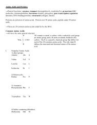
→ The major source of amino acids is the diet. Humans can only synthesize 11 of the <strong>20</strong> common amino acids.<br />
The other 9 (H I L K M F T W V) are essential. Arginine is essential only during growth. Tyr is not essential,<br />
but only because it can be readily synthesized from the essential Phe.<br />
→ No special storage compartment- all are in functional proteins-last to use as energy source<br />
→Many of the amino acids can be synthesized from pyruvate,PEP (glycolysis), 3-phosphoglycerate (glycolysis)<br />
or TCA cycle intermediates (see figure 16.14)<br />
Example: Pyruvate ⇔ alanine in 1 step<br />
a-ketoglutarate ⇔ Glu →→→Arg<br />
→ The amino acids are degraded by <strong>20</strong> different pathways that converge to just 7 metabolic intermediates (see<br />
figure 21.13). These intermediates enter the TCA cycle to produce energy or be converted into glucose.<br />
→Gly and three carbon to pyruvate<br />
→4 carbon to oxaloacetate<br />
→5 carbon to a-ketoglutarate<br />
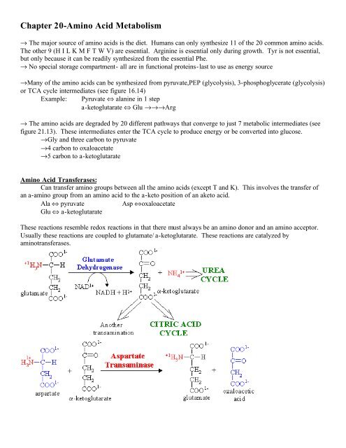
<strong>Amino</strong> <strong>Acid</strong> Transferases:<br />
Can transfer amino groups between all the amino acids (except T and K). This involves the transfer of<br />
an a-amino group from an amino acid to the a-keto position of an aketo acid.<br />
Ala ⇔ pyruvate<br />
Asp ⇔oxaloacetate<br />
Glu ⇔ a-ketoglutarate<br />
These reactions resemble redox reactions in that there must always be an amino donor and an amino acceptor.<br />
Usually these reactions are coupled to glutamate/ a-ketoglutarate. These reactions are catalyzed by<br />
aminotransferases.
Transport of nitrogen:<br />
→ Apoptosis- programmed cell death- and protein turnover (majority of proteins turn over every few<br />
days) lead to an excess of NH 3 as proteins are degraded. Free NH 3 is toxic . Most NH 3 is transported in blood<br />
as Gln or Ala (from the alanine cycle) to the liver.<br />
Urea Cycle<br />
Urea is produced in a 5 step process in the liver. The urea cycle was proposed by Krebs. Nitrogens in urea<br />
come from NH 3 and Asp. See figure 21.12 in book for the urea cycle. The urea cycle requires 4 ATP/cycle.<br />
→Step 1: Carbamoyl phosphate is formed from ammonia, bicarbonate and ATP.
→Step 2: citrulline is formed from ornithine and carbamoyl phosphate by the enzyme ornithine<br />
transcarbamoylase.<br />
→Step 3: :arginosuccinate is formed from citrulline and Aspartic acid via a citrullyl-AMP intermediate by the<br />
enzyme argininosuccinate synthetase. (This step requires 2 ATP)<br />
→Step 4: Argininosuccinate is converted to fumarate (TCA intermediate) and arginine by the enzyme<br />
argininosuccinate lyase.<br />
→Step 5: Arginine is hydrolyzed to form ornithine and urea by the enzyme arginase. The ornithine can then<br />
combine with carbmoyl phosphate to begin the cycle again.<br />
→ Note the "linking" of the Urea Cycle and the TCA cycle (the so-called Krebbs "bicycle").<br />
→Steps 1 and 2 occur in the mitochondria, and steps 3-5 occur in the cytosol. There is a citruline/ornithine<br />
exchange transpoter in the inner mitochondrial membrane.<br />
→The urea then travels to the kidneys for filtration and excretion.<br />
→The main regulation is controlled by need and diet. A high protein diet and starvation activate the cycle.<br />
N-acetylglutamate activates carbamoyl phosphate synthase.<br />
Metabolic Disorders:<br />
NH 3 is toxic because excess NH 3 converts a-ketoglutarate to Glu. This results in a break in the TCA<br />
cycle, reduced ATP, and leads to coma and death.<br />
→The therapy to treat any metabolic disorders that result in a decrease in the urea cycle are (1) limit<br />
protein intake, (2) remove excess NH3 with a compound the will bind the NH3 and be excreted, and (3) replace<br />
TCA cycle intermediates.<br />
Tyrosine Biosynthesis
Phe is converted to Tyr by phenylalanine hydroxylase. This is the biosynthetic pathway for making Tyr,<br />
and this is the first step in the degradation of Phe.<br />
→Phenylketonuria- autosommal recessive disorder in which phenylalanine hydroxylase is deficient.<br />
The Phe can be degraded so it is converted to phenylpyruvate (transaminase reaction) which build up in the<br />
body and causes irreversible brain damage.<br />
<strong>Amino</strong> <strong>Acid</strong> Derivatives<br />
Tyr → Dopamine (neurotransmitter)- excess dopamine is responsible for psychotic symptoms and<br />
schizophrenia, while lowered production of dopamine is a factor in Parkinson’s disease.<br />
→Norpinephrine (neurotransmitter)<br />
→Epinephrine (adrenaline) (neurotransmitter)<br />
These three processes occur in the brain (see figure 26.50 for structures)<br />
Tyr<br />
Trp<br />
→Melanin- skin pigment<br />
→thyroid hormone- stimulates β-oxidation in cells<br />
→Seratonin- neurotransmitter in brain- causes contraction of smooth muscles of arterioles and<br />
bronchioles (sleep inducing)<br />
→Melatonin –produced in the brain- sleep inducing<br />
→↑ amino acids in blood ↓ Trp to brain ↑ wakefulness<br />
→↑carbohydrates causes ↑ Trp to brain ↑ sleepiness<br />
Gly<br />
Glutamate<br />
His<br />
→inhibitory neurotransmitter<br />
→converted to GABA-inhibitory neurotransmitter<br />
→histamine (decarboxylation)