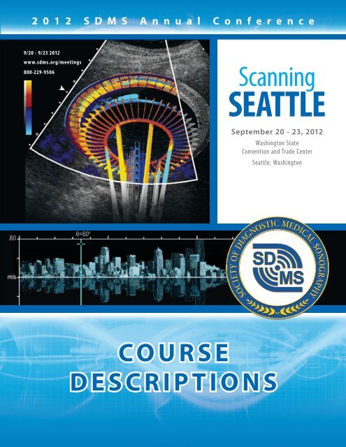
Course Descriptions - Society of Diagnostic Medical Sonography
Course Descriptions - Society of Diagnostic Medical Sonography
Course Descriptions - Society of Diagnostic Medical Sonography

Create successful ePaper yourself
Turn your PDF publications into a flip-book with our unique Google optimized e-Paper software.
2 0 1 2 S D M S A n n u a l C o n f e r e n c e<br />
9/20 - 9/23 2012<br />
www.sdms.org/meetings<br />
800-229-9506<br />
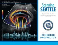
Scanning<br />
SEATTLE<br />
September 20 - 23, 2012<br />
Washington State<br />
Convention and Trade Center<br />
Seattle, Washington<br />
COURSE<br />
DESCRIPTIONS
Thursday Tutorials<br />
September 20, 2012<br />
8:00 am - 9:30 am<br />
Registry Review<br />
Registry Review: Physics (TH-7)<br />
SDMS CME Credits: 1.5 (SPI)<br />
Tammy Stearns, MS, RDMS, RVT, RT(R)<br />
Physics concepts will be simplified combining theory<br />
with application to current clinical practice. Key topics<br />
will include sound wave, pulsed sonography, artifacts<br />
and image resolution variables, transducers, and<br />
instrumentation. Models and experiments will be utilized<br />
to allow an active learning experience. Questions will<br />
be posed throughout the presentation to allow the<br />
participant to test themselves.<br />
9:45 am - 11:15 am<br />
Registry Review: Doppler, Hemodynamics & Quality<br />
Assurance (TH-14)<br />
SDMS CME Credits: 1.5 (SPI)<br />
Sheryl Goss, MS, RDMS, RDCS, RVT, RT(R)(S)<br />
This presentation will emphasize the relationship<br />
between circulatory hemodynamics, spectral waveforms,<br />
and color Doppler characteristics. Image optimization<br />
techniques and pitfalls will be illustrated to enhance<br />
clinical practice, and build confidence to take a national<br />
certification exam. Quality assurance phantom testing<br />
and implementing best practices to minimize potential<br />
bioeffects will also be discussed.<br />
Breakout: General Registry Review<br />
12:45 pm - 2:15 pm<br />
General Registry Review: Gynecology (TH-21)<br />
SDMS CME Credits: 1.5 (OB)<br />
Jill Trotter, BS, RDMS, RVT, RT(R)<br />
This review session will utilize an image-based<br />
approach to provide an overview <strong>of</strong> gynecological<br />
findings in sonography. In addition to the sonographic<br />
characteristics, the review will include risk factors, clinical<br />
signs and symptoms, basic pathophysiology and protocol<br />
modifications for facilitating diagnosis.<br />
2:30 pm - 4:00 pm<br />
General Registry Review: Obstetrics (TH-28)<br />
SDMS CME Credits: 1.5 (OB)<br />
Joie Burns, MS, RDMS, RVT , RT (R)(S)<br />
This review will utilize an image-based approach to<br />
provide an overview <strong>of</strong> fetal abnormalities. In addition<br />
to the sonographic characteristics, the review will<br />
include risk factors, clinical signs and symptoms,<br />
basic embryologic and genetic considerations,<br />
pathophysiology and protocol modifications for<br />
facilitating diagnosis.<br />
4:15 pm - 5:45 pm<br />
General Registry Review: Abdominal (TH-29)<br />
SDMS CME Credits: 1.5 (AB)<br />
Kathryn Kuntz, MEd, RDMS, RVT, RT(R), FSDMS<br />
This review will utilize an image-based approach to<br />
the pathophysiology <strong>of</strong> the major abdominal organs<br />
(hepatobiliary system, kidneys, spleen) and great vessels.<br />
Participants will be challenged to identify risk factors,<br />
clinical signs and symptoms, lab values, and to recognize<br />
sonographic characteristics for the topics presented.<br />
Breakout: Vascular Registry Review<br />
12:45 pm - 2:15 pm<br />
Vascular Registry Review: Cerebrovascular (TH-31)<br />
SDMS CME Credits: 1.5 (VT)<br />
Sheryl Goss, MS, RDMS, RDCS, RVT, RT(R)(S)<br />
This presentation will detail extracranial and intracranial<br />
circulatory assessment and associated pathological<br />
findings. Risk factors, signs and symptoms, sonographic<br />
techniques, and diagnostic criteria will be shared. Images<br />
demonstrating the various pathologic findings will be<br />
utilized to enhance the participant’s knowledge.<br />
2:30 pm - 4:00 pm<br />
Vascular Registry Review: Arterial (TH-32)<br />
SDMS CME Credits: 1.5 (VT)<br />
Marsha Neumyer, BS, RVT, FSDMS, FSVU, FAIUM<br />
This discussion <strong>of</strong> noninvasive peripheral arterial<br />
evaluations will address indirect physiologic testing and<br />
duplex sonographic assessment <strong>of</strong> native arteries and<br />
arterial bypass grafts. Specific attention will be given to<br />
technical applications, waveform analysis, and current<br />
interpretive criteria. Images will be utilized to illustrate<br />
risk factors, mechanisms <strong>of</strong> disease, signs and symptoms,<br />
treatment methods and the correlative studies that<br />
are most commonly associated with peripheral arterial<br />
disorders.<br />
4:15 pm - 5:45 pm<br />
Vascular Registry Review: Venous and Abdominal<br />
Vasculature (TH-33)<br />
SDMS CME Credits: 1.5 (VT)<br />
Joie Burns, MS, RDMS, RVT , RT (R)(S)<br />
This presentation will provide an overview <strong>of</strong> venous<br />
testing for thrombosis and insufficiency. Vascular<br />
assessment <strong>of</strong> the abdomen is also presented in<br />
2
this session. Clinical indications, scan techniques,<br />
and pathological conditions with their correlating<br />
sonographic appearances will be demonstrated through<br />
use <strong>of</strong> images.<br />
8:00 am - 9:30 am<br />
Student Conclave<br />
ARDMS Mock Discipline Hearing (TH-1)<br />
SDMS CME Credits: 1.5 (OT)<br />
Gwen Henderson<br />
Hilary Wilson<br />
Three mock discipline hearings will be held, each<br />
lasting about 15 minutes. The first hearing will involve<br />
a sonographer alleged to have cheated during an<br />
examination. The second hearing will involve a<br />
sonographer alleged to have misrepresented themselves<br />
as ARDMS certified (when in fact they were not), and<br />
the third hearing will involve a sonographer who has<br />
reported several criminal convictions related to substance<br />
abuse (alcohol, drugs).<br />
9:45 am - 11:15 am<br />
Signs in <strong>Sonography</strong> (TH-8)<br />
SDMS CME Credits: 1.5 (OT)<br />
Catherine Rienzo, MS, RDMS, RT(R)<br />
Marianna Desmond, MEd, RDMS, RT (R)<br />
What does Mickey Mouse, Yin-Yang and a lemon have<br />
in common? Certain diseases show classic sonographic<br />
signs that resemble various types <strong>of</strong> food items and<br />
inanimate objects. In this presentation various signs are<br />
discussed and correlated with in a pictorial way. These<br />
signs” are highly memorable.<br />
12:45 pm - 2:15 pm<br />
Mind Body & Soul, Transition from Academic to<br />
Clinical World (TH-15)<br />
SDMS CME Credits: 1.5 (OT)<br />
Patty Braga, MSEd, RT(R), RDMS, RDCS, RVT<br />
Stephanie Wilson, BS, RDMS, RVT<br />
This lecture will be an interactive session with students<br />
to help them transition from the academic world into<br />
the clinical world. We will begin with an interview <strong>of</strong><br />
pr<strong>of</strong>essionalism and how it impacts patient outcomes.<br />
The focus is to become aware <strong>of</strong> the whole picture<br />
when they present themselves to anyone in the facility<br />
from patients to physicians. Adequate communication<br />
is an essential aspect <strong>of</strong> healthcare and is the key<br />
to exceptional patient care. Determining how to<br />
communicate from the patients points <strong>of</strong> view and<br />
learning some good communication skill techniques can<br />
make not only the patients but your day a more pleasant<br />
experience.<br />
2:30 pm - 4:00 pm<br />
Q & A: Where Do We Go From Here? (TH-22)<br />
SDMS CME Credits: 1.5 (OT)<br />
Catherine Rienzo, MS, RDMS, RT(R),<br />
Alia Grattan, RDMS, RVT,<br />
Joy Guthrie, PhD, RDMS, RDCS, RVT, RCS, RCCS, RVS, FSDMS,<br />
and Patty Braga, MSEd, RT(R), RDMS, RDCS, RVT<br />
A panel <strong>of</strong> experienced educators will explore topics <strong>of</strong><br />
interest to students. This question and answer session<br />
will help students prepare for an exciting career in<br />
diagnostic medical sonography. The panel will discuss<br />
the students’ career path in the ever-changing medical<br />
environment.<br />
4:15 pm - 5:45 pm<br />
Interesting Cases (TH-30)<br />
SDMS CME Credits: 1.5 (OT)<br />
Phillip Bendick, PhD, RVT, FSDMS<br />
Margaret Park, BS, RDCS, RVT, FASE<br />
Robert DeJong, RDMS, RDCS, RVT, FSDMS<br />
Jill Trotter, BS, RDMS, RVT, RT(R)<br />
This presentation will include interesting cases<br />
presented by SDMS Faculty and Board Members.<br />
Cases will be presented in multiple specialties<br />
including: Abdominal, Cardiac, Ob/Gyn, and Vascular<br />
sonography.<br />
Note: This course will include attendees from all<br />
Thursday Tutorials except those attending the<br />
Registry Review Tutorial.<br />
8:00 am - 9:30 am<br />
Educators Tutorial<br />
Legal Issues Involved in Tough Academic Decisions<br />
about Students - Part I (TH-2)<br />
SDMS CME Credits: 1.5 (OT)<br />
Steve Milam, JD<br />
Educators at many levels are charged with the<br />
responsibility for the evaluation <strong>of</strong> student performance<br />
in clinical and pr<strong>of</strong>essional experiences. Decisions<br />
regarding satisfactory or unsatisfactory performance,<br />
especially when it results in academic dismissal, must be<br />
based on the faculty’s pr<strong>of</strong>essional judgment. Students<br />
must he apprised <strong>of</strong> their progress or lack there<strong>of</strong>.<br />
This program is designed to give participants a better<br />
understanding <strong>of</strong> how the courts view such decisions in<br />
clinical and pr<strong>of</strong>essional experience programs. Using a<br />
case study, the program will address the importance, use<br />
and role <strong>of</strong> “essential qualifications/ competencies” when<br />
using subjective pr<strong>of</strong>essional and academic judgment<br />
to assess students in each <strong>of</strong> the following difficult and<br />
tough academic decision making areas.<br />
3
9:45 am - 11:15 am<br />
Legal Issues Involved in Tough Academic Decisions<br />
about Students - Part II (TH-9)<br />
SDMS CME Credits: 1.5 (OT)<br />
Steve Milam, JD<br />
Educators at many levels are charged with the<br />
responsibility for the evaluation <strong>of</strong> student performance<br />
in clinical and pr<strong>of</strong>essional experiences. Decisions<br />
regarding satisfactory or unsatisfactory performance,<br />
especially when it results in academic dismissal, must be<br />
based on the faculty’s pr<strong>of</strong>essional judgment. Students<br />
must he apprised <strong>of</strong> their progress or lack there<strong>of</strong>.<br />
This program is designed to give participants a better<br />
understanding <strong>of</strong> how the courts view such decisions in<br />
clinical and pr<strong>of</strong>essional experience programs. Using a<br />
case study, the program will address the importance, use<br />
and role <strong>of</strong> “essential qualifications/ competencies” when<br />
using subjective pr<strong>of</strong>essional and academic judgment<br />
to assess students in each <strong>of</strong> the following difficult and<br />
tough academic decision making areas.<br />
12:45 pm - 2:15 pm<br />
Hot Topics in Education (TH-16)<br />
SDMS CME Credits: 1.5 (OT)<br />
Catherine Rienzo, MS, RDMS, RT(R)<br />
Steve Milam, JD<br />
Marianna Desmond, EdM, RDMS, RT(R)<br />
Carol Mitchell, PhD, RDMS, RDCS, RT, RVT, FSDMS, FASE<br />
A panel <strong>of</strong> experienced educators will answer questions<br />
from the audience. This interactive question and answer<br />
session will address issues raised by the audience<br />
members..<br />
2:30 pm - 4:00 pm<br />
Advanced Item Types on the ARDMS Exam (TH-23)<br />
SDMS CME Credits: 1.5 (OT)<br />
Pat Grier, BS, RT, RDMS<br />
Ellen Julian, PhD<br />
This presentation will include an interactive discussion <strong>of</strong><br />
the applications and advantages <strong>of</strong> items and test forms<br />
that utilize them. Demonstrations <strong>of</strong> the development<br />
<strong>of</strong> ‘interactive dashboard’ and ‘hotspot’ items will be<br />
presented. Examples <strong>of</strong> script concordance items will be<br />
discussed. The practical application <strong>of</strong> statistical analysis<br />
and psychometrics appropriate to these testing strategies<br />
will be discussed.<br />
4:15 pm - 5:45 pm<br />
Interesting Cases (TH-30)<br />
SDMS CME Credits: 1.5 (OT)<br />
See description on page 3.<br />
8:00 am - 9:30 am<br />
Abdominal <strong>Sonography</strong><br />
Optimizing Breast Imaging (TH-3)<br />
SDMS CME Credits: 1.5 (BR)<br />
Cindy Rapp, BS, RDMS, RDCS, FSDMS, FAIUM<br />
<strong>Sonography</strong> <strong>of</strong> the breast is extremely technique and<br />
operator dependent. We will discuss proper equipment,<br />
system optimization and proper scanning techniques.<br />
This session will also provide in-depth information on<br />
which women can benefit most from breast ultrasound,<br />
as an adjunct to mammography; illustrate the<br />
complementary roles <strong>of</strong> mammography and ultrasound;<br />
and discuss the latest developments in breast ultrasound<br />
technology and their applications.<br />
9:45 am - 11:15 am<br />
<strong>Sonography</strong> <strong>of</strong> the Shoulder (TH-10)<br />
SDMS CME Credits: 1.5 (AB)<br />
Veronica Rodriguez, RDMS<br />
This presentation will demonstrate basic hands-on<br />
scanning techniques <strong>of</strong> the shoulder. Attendees will learn<br />
to correctly identify normal musculoskeletal anatomy<br />
including: tendons, muscles, and ligaments <strong>of</strong> the<br />
shoulder.<br />
12:45 pm - 2:15 pm<br />
<strong>Sonography</strong> <strong>of</strong> Extremities (TH-17)<br />
SDMS CME Credits: 1.5 (AB)<br />
Veronica Rodriguez, RDMS<br />
This presentation will cover basic hands-on scanning<br />
techniques for the elbow, wrist, knee and ankle.<br />
Attendees will learn to correctly identify normal<br />
musculoskeletal anatomy <strong>of</strong> tendons, muscles, ligaments,<br />
within selected extremities.<br />
2:30 pm - 4:00 pm<br />
Inflammatory Conditions (TH-24)<br />
SDMS CME Credits: 1.5 (AB)<br />
Cindy Rapp, BS, RDMS, RDCS, FSDMS, FAIUM<br />
Inflammation can manifest itself in similar sonographic<br />
findings regardless <strong>of</strong> which organ is inflamed. We will<br />
discuss these findings in a wide spectrum <strong>of</strong> different<br />
inflammatory conditions such as appendicitis, Crohn’s<br />
disease, diverticulitis, acute cholecystitis, acute hepatitis,<br />
acute pyelonephritis, salpingitis, and acute epididymoorchitis.<br />
4:15 pm - 5:45 pm<br />
Interesting Cases (TH-30)<br />
SDMS CME Credits: 1.5 (OT)<br />
See description on page 3.<br />
4
8:00 am - 9:30 am<br />
Cardiac <strong>Sonography</strong><br />
Recommendations for Acquisition & Display Using 3-D<br />
Echocardiography (TH-4)<br />
SDMS CME Credits: 1.5 (AE)<br />
David Adams, RDCS, RCS, FSDMS<br />
This lecture and live scanning session will include how<br />
to assess and integrate 3-D/4-D imaging into clinical<br />
practice.<br />
9:45 am - 11:15 am<br />
Guidelines & Recommendations for the Assessment <strong>of</strong><br />
Right Heart Function (TH-11)<br />
SDMS CME Credits: 1.5 (AE)<br />
Kenneth Horton, RDCS, RCS, FASE<br />
This lecture and live scanning session will include how<br />
to integrate conventional and advanced measurements<br />
for right heart function. There will also be a discussion <strong>of</strong><br />
current guidelines.<br />
12:45 pm - 2:15 pm<br />
Guidelines & Recommendations for the Assessment <strong>of</strong><br />
Diastolic Function (TH-18)<br />
SDMS CME Credits: 1.5 (AE)<br />
Jeffrey Hill, BS, RDCS, FASE<br />
This lecture and live scanning session will include<br />
a comprehensive overview <strong>of</strong> diastolic function. A<br />
discussion <strong>of</strong> current guidelines and standards will also<br />
be part <strong>of</strong> this presentation.<br />
2:30 pm - 4:00 pm<br />
Guidelines & Recommendations for the Assessment <strong>of</strong><br />
Valvular Regurgitation (TH-25)<br />
SDMS CME Credits: 1.5 (AE)<br />
Carol Mitchell, PhD, RDMS, RDCS, RT, RVT, FSDMS, FASE<br />
This presentation will review the current accepted<br />
standards for evaluating and grading the severity <strong>of</strong> semilunar<br />
and atrioventricular valvular heart disease for both<br />
stenotic and regurgitant lesions.<br />
4:15 pm - 5:45 pm<br />
Interesting Cases (TH-30)<br />
SDMS CME Credits: 1.5 (OT)<br />
See description on page 3.<br />
8:00 am - 9:30 am<br />
Ob/Gyn <strong>Sonography</strong><br />
Interesting GYN Cases Solved with 3-D Imaging (TH-5)<br />
SDMS CME Credits: 1.5 (OB)<br />
Jill Trotter, BS, RDMS, RVT, RT(R)<br />
With the recent advances and increased utilization <strong>of</strong><br />
3-D sonographic technology, the scope <strong>of</strong> gynecological<br />
imaging is evolving. This presentation will present the use<br />
<strong>of</strong> this technology through interesting cases. From lost<br />
IUDs to uterine malformations and some unimaginable<br />
scenarios in between, the information presented will<br />
assist the sonographer in embracing this new technology<br />
as part <strong>of</strong> a routine GYN protocol.<br />
9:45 am - 11:15 am<br />
1-13 + 6 Weeks Scan: Nuchal Translucency, Nasal Bone<br />
Assessment & Beyond (TH-12)<br />
SDMS CME Credits: 1.5 (OB)<br />
Ben Buentipo, BS, RDMS<br />
This presentation will emphasize the importance <strong>of</strong> first<br />
trimester screening for chromosomal abnormalities.<br />
Maternal serum markers, diagnosis <strong>of</strong> major fetal<br />
abnormalities and other sonographic markers for<br />
aneuploidy and preeclampsia will also be discussed<br />
within the presentation.<br />
12:45 pm - 2:15 pm<br />
Advanced Fetal Abnormalities (TH-18)<br />
SDMS Credits: 1.5 (OB)<br />
Julia Solomon, MD<br />
This presentation will describe a more detailed evaluation<br />
<strong>of</strong> selected aspects <strong>of</strong> fetal anatomy with emphasis on<br />
common yet subtle abnormalities. The thought process<br />
behind further evaluation <strong>of</strong> these abnormalities is<br />
reviewed, along with the mechanism <strong>of</strong> arriving at a<br />
differential diagnosis. Finally, select case examples <strong>of</strong><br />
major malformation sequences that were missed on<br />
initial examinations are shown, along with images<br />
depicting the more detailed referral examination and<br />
ultimate diagnosis.<br />
2:30 pm - 4:00 pm<br />
Assessing Fetal Anatomy with 3-D/4-D (TH-26)<br />
SDMS CME Credits: 1.5 (OB)<br />
Cheryl Vance, MA, RT(R)(M), RDMS, RVT<br />
3-D technology has been around since 1989…23+ years!<br />
Volume imaging is finally being embraced into the<br />
routine imaging protocols <strong>of</strong> most departments. This<br />
presentation will review the latest 3-D/4-D technologies<br />
and what anatomy is most applicable for each feature.<br />
Additionally, attendees will learn how to simplify<br />
dataset manipulation to help ensure their success when<br />
interrogating volume datasets.<br />
4:15 pm - 5:45 pm<br />
Interesting Cases (TH-30)<br />
SDMS CME Credits: 1.5 (OT)<br />
See description on page 3.<br />
5
8:00 am - 9:30 am<br />
Vascular <strong>Sonography</strong><br />
Transcranial Doppler: Imaging Fundamentals (TH-6)<br />
SDMS CME Credits: 1.5 (VT)<br />
Stephanie Wilson, BS, RDMS, RVT<br />
This presentation will be an overview <strong>of</strong> what is required<br />
to perform a transcranial Doppler imaging examination.<br />
During the presentation, short segments <strong>of</strong> live scanning<br />
will be used to demonstrate the concepts presented such<br />
as patient positioning, transducer manipulation, realtime<br />
views <strong>of</strong> B-mode, color and spectral Doppler. The<br />
four transcranial imaging windows will be demonstrated<br />
along with the intracranial anatomy and presentation<br />
<strong>of</strong> pathology. A validated scanning protocol and<br />
applications for transcranial imaging will be presented.<br />
9:45 am - 11:15 am<br />
Duplex Ultrasound Abdominal Vascular Evaluation<br />
(TH-13)<br />
SDMS CME Credits: 1.5 (VT)<br />
Phillip Bendick, PhD, RVT, FSDMS<br />
This presentation will combine didactic lecture with a<br />
live demonstration <strong>of</strong> the techniques for a complete<br />
abdominal vascular examination. Emphasis will be placed<br />
on the aorto-iliac system and proper technique for<br />
aneurysmal evaluation. The lecture and demonstration<br />
will also address techniques for the mesenteric (celiac,<br />
SMA and IMA) vessels and the renal arteries using color<br />
Doppler and pulsed spectral Doppler evaluation. Normal<br />
characteristics and waveform morphology will be<br />
discussed followed by presentation <strong>of</strong> abnormal anatomy<br />
and waveforms as well as the possible causes for the<br />
abnormalities.<br />
12:45 pm - 2:15 pm<br />
Vascular Laboratory Markers <strong>of</strong> Cardiovascular Risk<br />
(TH-20)<br />
SDMS Credits: 1.5 (VT)<br />
Marsha Neumyer, BS, RVT, FSDMS, FSVU, FAIUM<br />
Historically, noninvasive vascular testing has been used<br />
to detect, localize and define the severity <strong>of</strong> arterial<br />
and venous disorders. In recent years, investigators<br />
have demonstrated that a number <strong>of</strong> these tests have<br />
predictive value for cardiovascular risk. This presentation<br />
will address the technical applications and use <strong>of</strong> intimalmedia<br />
thickness measurements, brachial reactivity<br />
studies and a calculated ankle-brachial index as markers<br />
for cardiovascular risk assessment.<br />
2:30 pm - 4:00 pm<br />
Duplex Arterial Evaluation for Lower Extremities<br />
(TH-27)<br />
SDMS CME Credits: 1.5 (VT)<br />
Kari Olmstead Campbell, BS, RVT<br />
This presentation will focus on sonographic techniques,<br />
interpretive criteria and the specific role <strong>of</strong> ultrasound<br />
for evaluating patients for suspected arterial<br />
occlusive disease prior to arterial intervention and<br />
revascularization.<br />
4:15 pm - 5:45 pm<br />
Interesting Cases (TH-30)<br />
SDMS CME Credits: 1.5 (OT)<br />
See description on page 3.<br />
Friday <strong>Course</strong> <strong>Descriptions</strong><br />
September 21, 2012<br />
7:45 am – 8:00 am<br />
President’s Address<br />
Plenary Sessions<br />
Joy Guthrie, PhD, RDMS, RDCS, RVT, RCS, RCCS, RVS, FSDMS<br />
8:00 am – 8:50 am<br />
Keynote Presentation:<br />
Turn Your Passion into Action! (FR-36)<br />
SDMS CME Credits: 1 (OT)<br />
Greg Bennick<br />
Greg Bennick speaks about Ideas, Impact and Innovation.<br />
He makes people laugh while inviting them to think. He<br />
speaks about getting involved in ideas, leadership, and<br />
creativity. Greg does more than just make people laugh:<br />
he empowers and inspires. He has been called upon to<br />
be a guest speaker before an enormous range <strong>of</strong> clients<br />
from all walks <strong>of</strong> life, including TEDxPugetSound (an idea<br />
conference devoted to sharing innovation), Fortune 500<br />
companies, major universities, and public audiences<br />
worldwide.<br />
9:00 am – 9:50 am<br />
McLaughlin Memorial Lecture:<br />
Where Have We Been? Where Are We Now?<br />
Where Are We Going? (FR-37)<br />
SDMS CME Credits: 1 (OT)<br />
Marsha Neumyer, BS, RVT, FSDMS, FSVU, FAIUM<br />
Together we will take a fun journey through the past<br />
and into the present and catch a glimpse <strong>of</strong> the future<br />
for the <strong>Society</strong> <strong>of</strong> <strong>Diagnostic</strong> <strong>Medical</strong> <strong>Sonography</strong> and<br />
6
ecall the people who have played key roles in our history.<br />
During the journey, we’ll explore the hills, the valleys and<br />
some <strong>of</strong> the rocky roads that brought our organization<br />
to its current place <strong>of</strong> leadership in the field <strong>of</strong> medical<br />
ultrasound. Along the way, we’ll capture a vision <strong>of</strong> what<br />
the future might have in store and the pr<strong>of</strong>essional<br />
colleagues who will take us there.<br />
10:40 am – 11:30 am<br />
General Session: Sonographer & Physicians Relations<br />
– A Working Partnership for Patient Care (FR-38)<br />
SDMS CME Credits: 1 (OT)<br />
Beverly Hashimoto, MD<br />
Winslow “Ted” Whitten, BA, RDMS, RVT<br />
This session will address the unique relationship between<br />
clinically practicing sonographers and the physicians<br />
with whom they work. Sonographers apply independent<br />
pr<strong>of</strong>essional judgment to adapt the diagnostic medical<br />
examination to optimize examination results. Results<br />
<strong>of</strong> the ultrasound examination are then provided to the<br />
physician in either written or oral form as preliminary<br />
findings. The physician is then responsible for making the<br />
medical diagnosis and initiating the plan <strong>of</strong> treatment.<br />
Sonographers provide their services working under the<br />
supervision <strong>of</strong> a referring or interpreting physician. Both<br />
parties, physician and sonographer, are dependent on<br />
each other performing their clinical responsibilities in<br />
a competent and thorough manner to insure the best<br />
possible care for their patients. Additionally, given the<br />
evolving nature <strong>of</strong> the American healthcare system, and<br />
in particular medical imaging, some physician specialty<br />
groups are now beginning to utilize some sonographers<br />
with advanced clinical skills in a physician extender role.<br />
These “Advance Practice Sonographers” or “Ultrasound<br />
Practitioners” are establishing a new and expanded<br />
relationship with their physician partners.<br />
1:30 pm - 2:20 pm<br />
Breakout Sessions<br />
Abdominal <strong>Sonography</strong> Track<br />
Abdominal Vascular Pathology (FR-39)<br />
SDMS CME Credits: 1 (AB)<br />
Edward Grant, MD<br />
Upon completion <strong>of</strong> this educational activity, the<br />
participant will demonstrate understanding <strong>of</strong> common<br />
and uncommon vascular pathology which occurs in the<br />
abdomen.<br />
Cardiac <strong>Sonography</strong> Track<br />
Methods Used in the Evaluation <strong>of</strong> Systolic Function<br />
(FR-40)<br />
SDMS CME Credits: 1 (AE)<br />
Kenneth Horton, RDCS, RCS, FASE<br />
This lecture will include basic and advanced methods <strong>of</strong><br />
assessing ventricular systolic function.<br />
Ob/Gyn <strong>Sonography</strong> Track<br />
Case Studies in High Risk Obstetrics (FR-41)<br />
SDMS CME Credits: 1 (OB)<br />
Charlotte Henningsen, MS, RDMS, RVT, RT(R), FSDMS, FAIUM<br />
This presentation will introduce a collection <strong>of</strong> unusual,<br />
rare, and/or complicated obstetrical anomalies involving<br />
the central nervous, cardiopulmonary, gastrointestinal,<br />
genitourinary, and/or skeletal systems. Each anomaly<br />
will be discussed within a case presentation format.<br />
Associated risk factors, pertinent laboratory data, and<br />
prognosis will also be explored.<br />
Vascular <strong>Sonography</strong> Track<br />
AAA - The Role <strong>of</strong> Duplex Ultrasound (FR-42)<br />
SDMS CME Credits: 1 (VT)<br />
R. Eugene Zierler, MD<br />
This session will review the clinical features <strong>of</strong> abdominal<br />
aortic aneurysms and the role <strong>of</strong> the vascular laboratory<br />
in diagnosis, treatment, and follow-up after open surgical<br />
or endovascular intervention. Case examples will be used<br />
to illustrate the key duplex findings after endovascular<br />
aortic aneurysm repair.<br />
2:30 pm - 3:20 pm<br />
Abdominal <strong>Sonography</strong> Track<br />
3-D <strong>Sonography</strong> in Pediatrics (FR-43)<br />
SDMS CME Credits: 1 (AB)<br />
Jennifer McDowell, MM, RDMS, RVT, RT(R)<br />
Audience will learn to articulate 3-D fundamentals. They<br />
will also learn how and when to apply 3-D to their current<br />
imaging protocol. We will also cover when to use 3-D<br />
imaging to help decrease their transducer time and ease<br />
<strong>of</strong> patient comfort..<br />
Cardiac <strong>Sonography</strong> Track<br />
Understanding Cardiac Embryology & Congenital<br />
Heart Disease (FR-44)<br />
SDMS CME Credits: 1 (AE)<br />
Carol Mitchell, PhD,RDMS, RDCS, RT, RVT, FSDMS, FASE<br />
This presentation will review the basic cardiac<br />
embryology events <strong>of</strong> looping, septation and aortic<br />
arch formation and discuss anomalies that occur due to<br />
abnormal development.<br />
Ob/Gyn <strong>Sonography</strong> Track<br />
First Trimester Fetal Echocardiography (FR-45)<br />
SDMS CME Credits: 1 (FE)<br />
Julia Solomon, MD<br />
With the earlier identification <strong>of</strong> at-risk fetuses, the need<br />
to accurately evaluate cardiac anatomy becomes more<br />
pressing at younger gestational ages. This presentation<br />
will outline the first trimester features associated with<br />
increased risks <strong>of</strong> congenital heart disease, describe<br />
7
how to accurately assess these parameters, and will<br />
demonstrate the evaluation <strong>of</strong> structural and functional<br />
cardiac parameters in the 12-14 week gestational age<br />
range. Attendees will learn to recognize which fetuses<br />
require further evaluation for congenital heart disease.<br />
Finally, the presentation will showcase some <strong>of</strong> the<br />
advantages <strong>of</strong> <strong>of</strong>fline volume dataset manipulation for<br />
the evaluation <strong>of</strong> first trimester cardiac anatomy.<br />
Vascular <strong>Sonography</strong> Track<br />
Strategic Use <strong>of</strong> Duplex Scanning for Patients with<br />
Suspected Arterial Occlusive Disease (FR-46)<br />
SDMS CME Credits: 1 (VT)<br />
R. Eugene Zierler, MD<br />
This session will cover current vascular laboratory<br />
approaches to the evaluation <strong>of</strong> peripheral arterial<br />
disease, with emphasis on the patient who may<br />
be a candidate for open surgical or catheter-based<br />
intervention. Use <strong>of</strong> vascular testing for surveillance after<br />
arterial interventions will also be discussed.<br />
4:10 pm - 5:00 pm<br />
Abdominal <strong>Sonography</strong> Track<br />
Case Studies in Abdominal <strong>Sonography</strong> (FR-47)<br />
SDMS CME Credits: 1 (AB)<br />
Charlotte Henningsen, MS, RDMS, RVT, RT(R), FSDMS, FAIUM<br />
Imaging <strong>of</strong> the adult abdomen can be very complex, as<br />
patient symptoms can be misleading which can lead<br />
to an unexpected diagnosis or unique presentation<br />
<strong>of</strong> a particular disease or pathology. This presentation<br />
will introduce a collection <strong>of</strong> unusual, rare, and/or<br />
complicated anomalies <strong>of</strong> the abdomen. Each anomaly<br />
will be discussed in a case presentation format.<br />
Correlative imaging, pertinent laboratory date, and<br />
prognosis will also be explored.<br />
Cardiac <strong>Sonography</strong> Track<br />
3 <strong>of</strong> a Kind Interactive Game (FR-48)<br />
SDMS CME Credits: 1 (AE)<br />
David Adams, RDCS, RCS, FSDMS<br />
This interactive game will allow sonographer to apply<br />
their critical thinking skills in specific cases. The game<br />
will be used as a learning tool to showcase a variety <strong>of</strong><br />
cardiovascular diseases.<br />
Ob/Gyn <strong>Sonography</strong> Track<br />
3-D Imaging in Gynecology (FR-49)<br />
SDMS CME Credits: 1 (OB)<br />
Ben Buentipo, BS, RDMS<br />
This presentation will emphasize the potential benefit <strong>of</strong><br />
the 3-D approach in gynecology. The attendee will learn<br />
a segmental approach to applying the 3-D technique and<br />
learn to assess normal gynecologic anatomy, common<br />
pelvic abnormalities, and normal variants with this<br />
method.<br />
Vascular <strong>Sonography</strong> Track<br />
Cerebrovascular <strong>Sonography</strong>: Beyond the Bifurcation<br />
(FR-50)<br />
SDMS CME Credits: 1 (VT)<br />
Edward Grant, MD<br />
Attendees will develop understanding <strong>of</strong> common and<br />
uncommon pathologic conditions and imaging features<br />
which occur in the carotid and vertebral arteries that are<br />
not related to internal carotid artery stenosis.<br />
Saturday <strong>Course</strong> <strong>Descriptions</strong><br />
September 22, 2012<br />
8:00 am - 8:50 am<br />
Breakout Sessions<br />
Abdominal <strong>Sonography</strong> Track<br />
Elastography <strong>of</strong> the Breast: The Basics (SA-51)<br />
SDMS CME Credits: 1 (BR)<br />
Dennis McDonald, MD<br />
Elastography is a new sonographic adjuvant<br />
to conventional breast sonography that aids in<br />
characterizing a variety <strong>of</strong> breast conditions. This<br />
presentation will review the basic principles and types<br />
<strong>of</strong> elastography commercially available. Information will<br />
be provided on how your breast center can utilize this<br />
technique in each breast sonography examination.<br />
Cardiac <strong>Sonography</strong> Track<br />
Fetal Echocardiography: Physiologic Complications in<br />
the Womb (SA-52)<br />
SDMS CME Credits: 1 (FE)<br />
Joy Guthrie, PhD, RDMS, RDCS, RVT, RCS, RCCS, RVS, FSDMS<br />
This presentation will take a unique look at circulation<br />
starting from the maternal uterus through the fetal<br />
circulation. Both normal and abnormal physiology in the<br />
fetal echocardiogram will be described using this lens.<br />
8
Ob/Gyn <strong>Sonography</strong> Track<br />
Look Alikes in the Pelvis (SA-53)<br />
SDMS CME Credits: 1 (OB)<br />
Sandra Allison, MD<br />
A variety <strong>of</strong> benign conditions that occur in the pelvis<br />
can mimic malignant pathology and vice versa. Pointers<br />
on distinguishing between these benign and malignant<br />
entities will be presented.<br />
Vascular <strong>Sonography</strong> Track<br />
Diagnosis <strong>of</strong> Venous Valvular Incompetence (SA-54)<br />
SDMS CME Credits: 1 (VT)<br />
Jennifer McDowell, MM, RDMS, RVT, RT(R)<br />
This presentation will help participants understand<br />
treatment options for the patient. Attendees will learn<br />
what the physician will need to treat the venous disease<br />
(depending upon treatment option being utilized).<br />
Evaluation <strong>of</strong> clinical signs <strong>of</strong> Venous reflux disease will<br />
also be covered.<br />
9:00 am - 9:50 am<br />
Abdominal <strong>Sonography</strong> Track<br />
3-D Breast <strong>Sonography</strong>: What Have We Been Missing?<br />
(SA-55)<br />
SDMS CME Credits: 1 (BR)<br />
Dennis McDonald, MD<br />
Historically breast sonography has had a limited role<br />
in the evaluation <strong>of</strong> mammographic and clinical breast<br />
abnormalities. This presentation will review breast<br />
sonography anatomy and will describe the technique <strong>of</strong><br />
3-D acquisition and display. We will demonstrate how 3-D<br />
breast sonography will not only improve malignant lesion<br />
characterization and staging, but also how it will improve<br />
breast sonography workflow and department efficiency.<br />
Cardiac <strong>Sonography</strong> Track<br />
What’s New in Contrast Imaging? Beyond the LV<br />
(SA-56)<br />
SDMS CME Credits: 1 (AE)<br />
J. Todd Belcik, BS, RDCS, RCS, FASE<br />
This lecture will feature a review <strong>of</strong> new research <strong>of</strong><br />
perfusion imaging using contrast-enhanced sonography..<br />
Ob/Gyn <strong>Sonography</strong> Track<br />
<strong>Sonography</strong> in Assisted Reproductive Technologies:<br />
It’s More Than Counting Follicles (SA-57)<br />
SDMS CME Credits: 1 (OB)<br />
Paul Zarutskie, MD<br />
This presentation will review both the diagnostic<br />
and therapeutic use <strong>of</strong> ultrasound technologies<br />
necessary to achieve a pregnancy. Additionally, the<br />
latest advancements in ultrasound technology will be<br />
presented to motivate the attendee to go beyond the<br />
basics to assess the potential <strong>of</strong> color flow modalities and<br />
state-<strong>of</strong>-the-art technologies to improve embryo transfer<br />
technologies. The attendees will be asked to share<br />
their visions <strong>of</strong> an idealistic/futuristic role <strong>of</strong> ultrasound<br />
technologies in reproductive medicine.<br />
Vascular <strong>Sonography</strong> Track<br />
Sonographic Evaluation <strong>of</strong> Liver & Kidney Transplants<br />
(SA-58)<br />
SDMS CME Credits: 1 (VT)<br />
Edward Grant, MD<br />
The attendee will develop an understanding <strong>of</strong><br />
sonographic techniques as they are applied to pathologic<br />
processes affecting the transplanted liver and kidney.<br />
The will also learn to image their findings.<br />
10:40 am - 11:30 am<br />
Abdominal <strong>Sonography</strong> Track<br />
Musculoskeletal <strong>Sonography</strong>: Applications for Upper<br />
Extremities (SA-59)<br />
SDMS CME Credits: 1 (AB)<br />
Sandra Allison, MD<br />
The use <strong>of</strong> ultrasound in evaluating common pathologic<br />
conditions affecting the upper extremity will be<br />
presented. This includes rotator cuff tears, ligamentous<br />
pathology about the elbow, and traumatic and<br />
inflammatory conditions that afflict the wrist and hands.<br />
Cardiac <strong>Sonography</strong> Track<br />
Catheterization 101: What Are We Measuring & Why<br />
Are We Measuring it? (SA-60)<br />
SDMS CME Credits: 1 (AE)<br />
Gerard Aurigemma, MD, FASE<br />
This lecture will provide insight to procedures and<br />
measurements performed in the catheterization lab.<br />
Ob/Gyn <strong>Sonography</strong> Track<br />
Fetal Anomalies Associated with Maternal Disease<br />
(SA-61)<br />
SDMS CME Credits: 1 (OB)<br />
Armando Fuentes, MD, MBA<br />
This presentation will cover maternal diseases which<br />
have an impact on pregnancy and fetal development.<br />
The diseases covered include, maternal diabetes,<br />
hypertension, thyroid disorders, hematologic disorders<br />
and infections. The assessment <strong>of</strong> these high-risk fetuses<br />
will be discussed to provide the sonographer with tips<br />
and techniques to properly evaluate the pregnancy.<br />
9
Vascular <strong>Sonography</strong> Track<br />
Hemodialysis Access: Pre and Post-Placement<br />
Assessment (SA-62)<br />
SDMS CME Credits: 1 (VT)<br />
Stephanie Wilson, BS, RDMS, RVT<br />
This presentation is designed to introduce the main<br />
concepts used for evaluation <strong>of</strong> patients in need <strong>of</strong><br />
hemodialysis. Research has proven that a thorough<br />
established program to evaluate patients both pre and<br />
post operatively increases the probability <strong>of</strong> a successful<br />
dialysis access and improved patient outcomes. The<br />
session will demonstrate the fundamentals for a preoperative<br />
patient assessment with ultrasound including a<br />
description <strong>of</strong> the various types <strong>of</strong> access conduits. Then<br />
the post-operative evaluation and common pathologies<br />
affecting hemodialysis dysfunction will be described<br />
including some sample cases.<br />
1:30 pm - 2:20 pm<br />
Abdominal <strong>Sonography</strong> Track<br />
Musculoskeletal <strong>Sonography</strong>: Applications for Lower<br />
Extremities (SA-63)<br />
SDMS CME Credits: 1 (AB)<br />
Sandra Allison, MD<br />
The use <strong>of</strong> ultrasound in evaluating common<br />
pathologic conditions affecting the lower extremity<br />
will be presented. This includes hip and knee effusions,<br />
tendinous and ligamentous pathology about the knee<br />
and ankle, and traumatic and inflammatory conditions<br />
that afflict the ankles and feet.<br />
Cardiac <strong>Sonography</strong> Track<br />
What Measurements Are Needed to Diagnose<br />
Pericardial Disease? (SA-64)<br />
SDMS CME Credits: 1 (AE)<br />
J. Todd Belcik, BS, RDCS, RCS, FASE<br />
This lecture will include basic and advanced methods <strong>of</strong><br />
assessing and diagnosing pericardial disease.<br />
Ob/Gyn <strong>Sonography</strong> Track<br />
Bioethics <strong>of</strong> Reproductive Medicine: From Stem Cells<br />
to Babies (SA-65)<br />
SDMS CME Credits: 1 (OB)<br />
Paul Zarutskie, MD<br />
Reproductive medicine is associated with much ethical<br />
controversy. This presentation will educate and inform<br />
attendees <strong>of</strong> the medical, political, and religious cross<br />
road we are facing. This presentation will review the<br />
pros and cons while not making political or emotional<br />
opinions. It will review what it means, as a pr<strong>of</strong>essional,<br />
to be a moral agent and maintain our roles as educators,<br />
whether we are physicians, instructors, or medical<br />
students.<br />
Vascular <strong>Sonography</strong> Track<br />
De-Mystifying Physiologic Arterial Testing (SA-66)<br />
SDMS CME Credits: 1 (VT)<br />
Robert Daigle, BA, RVT, FSVU<br />
Traditional, indirect physiologic arterial testing can<br />
determine the presence <strong>of</strong> peripheral arterial disease,<br />
and its severity. Although color duplex ultrasound, in the<br />
hands <strong>of</strong> an experienced operator, can detect occlusive<br />
disease and provide anatomic information, this method<br />
cannot assess the hemodynamic impact <strong>of</strong> the disease on<br />
distal limb perfusion. The objective <strong>of</strong> this presentation is<br />
to explain test methods, instrumentation and benefits <strong>of</strong><br />
indirect arterial testing.<br />
2:30 pm - 3:10 pm<br />
Abdominal <strong>Sonography</strong> Track<br />
Thyroid <strong>Sonography</strong> (SA-67)<br />
SDMS CME Credits: 1 (AB)<br />
Dennis McDonald, MD<br />
Patients are <strong>of</strong>ten referred for evaluation <strong>of</strong> a variety <strong>of</strong><br />
clinical presentations relating to the thyroid gland. This<br />
presentation will review normal thyroid sonographic<br />
anatomy and the common sonographic features <strong>of</strong><br />
malignant and benign nodules. We will review how<br />
optimizing the image and using the latest advancements<br />
in sonographic technology we can more accurately<br />
demonstrate, characterize, and display the pathology that<br />
is present.<br />
Cardiac <strong>Sonography</strong> Track<br />
Speckle Tracking Echocardiography for the<br />
Sonographer (SA-68)<br />
SDMS CME Credits: 1 (AE)<br />
Daniel Bourque, MS, RCS<br />
Speckle tracking is an advanced tool sonographers<br />
utilize to assess myocardial mechanics. Advances in<br />
the use <strong>of</strong> speckle tracking in both the atrium and<br />
ventricle are being researched and implemented in many<br />
echocardiography laboratories. This presentation will<br />
include an introduction to speckle tracking cincepts, case<br />
presentations, and technical tips.<br />
Ob/Gyn <strong>Sonography</strong> Track<br />
Fetal Gastrointestinal Anomalies (SA-69)<br />
SDMS CME Credits: 1 (OB)<br />
Armando Fuentes, MD, MBA<br />
This presentation will cover anomalies associated with<br />
the fetal gastrointestinal tract. Images will be used to<br />
demonstrate the sonographic appearance. Attendees<br />
will learn the abnormalities that are <strong>of</strong>ten seen in<br />
association with gastrointestinal anomalies. Information<br />
on the anticipated outcomes for the affected fetuses<br />
will be discussed to assist the attendee in assessing the<br />
severity <strong>of</strong> the abnormality identified.<br />
10
Vascular <strong>Sonography</strong> Track<br />
Duplex Evaluation <strong>of</strong> Arterial Stents (SA-70)<br />
SDMS CME Credits: 1 (VT)<br />
Kari Olmstead Campbell, BS, RVT<br />
This presentation will focus on sonographic techniques,<br />
latest interpretive criteria and key elements in scanning<br />
patients with and without stents in the various areas <strong>of</strong><br />
the vascular system.<br />
4:00 pm - 4:50 pm<br />
Abdominal <strong>Sonography</strong> Track<br />
Sonographic Imaging for Pediatric Abdominal<br />
Emergencies (SA-71)<br />
SDMS CME Credits: 1 (AB)<br />
Teresa Chapman, MD<br />
This presentation on pediatric abdominal emergencies<br />
will cover commonly seen diseases in the emergency<br />
setting affecting children and the appropriate<br />
sonographic evaluation for these processes.<br />
Cardiac <strong>Sonography</strong> Track<br />
Evaluation <strong>of</strong> Cardiac Masses, Thrombus & Other<br />
Things That Wiggle (SA-72)<br />
SDMS CME Credits: 1 (AE)<br />
Margaret Park, BS, RDCS, RVT, FASE<br />
This presentation will demonstrate a hodgepodge <strong>of</strong><br />
masses, tumors, thrombus, extra cardiac structures,<br />
artifact and normal anatomical variations using a casebased<br />
approach. Clinical symptoms and history paired<br />
with the echocardiographic characteristics and location<br />
will be discussed in determining the possible etiology or<br />
diagnosis.<br />
Ob/Gyn <strong>Sonography</strong> Track<br />
Newest Technologies & the Impact on the Ob/Gyn<br />
Sonographer (SA-73)<br />
SDMS CME Credits: 1 (OB)<br />
Arthur Fleischer, MD<br />
This presentation will discuss the newest technologies<br />
readily available to sonography departments including<br />
multiplanar imaging, volumetric imaging and matrix<br />
array technology. The application <strong>of</strong> these technologies<br />
to daily Ob/Gyn sonography exams will be demonstrated<br />
to <strong>of</strong>fer sonographers ways to enhance the exam by using<br />
them appropriately.<br />
Vascular <strong>Sonography</strong> Track<br />
Upper and Lower Extremity Vein Testing (SA-74)<br />
SDMS CME Credits: 1 (VT)<br />
Robert Daigle, BA, RVT, FSVU<br />
Venous duplex imaging has become the gold standard<br />
for the detection <strong>of</strong> deep venous thrombosis (DVT) in<br />
the upper and lower extremities. In addition to detecting<br />
venous thrombosis, the ability <strong>of</strong> duplex imaging to<br />
identify other causes <strong>of</strong> leg and arm pain and swelling<br />
is an important test modality. The purpose <strong>of</strong> this<br />
presentation is to demonstrate techniques and protocols<br />
for venous duplex imaging with emphasis on how to<br />
resolve difficult studies and make differential diagnoses.<br />
Sunday <strong>Course</strong> <strong>Descriptions</strong><br />
September 23, 2012<br />
8:00 am - 8:50 am<br />
Breakout Sessions<br />
Abdominal <strong>Sonography</strong> Track<br />
Interesting Pediatric <strong>Sonography</strong> Cases (SU-75)<br />
SDMS CME Credits: 1 (AB)<br />
Ramesh Iyer, MD<br />
The purpose <strong>of</strong> this presentation is to present challenging<br />
sonographic cases <strong>of</strong> varied diagnoses. There will be<br />
an emphasis on the pediatric population. Differential<br />
considerations and a brief discussion about the condition<br />
in question will be presented for each case. Learning<br />
points will be <strong>of</strong>fered for selected cases from both<br />
radiologist and sonographer perspectives.<br />
Cardiac <strong>Sonography</strong> Track<br />
Interesting Cases in Cardiac <strong>Sonography</strong> (SU-76)<br />
SDMS CME Credits: 1 (AE)<br />
Daniel Bourque, MS, RCS<br />
Rarely seen pathology can <strong>of</strong>ten be overlooked and<br />
difficult to assess. Taking the correct steps in recognizing<br />
rare and <strong>of</strong>ten life threatening pathologies is vital to<br />
patient care. This lecture will include case presentations,<br />
question and answer with a practicing panel, and<br />
audience participation.<br />
Ob/Gyn <strong>Sonography</strong> Track<br />
A Recent Advance in Gynecologic <strong>Sonography</strong>:<br />
Transperineal Imaging <strong>of</strong> the Pelvic Floor (SU-77)<br />
SDMS CME Credits: 1 (OB)<br />
Arthur Fleischer, MD<br />
This presentation will demonstrate the abnormalities<br />
11
commonly experienced in the female pelvic floor and the<br />
newest imaging methods utilized to evaluate the pelvic<br />
floor. The use <strong>of</strong> sonography in evaluating the anterior<br />
and posterior compartments <strong>of</strong> the female pelvis will be<br />
discussed, including the anal sphincter and rectovaginal<br />
septum. Imaging <strong>of</strong> the urogenital hiatus and the<br />
puborectalis muscle will be presented to demonstrate<br />
the role <strong>of</strong> sonography in evaluating and treating the<br />
abnormal conditions <strong>of</strong> the pelvic floor.<br />
Vascular <strong>Sonography</strong> Track<br />
Vascular Mapping Prior to Cardiac & Extremity Bypass<br />
Procedures (SU-78)<br />
SDMS CME Credits: 1 (VT)<br />
Alia Grattan, RDMS, RVT<br />
This presentation will address the examination <strong>of</strong> venous<br />
and arterial mapping for cardiac and vascular surgery<br />
including the state-<strong>of</strong>-the art percutaneous aortic valve<br />
replacement and robotic mitral valve repair. Venous<br />
mapping <strong>of</strong> the legs and arms for lower extremity<br />
bypass, mammary artery mapping, radial artery mapping<br />
for cardiac surgery, and arterial and venous mapping<br />
procedures for percutaneous AVR and robotic MV repair<br />
will be discussed.<br />
9:00 am - 9:50 am<br />
Abdominal <strong>Sonography</strong> Track<br />
Chronic Mesenteric Ischemia (SU-79)<br />
SDMS CME Credits: 1 (AB)<br />
Phillip Bendick, PhD, RVT, FSDMS<br />
This presentation will focus on the indications, techniques<br />
and diagnostic criteria for a complete mesenteric<br />
vascular examination, arterial and venous, as it relates to<br />
symptoms <strong>of</strong> chronic ischemia. Underlying etiologies to<br />
be presented will include both atherosclerotic and nonatherosclerotic<br />
disease, venous obstruction, and vascular<br />
compression syndromes, as well as touching on other less<br />
common pathologies.<br />
Cardiac <strong>Sonography</strong> Track<br />
Cross Modality Comparison in Patients with Heart<br />
Disease (SU-80)<br />
SDMS CME Credits: 1 (AE)<br />
Gerard Aurigemma, MD, FASE<br />
This lecture will include how different imaging modalities<br />
are used to determine cardiac disease.<br />
Ob/Gyn <strong>Sonography</strong> Track<br />
<strong>Sonography</strong> <strong>of</strong> the Cervix in Pre-Term Labor (SU-81)<br />
SDMS CME Credits: 1 (OB)<br />
Armando Fuentes, MD, MBA<br />
This presentation will introduce basic sonographic<br />
techniques for imaging the cervix in pregnancy while<br />
focusing on the changes that will be seen sonographically<br />
in pre-term labor. The role <strong>of</strong> sonography in diagnosing<br />
and monitoring the pre-term labor patient’s cervix will<br />
also be presented.<br />
Vascular <strong>Sonography</strong> Track<br />
Non-Vascular Pathology for RVT (SU-82)<br />
SDMS CME Credits: 1 (VT)<br />
Robert DeJong, RDMS, RDCS, RVT, FSDMS<br />
While scanning the carotid, renal, or other vessels, the<br />
vascular sonographer may notice pathology near the<br />
vessel being examined. The purpose <strong>of</strong> this presentation<br />
is to help the sonography unfamiliar with non-vascular<br />
pathology to develop a protocol to document the<br />
pathology as well as to learn to recognize pathologies<br />
that are suspicious for malignancy.<br />
10:10 am - 11:00 am<br />
Abdominal <strong>Sonography</strong> Track<br />
CSI: Clinical Sonographic Indications for Abdominal<br />
Imaging (SU-83)<br />
SDMS CME Credits: 1 (AB)<br />
Janette Wybo, BAS, RDMS, RDCS, RVT<br />
Attendees will learn to demonstrate understanding<br />
<strong>of</strong> common and uncommon abdominal pathologic<br />
conditions by correlating clinical indications with their<br />
sonographic appearances.<br />
Cardiac <strong>Sonography</strong> Track<br />
Cases from Outside the Echo Lab (SU-84)<br />
SDMS CME Credits: 1 (AE)<br />
Margaret Park, BS, RDCS, RVT, FASE<br />
Echocardiography has become an important tool in<br />
many specialized cardiac and surgical procedures taking<br />
the sonography team outside <strong>of</strong> the confined walls <strong>of</strong><br />
the echo laboratory areas. This case based presentation<br />
will demonstrate some innovative and interesting uses<br />
<strong>of</strong> echocardiography including transthoracic (TTE),<br />
transesophageal (TEE), and intracardiac (ICE) imaging.<br />
Ob/Gyn <strong>Sonography</strong> Track<br />
Is It Fetal Bronchopulmonary Sequestration or<br />
Congenital Cystic Adenomatoid Malformation? (SU-<br />
85)<br />
SDMS CME Credits: 1 (OB)<br />
Jennifer Durant, BS, RDMS, RDCS, RVT<br />
This presentation covers the basic pathology and<br />
scanning protocols for distinguishing between<br />
brohchopulmonary sequestration and congenital cystic<br />
adenomatoid malformation. Terminology specific to both<br />
pathology processes will be introduced. The sonographer<br />
will gain information and imaging techniques through<br />
fetal images.<br />
12
Vascular <strong>Sonography</strong> Track<br />
Lets Get Connected: <strong>Sonography</strong> <strong>of</strong> Peripheral<br />
Arterial Bypass Grafts (SU-86)<br />
SDMS CME Credits: 1 (VT)<br />
Kari Olmstead Campbell, BS, RVT<br />
This presentation will focus on ultrasound techniques<br />
and protocols, interpretive criteria and key elements<br />
to include during a vascular ultrasound examination <strong>of</strong><br />
peripheral arterial bypass grafts.<br />
11:10 am - 12:00 pm<br />
Abdominal <strong>Sonography</strong> Track<br />
Biopsy Techniques for the Sonographer (SU-87)<br />
SDMS CME Credits: 1 (AB)<br />
Robert DeJong, RDMS, RDCS, RVT, FSDMS<br />
<strong>Sonography</strong> is still underutilized as an imaging<br />
modality to guide various types <strong>of</strong> pathologies. Sadly,<br />
sonographers are also not routinely part <strong>of</strong> the biopsy<br />
team or process. The purpose <strong>of</strong> this presentation is<br />
to discuss the strengths <strong>of</strong> performing biopsies under<br />
sonography guidance, and the benefits <strong>of</strong> having a<br />
sonographer involved. Also to be discussed is the role <strong>of</strong><br />
new technologies, such as fusion imaging, and how they<br />
can be beneficial to improve biopsy outcomes.<br />
Cardiac <strong>Sonography</strong> Track<br />
My Most Challenging Diastolic Dysfunction Cases<br />
(SU-88)<br />
SDMS CME Credits: 1 (AE)<br />
Jeffrey Hill, BS, RDCS, FASE<br />
Challenging diastolic function cases will be presented<br />
with analysis on how to navigate through discrepant<br />
data.<br />
Ob/Gyn <strong>Sonography</strong> Track<br />
Basic Neonatal Head (SU-89)<br />
SDMS CME Credits: 1 (NE)<br />
Tammy Stearns, MS, RDMS, RVT, RT(R)<br />
Basic neonatal care along with an explanation <strong>of</strong><br />
commonly encountered equipment will assist in<br />
familiarizing the sonographer with the Neonatal Intensive<br />
Care Unit. Anatomy and scanning techniques will be<br />
presented along with commonly identified images.<br />
(No more trying to find the Batwings and Pac-man<br />
sign!) Commonly encountered pathologies will also be<br />
presented and discussed.<br />
Vascular <strong>Sonography</strong> Track<br />
Emerging Technologies (SU-90)<br />
SDMS CME Credits: 1 (VT)<br />
Cheryl Vance, MA, RT(R)(M), RDMS, RVT<br />
This presentation will review the technologies<br />
commonly over-looked and/or under-utilized. The<br />
latest advancements in ultrasound technology will<br />
be presented to motivate the attendee to go beyond<br />
the basics to assess potential pathology by using<br />
multi-modalities and state-<strong>of</strong>-the-art technologies.<br />
Additionally, emerging technologies will be investigated<br />
to give the attendee a glimpse in to the future <strong>of</strong><br />
sonography.<br />
13