Current Trends in Echocardiography - Society of Diagnostic Medical ...
Current Trends in Echocardiography - Society of Diagnostic Medical ...
Current Trends in Echocardiography - Society of Diagnostic Medical ...

Create successful ePaper yourself
Turn your PDF publications into a flip-book with our unique Google optimized e-Paper software.
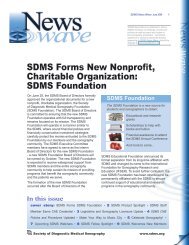
SDMS News Wave February 2010 1<br />
SDMS News Wave is published to <strong>in</strong>form<br />
SDMS members <strong>of</strong> meet<strong>in</strong>gs, events and<br />
policies as well as trends and issues <strong>in</strong><br />
the sonography pr<strong>of</strong>ession. Comments,<br />
questions or concerns about the articles<br />
appear<strong>in</strong>g <strong>in</strong> SDMS News Wave, should be<br />
directed to newswave@sdms.org.<br />
<strong>Current</strong> <strong>Trends</strong> <strong>in</strong> <strong>Echocardiography</strong><br />
Technological advances mean cardiac sonographers play a<br />
greater role <strong>in</strong> help<strong>in</strong>g to diagnose, treat heart disease<br />
By Beth W. Orenste<strong>in</strong><br />
Over the past 10 years, advances <strong>in</strong> transducer<br />
technology and image process<strong>in</strong>g have come fast<br />
and furious and have changed the practice <strong>of</strong><br />
echocardiography.<br />
Members <strong>of</strong> SDMS who specialize <strong>in</strong><br />
echocardiography say many <strong>of</strong> the recent<br />
advances are excit<strong>in</strong>g because they allow<br />
sonography to play an even greater role <strong>in</strong> the<br />
early detection and state-<strong>of</strong>-the-art treatments<br />
for heart disease and thus for patients to live<br />
longer, healthier lives.<br />
Here are some <strong>of</strong> the many trends <strong>in</strong> fetal,<br />
pediatric and adult echocardiography that SDMS<br />
members are excited about:<br />
Fetal <strong>Echocardiography</strong><br />
Sonography has been used s<strong>in</strong>ce the mid-1980s<br />
to help identify congenital heart defects dur<strong>in</strong>g<br />
the second trimester. The “basic” exam<strong>in</strong>ation<br />
<strong>of</strong> the fetal heart <strong>in</strong>volves a four-chamber view<br />
– evaluation <strong>of</strong> the right and left atrial and<br />
ventricular chambers and their respective valves.<br />
In the past several years, there has been a<br />
movement <strong>in</strong> the obstetric ultrasound community<br />
to encourage obstetric sonographers to perform<br />
In this issue<br />
cover story: <strong>Current</strong> <strong>Trends</strong> <strong>in</strong> <strong>Echocardiography</strong> • 6 Letter From the President • 7 Member Benefit<br />
Spotlight - SDMS Salary & Benefit Survey Reports • 8 SDMS Foundation Announces First 2010 Scholarship<br />
Recipients • February iPod Touch W<strong>in</strong>ner • 9 SDMS Announces the 2010 SDMS Annual Conference Sponsorship<br />
and Exhibit Program • SDMS Foundation Receives IRS Tax-Exempt Status • 10 New Member Scan • Upcom<strong>in</strong>g<br />
Web<strong>in</strong>ars • 11 SDMS Fellow Spotlight • SDMS Product Spotlight 12 SDMS Welcomes New Members<br />
LVOT<br />
“basic” exam –<br />
four-chamber view<br />
(pictured left)<br />
“extended basic”<br />
exam – cardiac<br />
outflow tracts<br />
(pictured below)<br />
RVOT<br />
the “extended basic” exam which <strong>in</strong>cludes the<br />
cardiac outflow tracts. The outflow tracts consist<br />
<strong>of</strong> the ma<strong>in</strong> pulmonary artery exit<strong>in</strong>g the right<br />
ventricle and the aorta exit<strong>in</strong>g the left ventricle.<br />
“With the proper tra<strong>in</strong><strong>in</strong>g and education,<br />
imag<strong>in</strong>g the outflow tracts and demonstrat<strong>in</strong>g<br />
their relationship to each other only <strong>in</strong>creases<br />
scann<strong>in</strong>g time by a few m<strong>in</strong>utes,” says Lisa<br />
M. Allen, BS, RDMS, RDCS, RVT, FAIUM, the<br />
<strong>Society</strong> <strong>of</strong> <strong>Diagnostic</strong> <strong>Medical</strong> Sonography<br />
www.sdms.org
Ultrasound Coord<strong>in</strong>ator for the Regional Per<strong>in</strong>atal<br />
Center at SUNY Upstate <strong>Medical</strong> University <strong>in</strong><br />
Syracuse, N.Y. “Once you obta<strong>in</strong> the apical fourchamber<br />
view <strong>of</strong> the heart, the outflow tracts can<br />
easily be obta<strong>in</strong>ed by angl<strong>in</strong>g or sweep<strong>in</strong>g the<br />
transducer toward the head <strong>of</strong> the fetus.”<br />
Add<strong>in</strong>g the outflow tracts to departmental protocol<br />
<strong>in</strong>creases the ability to detect congenital cardiac<br />
abnormalities that may not be apparent <strong>in</strong> the fourchamber<br />
view. “If you supplement the four-chamber<br />
view <strong>of</strong> the fetal heart with the addition <strong>of</strong> the outflow<br />
tracts, you can potentially <strong>in</strong>crease the detection rate<br />
<strong>of</strong> congenital heart defects from approximately 60%<br />
to 90%,” Allen says.<br />
The fetal echocardiography community also is abuzz<br />
over three-dimensional (3D) and four-dimensional<br />
(4D) ultrasound and the potential <strong>of</strong> this technology<br />
to significantly improve the evaluation <strong>of</strong> the fetal<br />
heart. Spatiotemporal Image Correlation (STIC), a 4D<br />
application, allows for evaluation and manipulation <strong>of</strong><br />
the heart <strong>in</strong> real-time, Allen says. Today’s ultrasound<br />
equipment allows sonographers to gather a volume <strong>of</strong><br />
data from which they can create detailed 3D images<br />
<strong>of</strong> the t<strong>in</strong>y fetal heart which is constantly <strong>in</strong> motion.<br />
With computer manipulation <strong>of</strong> the data sets, the<br />
images also can be viewed <strong>in</strong> 4D, allow<strong>in</strong>g physicians<br />
to see the entire fetal heart cycle from start to f<strong>in</strong>ish.<br />
The 4D image <strong>in</strong>cludes the work<strong>in</strong>gs <strong>of</strong> the heart<br />
chambers, the flutter<strong>in</strong>g <strong>of</strong> the heart valves, and<br />
the flow <strong>of</strong> blood <strong>in</strong> the heart and its vessels. “This<br />
fasc<strong>in</strong>at<strong>in</strong>g technology really gives you a whole new<br />
perspective,” Allen says.<br />
Today, the ultrasound mach<strong>in</strong>es equipped with<br />
3D and 4D capabilities are found mostly <strong>in</strong> major<br />
centers, but Allen predicts that eventually most, if<br />
not all, obstetric facilities will <strong>of</strong>fer the technology<br />
– even the private doctors’ <strong>of</strong>fices. “I suspect that<br />
3D and 4D evaluation <strong>of</strong> the heart <strong>in</strong>clud<strong>in</strong>g STIC<br />
may someday become the standard <strong>of</strong> care for fetal<br />
echocardiography,” she says.<br />
Congenital heart defects are the most common <strong>of</strong> all<br />
birth defects with a reported <strong>in</strong>cidence <strong>of</strong> 8 per 1,000<br />
births. “When heart disease is diagnosed prenatally,<br />
it allows for proper counsel<strong>in</strong>g <strong>of</strong> the parents, delivery<br />
at a tertiary care center, and <strong>in</strong>volvement <strong>of</strong> the<br />
appropriate medical pr<strong>of</strong>essionals to optimize the<br />
outcome,” Allen says. “An unexpected congenital<br />
heart defect can be life-threaten<strong>in</strong>g. If the defect is<br />
diagnosed prenatally, there is a significant decrease<br />
<strong>in</strong> <strong>in</strong>fant morbidity and mortality.”<br />
SDMS News Wave February 2010 2<br />
There’s also a new registry for fetal echocardiography.<br />
The American Registry <strong>of</strong> <strong>Diagnostic</strong> <strong>Medical</strong><br />
Sonography (ARDMS) has <strong>in</strong>troduced the Registered<br />
<strong>Diagnostic</strong> Cardiac Sonographer (RDCS) <strong>in</strong> fetal<br />
echocardiography. The new registry reflects “a<br />
grow<strong>in</strong>g trend to <strong>in</strong>crease the knowledge and<br />
educational opportunities for sonographers,”<br />
Allen says. Multiple <strong>in</strong>teractive tutorials, dedicated<br />
conferences, and educational products have emerged<br />
to support this grow<strong>in</strong>g specialty, she says. Recently,<br />
SDMS released a National Certification Review <strong>in</strong><br />
Fetal Cardiac Sonography.<br />
Pediatric <strong>Echocardiography</strong><br />
One <strong>of</strong> the most significant advances <strong>in</strong> pediatric<br />
echo over the last decade is mov<strong>in</strong>g to a digital<br />
format, says Joy Guthrie, DHSc, RDMS, RDCS, RVT,<br />
RCS, RVS, ROUB, Ultrasound Supervisor/Technical<br />
Director for Community Regional <strong>Medical</strong> Center <strong>in</strong><br />
Fresno, California. “This is no different from adult<br />
echo <strong>in</strong> some sense,” she says. “But it is a significant<br />
advance <strong>in</strong> pediatrics because the digital format<br />
allows for <strong>in</strong>stantaneous comparison <strong>of</strong> previous<br />
studies. That’s critical to pediatrics because there<br />
are so many conditions that you need to follow<br />
sequentially – pre- and post-repair. With videotapes,<br />
comparisons were subjective at best.”<br />
The advent <strong>of</strong> pediatric transesophageal (TEE)<br />
probes also has been a big advance for pediatric<br />
echo, Guthrie says. “Transesophageal probes provide<br />
an unimpeded sonographic visualization <strong>of</strong> the<br />
<strong>in</strong>tracardiac structures. They’re useful <strong>in</strong> visualiz<strong>in</strong>g<br />
very f<strong>in</strong>e valvular abnormalities, the thoracic aorta,<br />
vegetations and thrombus <strong>in</strong> the atrial appendage. It<br />
is also useful <strong>in</strong> detect<strong>in</strong>g subtle anatomic variations<br />
seen <strong>in</strong> congenital heart disease.”<br />
Pediatric-sized TEE probes make the exams possible,<br />
Guthrie says. “In the past it wasn’t possible because<br />
the adult probe was just too big to put down the<br />
baby’s or child’s esophagus. Now the probe is small<br />
enough to successfully manipulate it <strong>in</strong> children and<br />
get superb images.”<br />
Because the new sonographic imag<strong>in</strong>g equipment<br />
and better surgical techniques allow cl<strong>in</strong>icians<br />
to detect and repair more cardiac abnormalities<br />
earlier, patients are liv<strong>in</strong>g longer. As a result, “as a<br />
sonographer, you’re do<strong>in</strong>g a lot more work with young<br />
adult patients who have had a repair for some form<br />
<strong>of</strong> congenital heart disease,” says Brooke K. Harland,<br />
Med, RDCS, RVT, an echo and vascular sonographer<br />
with Cardiology Associates <strong>of</strong> Kentucky <strong>in</strong> Lex<strong>in</strong>gton.
In the past, Harland says, pediatric cardiac<br />
sonographers worked mostly with younger children<br />
because many didn’t survive <strong>in</strong>to adulthood. Today,<br />
she says, improved surgical techniques have created<br />
a grow<strong>in</strong>g population <strong>of</strong> adults with congenital<br />
heart disease that require specialized follow up. For<br />
example, <strong>in</strong> the past, patients born with hypoplastic<br />
left heart syndrome – a congenital condition where<br />
the left side <strong>of</strong> the heart (mitral valve, left ventricle,<br />
and aorta) does not develop completely – were<br />
palliated with a series <strong>of</strong> very complex surgeries that<br />
served as a bridge to an eventual heart transplant.<br />
Today, improved techniques and different approaches<br />
such as a “hybrid” repair have elim<strong>in</strong>ated the need<br />
for one <strong>of</strong> the surgeries thereby reduc<strong>in</strong>g the risk<br />
<strong>of</strong> cardiac bypass and allow<strong>in</strong>g the patients to<br />
grow. “Patient care has improved because <strong>of</strong> these<br />
advanced techniques,” Harland says. The orig<strong>in</strong>al<br />
pathways used for the repairs – the Norwood, Glenn,<br />
and Fontan operations – are still used today and have<br />
allowed patients to survive <strong>in</strong>to their 20s and 30s.<br />
Pediatric cardiac sonographers also may be see<strong>in</strong>g<br />
more teenagers who are plann<strong>in</strong>g gastric bypass<br />
surgery – as childhood obesity has become an<br />
epidemic. “A lot <strong>of</strong> centers are perform<strong>in</strong>g gastric<br />
bypass procedures on teenagers and they want a<br />
basel<strong>in</strong>e assessment <strong>of</strong> cardiac anatomy and function<br />
to rule out structural heart disease or cardiomyopathy<br />
as well as assess the teen’s cardiac function,”<br />
Harland says.<br />
Adult <strong>Echocardiography</strong><br />
The adult echo world is abuzz about 2D and 3D<br />
SDMS News Wave February 2010 3<br />
speckle track<strong>in</strong>g, relatively new techniques for<br />
the assessment <strong>of</strong> heart function and stra<strong>in</strong><br />
imag<strong>in</strong>g, says Jeffrey Hill, BS, RDCS, FASE, a<br />
cl<strong>in</strong>ical and research sonographer at the University<br />
<strong>of</strong> Massachusetts <strong>Medical</strong> Center <strong>in</strong> Worcester,<br />
Massachusetts. Speckle track<strong>in</strong>g evaluates both<br />
regional and global function by measur<strong>in</strong>g the<br />
deformation <strong>of</strong> the heart muscle. “To give a historical<br />
perspective, <strong>in</strong> the early 2000s, color Doppler stra<strong>in</strong><br />
imag<strong>in</strong>g was successfully applied <strong>in</strong> different disease<br />
states such as ischemic, <strong>in</strong>filtrative and hypertrophic<br />
cardiomyopathy. For the first time, the use <strong>of</strong> stra<strong>in</strong><br />
imag<strong>in</strong>g was able to reduce the subjectivity <strong>of</strong> visual<br />
assessment for ventricular function, by apply<strong>in</strong>g<br />
direct measurement to the myocardium,” Hill says.<br />
“In 2003, Yang and coworkers were able to identify<br />
<strong>in</strong>tr<strong>in</strong>sic dysfunction <strong>in</strong> regions <strong>of</strong> the heart muscle<br />
that appeared normal by visual <strong>in</strong>spection. These<br />
abnormalities were most apparent <strong>in</strong> hypertrophic<br />
cardiomyopathy where the stra<strong>in</strong> patterns were<br />
virtually opposite <strong>of</strong> what we would have expected. It<br />
was a real eye-opener,” Hill says.<br />
However, there are technical issues and limitations<br />
with color Doppler stra<strong>in</strong> imag<strong>in</strong>g. The technology is<br />
based on Doppler pr<strong>in</strong>ciples, which <strong>in</strong>clude angle and<br />
frame rate dependency. In addition, color Doppler<br />
stra<strong>in</strong> imag<strong>in</strong>g is limited to the apical views, which<br />
represents only one <strong>of</strong> three “vectors” (longitud<strong>in</strong>al<br />
shorten<strong>in</strong>g) <strong>of</strong> myocardial deformation. “The advent<br />
<strong>of</strong> 2D speckle track<strong>in</strong>g around 2004, 2005 was a<br />
sem<strong>in</strong>al breakthrough for quantification <strong>of</strong> myocardial<br />
function and stra<strong>in</strong> imag<strong>in</strong>g,” Hill says. Unlike color<br />
Doppler stra<strong>in</strong> imag<strong>in</strong>g, 2D speckle track<strong>in</strong>g is<br />
“The adult echo<br />
world is abuzz<br />
about 2D and 3D<br />
speckle track<strong>in</strong>g,<br />
relatively new<br />
techniques for the<br />
assessment <strong>of</strong><br />
heart function and<br />
stra<strong>in</strong> imag<strong>in</strong>g”<br />
– Jeffrey Hill, BS,<br />
RDCS, FASE.
ased on conventional B-mode acoustic backscatter,<br />
permitt<strong>in</strong>g track<strong>in</strong>g <strong>of</strong> myocardial reflectivity or<br />
“speckles” frame-by-frame throughout the cardiac<br />
cycle. This technique is non-angle dependant and<br />
less frame rate dependant mak<strong>in</strong>g it easier to acquire<br />
compared to color Doppler stra<strong>in</strong> imag<strong>in</strong>g. “For the<br />
first time we are able to quantify myocardial function<br />
from the three vectors <strong>of</strong> myocardial deformation,<br />
which <strong>in</strong>cludes radial thicken<strong>in</strong>g, circumferential<br />
shorten<strong>in</strong>g and longitud<strong>in</strong>al shorten<strong>in</strong>g, by<br />
echocardiography. Keep <strong>in</strong> m<strong>in</strong>d that previously<br />
these vectors could only be evaluated by cardiac MRI<br />
tagg<strong>in</strong>g, the gold standard for the assessment <strong>of</strong><br />
myocardial function,” Hill says.<br />
Speckle track<strong>in</strong>g is readily available on and <strong>of</strong>f-cart<br />
on the latest ultrasound systems. Most importantly,<br />
Hill adds, speckle track<strong>in</strong>g technology is much easier<br />
to use for the sonographer compared to the color<br />
Doppler technology.<br />
Still, a major shortcom<strong>in</strong>g <strong>of</strong> 2D speckle track<strong>in</strong>g is<br />
acquir<strong>in</strong>g 2D <strong>in</strong>formation from a 3D structure - the<br />
heart is a three-dimensional dynamic structure that<br />
demonstrates a 40% change <strong>in</strong> deformation from the<br />
short axis (radial thicken<strong>in</strong>g) and 20% deformation<br />
<strong>in</strong> the long axis (longitud<strong>in</strong>al shorten<strong>in</strong>g) on average,<br />
Hill says. “What we have realized is that some <strong>of</strong> the<br />
speckles can be potentially lost by mov<strong>in</strong>g out <strong>of</strong><br />
ultrasound plane <strong>in</strong>to the third dimension, which can<br />
limit the evaluation <strong>of</strong> myocardial deformation by 2D<br />
speckle track<strong>in</strong>g. We now have 3D speckle track<strong>in</strong>g<br />
derived from the 3D volumes (See figure 1).” This<br />
technology has revolutionized echocardiography and<br />
myocardial analysis by captur<strong>in</strong>g all the speckles<br />
with<strong>in</strong> the 3D dataset, Hill says.<br />
“The ‘sky’s the limit’ with new 3D technology, as we<br />
are able to assess ejection fraction (EF) volumes and<br />
myocardial deformation from a s<strong>in</strong>gle 3D dataset,”<br />
Hill says. An <strong>in</strong>herent limitation <strong>of</strong> 3D is the frame<br />
or “volume” rates are fairly low and typically do not<br />
exceed 30 voxels per second. Although it is unknown<br />
how low <strong>of</strong> frame or volume rates are needed for<br />
accurate speckle track<strong>in</strong>g, greater than 40 frames<br />
per second are recommended for 2D speckle<br />
track<strong>in</strong>g, “and we would use the same frame/volume<br />
rates for 3D speckle imag<strong>in</strong>g as well,” Hill says. Also<br />
3D speckle track<strong>in</strong>g appears to be more dependent<br />
on image quality than 2D speckle track<strong>in</strong>g.<br />
“Where are we head<strong>in</strong>g?” Hill asks. “To a world <strong>of</strong> 3D<br />
imag<strong>in</strong>g,” he answers. However, he says, “The only<br />
way we can move forward is by improv<strong>in</strong>g 3D image<br />
SDMS News Wave February 2010 4<br />
quality and volume rates, along with improvements<br />
<strong>in</strong> 3D assessment <strong>of</strong> valvular regurgitation and<br />
stenosis. I do th<strong>in</strong>k someday, the echocardiogram<br />
that takes 45 m<strong>in</strong>utes to complete will only take 5<br />
m<strong>in</strong>utes to complete and the sonographer will then<br />
move to a workstation and work <strong>of</strong>fl<strong>in</strong>e with the 3D<br />
model. It will be ‘m<strong>in</strong>d over muscle’ for cardiovascular<br />
sonographers <strong>in</strong> the future as there will be more<br />
sophisticated s<strong>of</strong>tware to process and less scann<strong>in</strong>g<br />
time.”<br />
Another area where cardiac sonography is play<strong>in</strong>g a<br />
new and important role is <strong>in</strong> cardiac resynchronization<br />
therapy (CRT.) In CRT, a stopwatch-sized device is<br />
implanted <strong>in</strong>to the chest and connected by leads to<br />
the heart’s left and right ventricles. Through electrical<br />
impulses, the device resynchronizes heartbeats,<br />
allow<strong>in</strong>g blood to be pumped more effectively through<br />
the body. The cardiac sonographer can provide data<br />
from imag<strong>in</strong>g that first tells the cardiologist whether<br />
the patient is a candidate for resynchronization<br />
therapy and, second, if the patient is, how the<br />
cardiologist can best optimize the pac<strong>in</strong>g <strong>of</strong> the heart,<br />
Hill says.<br />
Another trend <strong>in</strong> pediatric and adult echocardiography,<br />
Harland says, is sonographers spend<strong>in</strong>g more time<br />
<strong>in</strong> the cardiac catheterization lab. “It used to be if<br />
you had a hole between the two top chambers <strong>of</strong> the<br />
heart, you had to have open heart surgery,” Harland<br />
says. Today, cardiologists might be able to do the<br />
repair percutaneously with a transcatherer device. An<br />
echocardiogram is required at the same time to guide<br />
the procedure. So cardiac sonographers and cath lab<br />
technologists work together, she says.<br />
Likewise, Harland says, cardiac sonographers are<br />
spend<strong>in</strong>g more time <strong>in</strong> the MR suite. “With the advent<br />
<strong>of</strong> 3D echo, there’s a lot <strong>of</strong> crossover between those<br />
two fields.” The crossover has meant additional<br />
tra<strong>in</strong><strong>in</strong>g for sonographers, especially those who didn’t<br />
have radiology <strong>in</strong> their backgrounds, she says.<br />
Cardiac sonographers also may f<strong>in</strong>d themselves<br />
work<strong>in</strong>g <strong>in</strong> satellite <strong>of</strong>fices, Harland says. Thanks to<br />
advances that have made ultrasound equipment<br />
smaller and more portable, “you may f<strong>in</strong>d yourself<br />
tak<strong>in</strong>g a laptop-sized mach<strong>in</strong>e and driv<strong>in</strong>g 100 miles<br />
to where there is no hospital or cardiologist and you<br />
spend your day there. I’ve heard <strong>of</strong> places that want<br />
cardiac sonographers to spend two days at a satellite<br />
site or to fly somewhere to do echo for the day or<br />
the week.” When the portable ultrasound mach<strong>in</strong>es<br />
first came on the scene, they were expensive and
didn’t <strong>of</strong>fer high quality imag<strong>in</strong>g, Harland says. But as<br />
they have improved, and as more probes have been<br />
developed for them, they have had a big impact on<br />
how echocardiography as well as other sonographic<br />
specialties is practiced.<br />
Still another trend, says Margaret Park, BS, RDCS,<br />
RVT, FASE, a research sonographer, at the Cleveland<br />
Cl<strong>in</strong>ic Heart and Vascular Institute, Cleveland, is the<br />
return <strong>of</strong> contrast <strong>in</strong> echocardiography. “We’ve just<br />
started to use contrast aga<strong>in</strong>,” she says. “It’s not new,<br />
but it’s com<strong>in</strong>g back <strong>in</strong>to the field as cutt<strong>in</strong>g-edge<br />
aga<strong>in</strong>.” When the FDA placed a black-box warn<strong>in</strong>g on<br />
echocardiography contrast agents <strong>in</strong> October 2007,<br />
their use dropped tremendously. However, the FDA<br />
has s<strong>in</strong>ce found the agents to be safe and relaxed<br />
its restrictions. “Everyone quit us<strong>in</strong>g them when they<br />
heard about the black-box warn<strong>in</strong>g,” Park says. “But<br />
now there’s a push to use them aga<strong>in</strong> – not just at<br />
major <strong>in</strong>stitutions but also at local hospitals and even<br />
<strong>in</strong> doctor’s <strong>of</strong>fices.”<br />
Because <strong>of</strong> the technologic advances, procedure<br />
volumes and cont<strong>in</strong>ued need for proper supervision<br />
<strong>in</strong> the field <strong>of</strong> sonography, Carol Mitchell, PhD, RT(R),<br />
RDMS, RDCS, RVT, program director at the University<br />
<strong>of</strong> Wiscons<strong>in</strong> Hospital and Cl<strong>in</strong>ic School <strong>of</strong> <strong>Diagnostic</strong><br />
<strong>Medical</strong> Sonography <strong>Echocardiography</strong>/Vascular<br />
Option <strong>in</strong> Madison, hopes to see the pr<strong>of</strong>ession<br />
establish<strong>in</strong>g new roles for advanced practice.<br />
Advanced practice is not a new concept for diagnostic<br />
medical sonography, Mitchell says. In 1999, Hall<br />
et al published an article entitled, “The Ultrasound<br />
Practitioner: A Proposal: Response to the SDMS<br />
for the Development <strong>of</strong> a Middle Care Provider <strong>in</strong><br />
Ultrasound Imag<strong>in</strong>g,” <strong>in</strong> the Journal <strong>of</strong> <strong>Diagnostic</strong><br />
<strong>Medical</strong> Sonography. “The roles for sonographers<br />
proposed <strong>in</strong> these publications could open the door<br />
for the discussion on a career ladder for the cl<strong>in</strong>ical<br />
sonographer,” she says.<br />
Beth W. Orenste<strong>in</strong> is a writer for SDMS.<br />
References<br />
SDMS News Wave February 2010 5<br />
1. Suchet I. Fetal <strong>Echocardiography</strong> – beyond the fourchamber<br />
view <strong>in</strong>to the next millennium. Can Assoc<br />
Radiol J 2003;54 (1):56-60.<br />
2. AIUM Technical Bullit<strong>in</strong>: Performance <strong>of</strong> the Basic Fetal<br />
Cardiac Ultrasound Exam<strong>in</strong>ation. J Ultrasound Med<br />
1988;17:601-607..<br />
3. Guidel<strong>in</strong>es: Cardiac screen<strong>in</strong>g exam<strong>in</strong>ation <strong>of</strong> the fetus:<br />
guidel<strong>in</strong>es for perform<strong>in</strong>g the ‘basic’ and ‘extended’<br />
basic cardiac scan. Ultrasound Obstet Gynecol 2006;<br />
27:107-113.<br />
4. American <strong>Society</strong> <strong>of</strong> <strong>Echocardiography</strong> Report:<br />
American <strong>Society</strong> <strong>of</strong> Echocardiograohy Guidel<strong>in</strong>es<br />
and Standards for the Performance <strong>of</strong> the Fetal<br />
Echocardiogram. Journal <strong>of</strong> the American <strong>Society</strong> <strong>of</strong><br />
<strong>Echocardiography</strong> 2004;17:803-10.<br />
5. Hurlburt HM, Aurigemma GP, Hill JC, et al.<br />
Direct ultrasound measurement <strong>of</strong> longitud<strong>in</strong>al,<br />
circumferential, and radial stra<strong>in</strong> us<strong>in</strong>g 2-dimensional<br />
stra<strong>in</strong> imag<strong>in</strong>g <strong>in</strong> normal adults. <strong>Echocardiography</strong><br />
2007;24:723-31.<br />
6. Bogaert J, Rademakers FE. Regional nonuniformity <strong>of</strong><br />
normal adult human left ventricle J Physiol Heart Circ<br />
Physiol. 280: H610–H620, 2001.<br />
7. Nesser HJ, Mor-Avi V, Gorissen W, et al. Quantification<br />
<strong>of</strong> left ventricular volumes us<strong>in</strong>g three-dimensional<br />
echocardiographic speckle track<strong>in</strong>g: comparison with<br />
MRI. Eur Heart J 2009;30:1565-73.<br />
8. Yang H, Sun JP, Lever HM, et al. Use <strong>of</strong> stra<strong>in</strong> imag<strong>in</strong>g<br />
<strong>in</strong> detect<strong>in</strong>g segmental dysfunction <strong>in</strong> patients with<br />
hypertrophic cardiomyopathy. Journal <strong>of</strong> the American<br />
<strong>Society</strong> <strong>of</strong> <strong>Echocardiography</strong> 2003;16:233-9.<br />
9. Advanced Cardiovascular Sonographer: A Proposal <strong>of</strong><br />
the American <strong>Society</strong> <strong>of</strong> <strong>Echocardiography</strong> Advanced<br />
Practice Sonographer Task Force<br />
Carol Mitchell, Fletcher A. Miller, S. Michelle Bierig,<br />
Merri L. Bremer, Donna Ehler, Timothy Hanlon, Daren<br />
Keller, Claudia E. Korcarz, Judy R. Mangion, Jane E.<br />
Marshall, Marti L. McCulloch, Brad Mehl, Rick Rigl<strong>in</strong>g,<br />
Cassie Robb<strong>in</strong>s, Liza Sanchez, Matt M. Umland.<br />
Journal <strong>of</strong> the American <strong>Society</strong> <strong>of</strong> <strong>Echocardiography</strong>,<br />
December 2009; pp. 1409-13<br />
10. Ultrasound Practitioner Master’s Degree Curriculum<br />
and Questionnaire Response by the SDMS<br />
Membership. R. Hall, M. Bierig, C. C<strong>of</strong>f<strong>in</strong>, C. Ismail, A.<br />
Jones, D. Kawamura, W. Persutte, D. Roberts, and J. L.<br />
Spitz. Journal <strong>of</strong> <strong>Diagnostic</strong> <strong>Medical</strong> Sonography, May<br />
2001; vol. 17: pp. 154 - 161.
Letter From the President<br />
By Charlotte Henn<strong>in</strong>gsen, MS, RT(R), RDMS, RVT, FSDMS<br />
SDMS News Wave February 2010 6<br />
SDMS: Many Activities, One Message<br />
There are any number <strong>of</strong> activities that the <strong>Society</strong> is either engaged <strong>in</strong> currently,<br />
or plann<strong>in</strong>g, dur<strong>in</strong>g 2010. We have a new SDMS Foundation that <strong>in</strong> its first six<br />
months <strong>of</strong> operation approved over $65,000 <strong>in</strong> grants and scholarships to<br />
deserv<strong>in</strong>g members. Our Foundation is look<strong>in</strong>g to exceed that number this year,<br />
through its Annual Conference grants, Educational Scholarships, and Certification<br />
Exam<strong>in</strong>ation Grant program. All <strong>of</strong> these Foundation programs support either<br />
directly or <strong>in</strong>directly the primary focus <strong>of</strong> all SDMS activities program…support<strong>in</strong>g<br />
the establishment <strong>of</strong> certification standards. Our primary objective is a simple<br />
one: motivate congressional decision-makers to establish mandatory certification.<br />
The Foundation’s Certification Exam<strong>in</strong>ation Review Grant program is our attempt<br />
to assist sonographers <strong>in</strong> meet<strong>in</strong>g their goals by mak<strong>in</strong>g these funds available to<br />
SDMS members who are prepar<strong>in</strong>g to take one <strong>of</strong> the national certification exams.<br />
In recent months GE has announced the marketplace release <strong>of</strong> a hand-held ultrasound scanner that has<br />
been described as the “new stethoscope <strong>of</strong> the 21st century.” While we applaud the cont<strong>in</strong>ued evolution <strong>of</strong> the<br />
technology, and what it can mean for our patients, we also understand a fundamental truth about the importance<br />
<strong>of</strong> how this technology will be employed; anyone who holds a transducer to provide ultrasound exam<strong>in</strong>ations<br />
needs to be tra<strong>in</strong>ed and certified. The patients we serve deserve that this quality control standard be put <strong>in</strong><br />
place by those persons who have the oversight responsibility to ensure patient safety. Taxpayers who foot the bill<br />
for federal healthcare programs deserve that their tax dollars be effectively applied by limit<strong>in</strong>g reimbursement for<br />
ultrasound exam<strong>in</strong>ations to those who have met the certification standard. We do not object to others us<strong>in</strong>g the<br />
benefits generated from ultrasound technology; we just believe that the same standard should be applied to all<br />
providers whether they be physicians, nurses, EMT’s, or sonographers…education and certification.<br />
The SDMS Annual Conference (AC) <strong>in</strong> 2010 is scheduled for Denver October 14-17. We extend an <strong>in</strong>vitation to<br />
all SDMS members to jo<strong>in</strong> us <strong>in</strong> Denver. Regardless <strong>of</strong> your sonography specialty area, whether it is OB/GYN,<br />
Cardiac, Vascular, or Abdom<strong>in</strong>al, the cutt<strong>in</strong>g edge <strong>in</strong>formation on cl<strong>in</strong>ical updates and applications will be <strong>of</strong>fered<br />
at the Denver meet<strong>in</strong>g. The SDMS AC is a wonderful opportunity to learn, swap ideas, and expand your network<br />
<strong>of</strong> sonographer colleagues from throughout the country. Once aga<strong>in</strong>, the SDMS Foundation can help. There are<br />
Foundation Annual Conference grants available after June 1, 2010 to SDMS members specifically designed<br />
to help SDMS members have the AC experience. We encourage you to explore these options listed on the<br />
Foundation’s website: http://www.sdmsfoundation.org/programs.aspx<br />
Dur<strong>in</strong>g the SDMS AC you will also hear about the association’s primary objective; support for federal-level<br />
standards for mandatory certification. It is our center piece <strong>of</strong> activity. It is our focus. It is the keystone <strong>of</strong> our<br />
strategic plan. We welcome your voice and support to help get the message out…anyone who holds a transducer<br />
should be certified and have demonstrated m<strong>in</strong>imal level competency by hav<strong>in</strong>g successfully completed a<br />
credential<strong>in</strong>g process through one <strong>of</strong> the national sonography registry bodies.<br />
On Friday, February 26 the U.S. House <strong>of</strong> Representative’s Energy & Commerce Committee convened congressional<br />
hear<strong>in</strong>gs to take testimony on issues related to patient safety and medical imag<strong>in</strong>g. While the focus <strong>of</strong> the<br />
hear<strong>in</strong>gs was primarily address<strong>in</strong>g the issue <strong>of</strong> ioniz<strong>in</strong>g radiation and recent <strong>in</strong>cidents <strong>of</strong> over-radiat<strong>in</strong>g patients<br />
that had been picked up <strong>in</strong> the media, we were able to have our written testimony submitted by the Chair Emeritus<br />
<strong>of</strong> the Committee, Representative D<strong>in</strong>gell <strong>of</strong> Michigan. Our message was simple and to the po<strong>in</strong>t, not all patient<br />
safety issues are a matter <strong>of</strong> ioniz<strong>in</strong>g radiation; patients can be and are harmed because the <strong>in</strong>dividual perform<strong>in</strong>g<br />
their ultrasound exam<strong>in</strong>ation is neither educated, nor certified to perform the work required. The members <strong>of</strong> the<br />
committee have the ability to change that environment now by establish<strong>in</strong>g federal standards to require appropriate<br />
certification and education for providers <strong>of</strong> medical imag<strong>in</strong>g services, <strong>in</strong>clud<strong>in</strong>g sonographers.<br />
S<strong>in</strong>cerely,<br />
Charlotte Henn<strong>in</strong>gsen, MS, RT, RDMS, RVT, FSDMS<br />
SDMS President
SDMS News Wave February 2010 7<br />
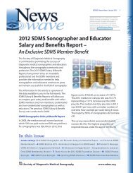
SDMS Member Benefit Spotlight -<br />
SDMS Sonographer Salary and Benefits Survey Report<br />
and SDMS Sonography Educator Salary & Benefits<br />
Survey Report<br />
What is the annual <strong>in</strong>come for the<br />
average sonographer?<br />
What do sonographers who hold more<br />
than one credential get paid?<br />
What is the average number <strong>of</strong> scans<br />
be<strong>in</strong>g performed per day and by what<br />
specialties?<br />
What benefits are employers pay<strong>in</strong>g for?<br />
Do you want the answers to these questions and<br />
more? SDMS has the answers.<br />
On a daily basis, SDMS staff would receive calls<br />
from SDMS members ask<strong>in</strong>g these questions and<br />
at the time, we did not have the answers they<br />
needed, you needed. So the <strong>Society</strong> decided<br />
to conduct a survey <strong>of</strong> its members and get the<br />
answers. We wanted the capability <strong>of</strong> provid<strong>in</strong>g<br />
our members with useful and broad-based<br />
data cover<strong>in</strong>g all relevant areas associated with<br />
sonographer <strong>in</strong>come and benefits, and related<br />
practice issues.<br />
In May 2008, SDMS released the SDMS<br />
Sonographer Salary and Benefits Survey Report<br />
and SDMS Educator Salary and Benefits Survey<br />
Report. The reports provide the answers<br />
you need and even better are FREE to SDMS<br />
members.<br />
SDMS members have the capability to download<br />
their personalized SDMS Sonographer Salary and<br />
Benefits Survey Report or SDMS Educator Salary<br />
and Benefits Survey Report <strong>in</strong> the Members Only<br />
area <strong>of</strong> the website.<br />
You can access your copy <strong>of</strong> the SDMS<br />
Sonographer or Educator Salary & Benefits<br />
Survey report at http://www.sdms.org/members/<br />
salary.asp.<br />
Access to the SDMS Sonographer Salary and<br />
Benefits Survey Report and SDMS Educator<br />
Salary and Benefits Survey Report is an exclusive<br />
SDMS member benefit and are not available to<br />
non-members.
SDMS News Wave February 2010 8<br />
SDMS Foundation Announces<br />
First 2010 Scholarship Recipients<br />
The <strong>Society</strong> <strong>of</strong> <strong>Diagnostic</strong> <strong>Medical</strong> Sonography<br />
(SDMS) Foundation’s Board <strong>of</strong> Directors is pleased<br />
to announce the first 2010 SDMS Foundation<br />
Scholarship Program recipients. The SDMS<br />
Foundation’s scholarship program provides a $2,500<br />
scholarship to a deserv<strong>in</strong>g sonography student<br />
just beg<strong>in</strong>n<strong>in</strong>g their career <strong>in</strong> diagnostic medical<br />
sonography. Another $2,500 scholarship is awarded<br />
to a deserv<strong>in</strong>g experienced sonographer who is<br />
cont<strong>in</strong>u<strong>in</strong>g their education by pursu<strong>in</strong>g an advanced<br />
sonography-related degree.<br />
Constance Besaw from Middleton, Wiscons<strong>in</strong> is a<br />
2010 SDMS Foundation scholarship recipient. Ms.<br />
Besaw is enrolled <strong>in</strong> general/vascular program at the<br />
University <strong>of</strong> Wiscons<strong>in</strong> Hospital and Cl<strong>in</strong>ics School <strong>of</strong><br />
<strong>Diagnostic</strong> <strong>Medical</strong> Sonography. She will graduate <strong>in</strong><br />
August 2011. The accredited program <strong>of</strong>fers general,<br />
vascular, and cardiac sonography programs and is<br />
based <strong>in</strong> Madison, Wiscons<strong>in</strong>.<br />
Rhonda Keller from Greenville, South Carol<strong>in</strong>a<br />
is also the recipient <strong>of</strong> a 2010 SDMS Foundation<br />
scholarship. Ms. Keller is enrolled <strong>in</strong> the Northcentral<br />
University’s Master <strong>of</strong> Education Degree <strong>in</strong> Higher<br />
Education Leadership and expects to graduate <strong>in</strong><br />
December 2010. Northcentral University is based <strong>in</strong><br />
Prescott Valley, Arizona. Ms. Keller has worked as a<br />
sonographer for more than 18 years and found she<br />
loved teach<strong>in</strong>g along the way. She hopes to use the<br />
Master’s degree she earns to help her better address<br />
the different learn<strong>in</strong>g philosophies among college<br />
students.<br />
The SDMS Foundation received more than 50<br />
applications for this round <strong>of</strong> its scholarship program.<br />
Each application was objectively scored based on the<br />
responses provided on the scholarship application.<br />
In addition, each applicant submitted an essay that<br />
addressed why they were an excellent candidate for<br />
the scholarship, their three to five year pr<strong>of</strong>essional<br />
plan, and how they planned to use their education<br />
for the betterment <strong>of</strong> the sonography and healthcare<br />
community. The essays were scored anonymously<br />
by a panel <strong>of</strong> sonographers and the scholarship<br />
recipients were selected based on their comb<strong>in</strong>ed<br />
total scores.<br />
The SDMS Foundation will award another round<br />
<strong>of</strong> scholarships later this year. The application<br />
deadl<strong>in</strong>e for the next round is July 31, 2010.<br />
Additional <strong>in</strong>formation and applications for 2010<br />
SDMS Foundation grants and scholarships is also<br />
available on the SDMS Foundation website (www.<br />
sdmsfoundation.org/programs.aspx). The SDMS<br />
Foundation charitable programs depends on<br />
donations from <strong>in</strong>dividual SDMS members and the<br />
sonography community. To make a donation to the<br />
SDMS Foundation, visit:<br />
www.sdmsfoundation.org/donor.aspx<br />
Congratulations to the<br />
February iPod Touch W<strong>in</strong>ner!<br />
Congratulations to Mary O’Connor <strong>of</strong> Houston, TX. Mary is the<br />
w<strong>in</strong>ner for our Membership Renewal Campaign for February 2010!<br />
Her name was picked randomly from all February members that<br />
renewed membership onl<strong>in</strong>e from January 1 to January 31.<br />
Any member that renews their membership ONLINE with<strong>in</strong><br />
30 days <strong>of</strong> receiv<strong>in</strong>g their FIRST renewal notice email will be<br />
automatically entered <strong>in</strong>to a draw<strong>in</strong>g to w<strong>in</strong> an Apple ® iPod Touch.<br />
• Members must renew onl<strong>in</strong>e to eligible for draw<strong>in</strong>g<br />
• Draw<strong>in</strong>gs will occur on the first <strong>of</strong> each month<br />
• Promotion valid January 1 through December 31, 2010
SDMS News Wave February 2010 9<br />
SDMS is excited to announce the 2010 SDMS Annual<br />
Conference Sponsorship and Exhibit Program<br />
2010 SDMS Annual Conference Exhibitor Prospectus<br />
SDMS is October the 14 largest – 17, 2010 • Hyatt and Regency Denver/Colorado fastest Convention grow<strong>in</strong>g Center • Denver, association<br />
Colorado<br />
<strong>in</strong> the world dedicated to all specialty areas <strong>of</strong><br />
sonography. The 2010 Annual Conference will be<br />
held October14-17, 2010 <strong>in</strong> Denver Colorado. We<br />
project an early sellout <strong>of</strong> exhibit and hotel space for<br />
the meet<strong>in</strong>g.<br />
The conference <strong>of</strong>fers over fourteen exhibit hall<br />
hours, six hours <strong>of</strong> dedicated exhibit hall time, which<br />
<strong>in</strong>cludes the Exhibit Hall Open<strong>in</strong>g Reception and<br />
refreshment breaks.<br />
Make sure your company has a ‘presence’ at<br />
this prestigious event. If decision makers <strong>in</strong> the<br />
sonography <strong>in</strong>dustry are your target audience, you<br />
can’t afford to pass up this opportunity!<br />
We hope your company will take advantage <strong>of</strong> this<br />
opportunity. Sponsorships and booth space will<br />
be processed and assigned <strong>in</strong> the order that forms<br />
(<strong>in</strong>clud<strong>in</strong>g payment) are received. We recommend an<br />
expeditious response to <strong>in</strong>sure your premiere exhibit<br />
hall space.<br />
For more <strong>in</strong>formation please go to:<br />
http://www.sdms.org/meet<strong>in</strong>gs/exhibitors/<br />
SDMS Foundation Receives IRS Tax-Exempt Status<br />
The <strong>Society</strong> <strong>of</strong> <strong>Diagnostic</strong> <strong>Medical</strong> Sonography (SDMS)<br />
Foundation has been notified that its application<br />
to the U.S. Internal Revenue Service (IRS) for taxexempt<br />
status under the Internal Revenue Code as a<br />
public charity (aka 501(c)(3) organization) has been<br />
approved. The designation by the IRS is retroactive to<br />
the SDMS Foundation’s formation on June 30, 2009.<br />
The IRS designation as a public charity ensures that<br />
<strong>in</strong>dividual and corporate donations made to the SDMS<br />
Foundation may be deductible to the extent permitted<br />
by law for federal <strong>in</strong>come tax purposes (consult your<br />
tax advisor for more <strong>in</strong>formation). Each donor to the<br />
SDMS Foundation receives a receipt that can be used<br />
as pro<strong>of</strong> <strong>of</strong> donation for tax purposes.<br />
The mission <strong>of</strong> the SDMS Foundation is to provide<br />
educational and scientific research support<br />
through grants, scholarships, and other charitable<br />
programs to the medical community and public,<br />
relat<strong>in</strong>g to <strong>Diagnostic</strong> <strong>Medical</strong> Sonography. The<br />
SDMS Foundation also fosters pr<strong>of</strong>essional learn<strong>in</strong>g<br />
and excellence by work<strong>in</strong>g to improve the field <strong>of</strong><br />
<strong>Diagnostic</strong> <strong>Medical</strong> Sonography. For further details on<br />
the SDMS Foundation’s grants and scholarships, visit<br />
http://www.sdmsfoundation.org/programs.aspx.<br />
It’s simple to make a donation to the SDMS<br />
Foundation:<br />
1. As part <strong>of</strong> your annual SDMS membership renewal<br />
(onl<strong>in</strong>e or by mail)<br />
2. Onl<strong>in</strong>e: http://www.sdmsfoundation.org/donate.aspx<br />
3. By mail: SDMS Foundation, 2745 Dallas Pkwy Ste<br />
350, Plano, TX 75093-8730.
New Member<br />
SCAN<br />
Kathryn Zale is currently<br />
a senior sonography<br />
student at The Ohio<br />
State University. She<br />
graduated from Miami<br />
University <strong>of</strong> Oxford, Ohio<br />
<strong>in</strong> 2004 with a Bachelor<br />
<strong>of</strong> Arts degree <strong>in</strong> English/<br />
journalism and another<br />
<strong>in</strong> Russian. Between<br />
2004 and 2005 she worked <strong>in</strong> Moscow, Russia. In<br />
addition to Kathryn’s <strong>in</strong>terest <strong>in</strong> travel and different<br />
cultures, another passion <strong>of</strong> hers is martial arts. In<br />
2004, she obta<strong>in</strong>ed her black belt <strong>in</strong> taekwondo and<br />
s<strong>in</strong>ce had the opportunity to study many different<br />
k<strong>in</strong>ds <strong>of</strong> martial arts <strong>in</strong>clud<strong>in</strong>g judo, hapkido and<br />
capoeira. In the fall <strong>of</strong> 2007 Kathryn married and<br />
moved to Columbus, Ohio. Her husband is also a<br />
student at Ohio State, earn<strong>in</strong>g his PhD <strong>in</strong> Horticulture<br />
specializ<strong>in</strong>g <strong>in</strong> breed<strong>in</strong>g and genetics <strong>of</strong> orchids, phlox<br />
and magnolias. They enjoy photography, hik<strong>in</strong>g and<br />
travel<strong>in</strong>g together.<br />
When asked what <strong>in</strong>spired Kathryn to choose a<br />
SDMS News Wave February 2010 10<br />
career <strong>in</strong> sonography, she replied “I grew up around<br />
ultrasound.” Her father has sold ultrasound equipment<br />
for over 30 years and grow<strong>in</strong>g up she loved ‘Take<br />
your Daughter to Work’ days. Unfortunately, she just<br />
never felt encouraged dur<strong>in</strong>g school to study science.<br />
It was not until years after graduat<strong>in</strong>g from Miami<br />
did she f<strong>in</strong>ally heed her parent’s advice and signed<br />
up for an anatomy course. It turns out parents do<br />
know best! Kathryn <strong>in</strong>stantly fell <strong>in</strong> love with anatomy<br />
and physiology and knew then that sonography was<br />
a perfect fit. S<strong>in</strong>ce be<strong>in</strong>g <strong>in</strong> the ultrasound program<br />
at Ohio State, her pr<strong>of</strong>essor, Dr. Kev<strong>in</strong> Evans, fellow<br />
classmates and cl<strong>in</strong>ical <strong>in</strong>structors have been a<br />
constant source <strong>of</strong> support and <strong>in</strong>spiration.<br />
Kathryn expressed her favorite benefit <strong>of</strong> the SDMS<br />
has been the JDMS and onl<strong>in</strong>e access to archived<br />
journals. It has been a tremendous help for writ<strong>in</strong>g<br />
class papers and just to learn more about what is new<br />
and upcom<strong>in</strong>g <strong>in</strong> the field. Other benefits she enjoys<br />
<strong>in</strong>clude updated <strong>in</strong>formation on legislative issues<br />
perta<strong>in</strong><strong>in</strong>g to sonography, as well as the national<br />
certification exam<strong>in</strong>ation reviews, which was helpful<br />
prior to tak<strong>in</strong>g the physics registry. She enjoyed the<br />
annual meet<strong>in</strong>g <strong>in</strong> Nashville and the opportunity to<br />
present her research through the student poster<br />
competition. Kathryn also mentioned that once she<br />
is credentialed, the CME onl<strong>in</strong>e tests and SDMS CME<br />
Tracker will be beneficial.<br />
Kathryn, welcome to the SDMS!<br />
Upcom<strong>in</strong>g SDMS Web<strong>in</strong>ars<br />
Participate <strong>in</strong> live presentations<br />
or watch the record<strong>in</strong>gs at your<br />
convenience. Then take the test for<br />
<strong>in</strong>stant CME credit, absolutely free<br />
for SDMS members.<br />
If you are unable to participate <strong>in</strong> these live web<strong>in</strong>ars,<br />
visit http://www.sdms.org/members/web<strong>in</strong>ars.asp<br />
for <strong>in</strong>formation on view<strong>in</strong>g a record<strong>in</strong>g <strong>of</strong> the web<strong>in</strong>ar.<br />
Registration: The SDMS Web<strong>in</strong>ar Series is FREE to<br />
current SDMS members and is not available to nonmembers<br />
(For <strong>in</strong>formation on jo<strong>in</strong><strong>in</strong>g SDMS, visit<br />
http://www.sdms.org/membership/ )<br />
All SDMS Web<strong>in</strong>ars are tracked by SDMS CME Tracker.<br />
http://www.sdms.org/members/web<strong>in</strong>ars.asp<br />
Understand<strong>in</strong>g the Other Imag<strong>in</strong>g Modalities<br />
Date: Thursday, March 11, 2010<br />
Time: 8:00 pm (Eastern); 7:00 pm (Central);<br />
6:00 pm (Mounta<strong>in</strong>); 5:00 pm (Pacific)<br />
CME Credits: 1.0 SDMS CME Credit (OT)<br />
FEATURED SPEAKER: Salvatore LaRusso, M.Ed. RDMS, RT(R)<br />
Contrast <strong>in</strong> the Echo Lab<br />
Date: Thursday, April 8, 2010<br />
Time: 8:00 pm (Eastern); 7:00 pm (Central);<br />
6:00 pm (Mounta<strong>in</strong>); 5:00 pm (Pacific)<br />
CME Credits: 1.0 SDMS CME Credit (AE)<br />
FEATURED SPEAKER: Allen Borowski, RDCS, FASE
SDMS Fellow<br />
Spotlight<br />
SDMS News Wave February 2010 11<br />
This is a cont<strong>in</strong>u<strong>in</strong>g series <strong>of</strong> <strong>in</strong>terviews <strong>of</strong><br />
our dist<strong>in</strong>guished SDMS Fellow members.<br />
Stephen M. McLaughl<strong>in</strong>,<br />
BS, RT(R), RDMS, FSDMS<br />
Year awarded fellow<br />
status: 1999<br />
On January 8, 2005,<br />
Steve, a colleague<br />
and friend, died after a<br />
19-month battle with bra<strong>in</strong><br />
cancer. Steve served on<br />
the SDMS Board <strong>of</strong> Directors for over ten years<br />
as a Regional Director, President-Elect, President,<br />
and Past President.<br />
In addition to his Board service, he served the<br />
association and sonography community <strong>in</strong> a<br />
multitude <strong>of</strong> roles: committee chair, Board liaison,<br />
national certification exam proctor, local ultrasound<br />
society president, and counselor.<br />
Steve’s goals as SDMS President were to grow<br />
membership and have <strong>Diagnostic</strong> <strong>Medical</strong><br />
Sonography recognized as a pr<strong>of</strong>ession by the<br />
United States Department <strong>of</strong> Labor. In 2001, Steve<br />
achieved that goal. Once <strong>Diagnostic</strong> <strong>Medical</strong><br />
Sonography was recognized as a pr<strong>of</strong>ession, Steve<br />
began to educate students and sonographers on the<br />
importance <strong>of</strong> be<strong>in</strong>g recognized as a pr<strong>of</strong>ession.<br />
When Steve passed away, the SDMS Board <strong>of</strong><br />
Directors felt it was important to keep Steve’s<br />
legacy alive so <strong>in</strong> 2006 they created the Stephen<br />
M. McLaughl<strong>in</strong> Memorial Lectureship to honor<br />
sonographers whose primary focus is on<br />
sonographer pr<strong>of</strong>essionalism. The lectureship is<br />
presented at the SDMS Annual Conference each<br />
year. The recipient is recognized at the SDMS<br />
Awards D<strong>in</strong>ner by the SDMS Board <strong>of</strong> Directors,<br />
colleagues and friends.<br />
Recipients <strong>of</strong> the lectureship must be an Active<br />
SDMS member <strong>in</strong> good stand<strong>in</strong>g, actively <strong>in</strong>volved<br />
<strong>in</strong> the pr<strong>of</strong>ession and a lum<strong>in</strong>ary <strong>in</strong> the field <strong>of</strong><br />
sonography with a dist<strong>in</strong>guished and notable career.<br />
To date, we have had the honor <strong>of</strong> present<strong>in</strong>g the<br />
Stephen M. McLaughl<strong>in</strong> Lectureship to:<br />
Donald Milburn, RDCS, RVT, FSDMS<br />
Terry DuBose, MS, RDMS, FSDMS, FAIUM<br />
Joan Baker, MSR, RDMS, RDCS, FSDMS<br />
Kev<strong>in</strong> Evans, PhD, RT(R)(M)(BD), RDMS, RVS, FSDMS<br />
SDMS Product Spotlight –<br />
SDMS now <strong>of</strong>fers MedLearn’s recently<br />
updated 2010 Ultrasound Coder!<br />
Are you mystified by<br />
the cod<strong>in</strong>g and bill<strong>in</strong>g<br />
requirements for diagnostic<br />
ultrasound procedures? Fear<br />
not, MedLearn’s Ultrasound<br />
Coder book will guide you<br />
through the <strong>in</strong>tricacies! This<br />
timely resource summarizes<br />
Medicare coverage policies<br />
and bill<strong>in</strong>g guidel<strong>in</strong>es.<br />
• Guidance related to biopsy, aspiration, needle<br />
localization and vascular-access procedures<br />
• Covers non-<strong>in</strong>vasive peripheral vascular cod<strong>in</strong>g<br />
• Divided <strong>in</strong>to diagnostic ultrasound, ultrasound<br />
guidance procedures, echocardiography (fetal<br />
and non-fetal), non-<strong>in</strong>vasive vascular diagnostic<br />
studies and <strong>in</strong>travascular ultrasound sections,<br />
each with a list <strong>of</strong> CPT codes and bill<strong>in</strong>g tips<br />
• Addresses recent code additions, <strong>in</strong>clud<strong>in</strong>g new<br />
codes for ultrasound and screen<strong>in</strong>g for aortic<br />
aneurysm<br />
To purchase this product, please go to: http://www.<br />
sdms.org/store/ProductDetails.aspx?ProductID=45
SDMS News Wave February 2010 12<br />
SDMS Welcomes New Members<br />
January 2010<br />
Amy Acuncius<br />
Debra Adams, RDMS<br />
Robbie Adams<br />
L<strong>in</strong>da Ahlborn, RT(R), RDMS<br />
Dersana Ajit, RDMS, RVT<br />
Marion Allen, RDMS<br />
Tanya Amos, RCS<br />
Mart<strong>in</strong> Andrzejewski, RT(N), RDMS, CNMT<br />
Taeko Aoki<br />
Carmen Arguelles, RT(M), RDMS<br />
Shannon Aust<strong>in</strong>, RDCS, RVT, CCT<br />
Colleen Baca, RDMS<br />
Mela<strong>in</strong>e Barco, RT(R)(M), RDMS, RVT<br />
Ela<strong>in</strong>e Baroni, RDMS<br />
Edgar Barros, RDMS<br />
Athena Basham, RDMS<br />
Bridie Baucum, RDMS<br />
Laura Becker, RT(R)(M), RDMS, RVT<br />
Candice Bee, RT(R)<br />
Mirela Belecciu, RDCS<br />
Michelle Bell, RT(R)(M), RDMS<br />
Constance Besaw<br />
Pamela Besse, RT(R), RDMS<br />
Tracy Biegel, RT(R), RDMS<br />
Malgorzata Bigos, RDMS<br />
Douglas Bisig, BA, RT(R), RDMS<br />
Christopher Blair, RT(R), RDMS, RVT<br />
John Blomeyer, RVT<br />
Kathryn Blosser<br />
Kathleen Boisseau<br />
Mojtaba Borairi, RT(R), RDCS<br />
Kristen Bottiglier, RDCS<br />
Zouliath Bouraima<br />
Patricia Bratus, RDCS<br />
Kathleen Brocar, RT(R), RDMS<br />
Vanessa Brown, RT(R), RDMS<br />
Phyllis Buch, RT(R)(M), RDMS<br />
Marci Bunt<strong>in</strong>g, RT(R)<br />
Heather Burlew, RDMS<br />
Michelle Burs<strong>in</strong>g, RDCS<br />
Lisa Burton, RT(R), RDMS<br />
Ariel Cagigas, RCS, RVS<br />
Susan Camp, BS, RT(R), RDMS.RVT<br />
Alane Campbell, RT(R)(CT), RDMS<br />
Anita Campbell<br />
Kimberly Canova, RDMS<br />
William Cardus, BS, RN, RDCS<br />
Miriam Carey<br />
Jean<strong>in</strong>e Carroll, RDMS<br />
La Tosha Carter, RDMS<br />
Marrey Carter, RDMS, RVT<br />
Portia Carver, RDMS, RVT<br />
Constance Casey, BS, RDMS, RDCS, RVT<br />
Herman Celest<strong>in</strong>e, RDMS, RVT<br />
Kathleen Chase, RDMS, RVT<br />
Jie Chen<br />
Judith Cho, RDMS<br />
Amzad Chowdhury<br />
Kalam Chowdhury<br />
Michele Christensen, RDMS<br />
Erica Cissell, RT(R), RDMS<br />
Shelley Clark, BS<br />
Tonja Clemons, RCS, RVS<br />
Gloria Clover, RDMS<br />
Audrey Clunie<br />
Holly Cohick, RT(R), RDMS, RVT<br />
Jill Coleman, RDMS<br />
Paul Coll<strong>in</strong>s, RDMS, RVT<br />
Mary Cooper<br />
Stacy Countryman, RT(R), RDMS<br />
Pamela Coyer, RDMS<br />
Skyleen Crawford-May, BS<br />
Roxie Crump<br />
Jennifer Culler, CMA<br />
Andrea Daniel, MS<br />
Terry Daniels, RT(R), RDCS<br />
Beth Darby, RDMS<br />
Peggy Davidson, RT(R)(M)(CT), RDMS<br />
Renee Deal, RT(R), RDMS, RVT<br />
Margaret Dean, RT(R), RDMS<br />
Vera Dega, BS, RDMS<br />
Debbie Denney, RDMS<br />
Nancy Denny, RT(R), RDMS<br />
Ken DeRoche<br />
Maria Di Cicco, RVT<br />
Stefanie Dibell, RT(R), RDMS<br />
Lisa Dilauri, RDCS, RCS, RVS, CCT, FASE<br />
Roberta Dobbs, BS<br />
Patricia Donl<strong>in</strong>, RT(R), RDMS<br />
Julie Dorman, RDMS, RVT<br />
Cherylann Doucette, RDMS<br />
Lacey Dube, BS, RDMS<br />
John Dubocq, RDMS<br />
Phyllis Dunlap, RDCS<br />
Marie Duval, RDMS<br />
Melissa Easterday<br />
Theresa Ellis, BS<br />
Sara Emery, BS<br />
Monica Fisher, RT(R), RDMS<br />
Lise Fishman<br />
G<strong>in</strong>a Flores<br />
Traci Flowers<br />
Timothy Gahara, MS, RDCS<br />
Tammy Galang, RDMS<br />
Jocell Garcia, RT(R), RDMS<br />
Carmen Gardner<br />
Daisy Garza, RT(R)<br />
Dawn Gawell, RT(R), RDMS<br />
Pamela Giacalone<br />
Vanessa Giannamore, RDCS<br />
Simone Gifford-Allen, RDMS<br />
Sarah Giszewski<br />
Juliana Gnade, RT(R), RDMS<br />
Forouzan Godarzi, BS, RT(R)(M)(CT)<br />
(MR), RDMS<br />
Sheila Gold, RT(R), RDMS, RVT<br />
Richard Goldsmith, RDMS, RVT<br />
Carlos Golston, RDCS<br />
Janene Good, BS, RN, RDCS<br />
Divya Goyal, RDCS<br />
Melanie Green, RDMS<br />
Kelli Griff<strong>in</strong><br />
Leslie Griff<strong>in</strong><br />
Wendi Grosch, BS, RT(R), RDMS<br />
Diane Gulbrandsen, RDMS, RVT<br />
Tracy Guzzo, RDMS<br />
George Gvazava<br />
Mary Claire Haenle<strong>in</strong>, RDMS<br />
Krist<strong>in</strong>a Hall, RDMS<br />
Stephen Harasty<br />
Fabiola Harden, RCS<br />
Kelly Harris, BS, RDMS, RDCS, RVT<br />
Ashley Harrison, RT(R)<br />
Kara Hatch<br />
Kathy Haxton<br />
Jennifer Hayford, RDMS<br />
Susan Haynes, RT(R), RDMS<br />
Brenda Heimer, RT(R), RDMS, RVT<br />
Rebecca Hellman, RT(R)<br />
Miranda Hemm<strong>in</strong>g<br />
Kev<strong>in</strong> Henderson<br />
Cheryl Henn<strong>in</strong>ger<br />
Amanda Henry, RT(R), RDMS, RVT<br />
Kather<strong>in</strong>e Hensley, RT(R)(M)(CT)<br />
Maria Hernandez, RT(R)(M), RDMS, RVT<br />
Paula Hill, RT(R), RDMS<br />
Jeremy H<strong>in</strong>ojosa<br />
Patsy Hobson, RDMS<br />
Lee H<strong>of</strong>fer, RDMS, RVT<br />
Andriah Holley-Smith<br />
Stephanie Hollis<br />
Andrea Hooks, BS, RT(R), RDMS<br />
Nathan Hughes, BS, RVT<br />
C<strong>in</strong>dy Jarvis, RT(R)(M), RDMS<br />
Charmel Jeffery, BS<br />
Charissa Johnson<br />
Robert Johnson, RDMS, RDCS, RVT<br />
Sheila Johnson, RDMS<br />
Eloise Jones, BS<br />
Myra Jones, RT(R), RDMS<br />
Heather Jopl<strong>in</strong><br />
Brooke Kangas<br />
Jennifer Keegan, BS, RDMS<br />
John Kelleher, RVT<br />
Sheri Kempfer, RT(R)(CT), RDMS, RVT<br />
Jennifer Kennedy, RT(R)(M)(CT), RDMS, RVT<br />
Jennifer Kennell<br />
Michele Kim, BS, RDMS<br />
Dana Kirby, RT(R), RDMS<br />
Cor<strong>in</strong>ne Klimek, RT(R)(M), RDMS<br />
Jessica Kolb, RDCS, RVT<br />
Roland Krahn, BS<br />
Heather Krot<strong>of</strong>il<br />
Connie Kruger, RN<br />
Tatyana Kumanovskaya, RDCS, RVT<br />
Mary Grace Kunda, RDMS<br />
Jessica Kyer<br />
Heather Kysl<strong>in</strong>ger<br />
Mark Lacasella, RDCS, RVT<br />
Emily Lamb<br />
Meredith Laney, RT(R)(S)<br />
Jennifer Lanier<br />
Elise Lansford<br />
Kimberly Larson-Curtis, RDMS, RVT, CRT<br />
Ann Laue, RT(R)(M), RDMS<br />
Allan Lawson, RT(R), RDMS<br />
Huong Le, BS<br />
Jennifer Lee, BS, RT(R), RDMS<br />
Reg<strong>in</strong>a Lee<br />
Marci Lehl, RT(R), RDMS, RVT<br />
Tiffany Letnes<br />
Rebecca Lewis
SDMS News Wave February 2010 13<br />
Martha Lickteig, RDMS<br />
Barbara L<strong>in</strong>dstrom, RT(R), RDMS<br />
Sujatha L<strong>in</strong>gala, BS, RDMS<br />
Kelly Liriano, RDMS<br />
Sara Livengood, RT(R), RDMS<br />
Maly Lo, RDMS<br />
Theresa Logan, BS<br />
Mary Lohmann, RDMS<br />
Traci Lombardi<br />
Jessica Longshore<br />
Cor<strong>in</strong>na Lopez<br />
Dom<strong>in</strong>go Lopez, RDMS<br />
Annmarie Lovemore, RDMS<br />
Victoria Luca, RDMS<br />
Melissa Lumbarkovski<br />
Thomas Ma<strong>in</strong>er<br />
Benjam<strong>in</strong> Makamson, DO, RPVI<br />
Angel Mart<strong>in</strong>ez, RDMS, RVT<br />
Jennifer Mart<strong>in</strong>ez, BS<br />
Donna Mart<strong>in</strong>i, BS, RT(R), RDMS<br />
Tissy Mathew, RDCS, RCS<br />
Raejean Mattison, BS, RDMS<br />
Donna Mattner, BS, RDMS<br />
Sjhanna Mayne<br />
Amanda McCarty<br />
Lee McDaniels, RDMS<br />
Roger McGee, RDMS, RVT<br />
Connie McGuire, BS, RVT<br />
Cora McIntyre, RT(R)(M), RDMS<br />
Julie McLaughl<strong>in</strong>, RT(R), RDMS<br />
Betre Melaku<br />
Christ<strong>in</strong>e Meslany, RT(R), RDMS<br />
Lisa Miguel, BS<br />
Dana Miller, RDMS<br />
Kimberly Miller, RDMS<br />
Rhonda Miller, RDMS<br />
Jessica Mitic<br />
Amal Mohammed, BS, RDMS<br />
Kimberly Moore, RVT<br />
Michael Morgan, RCS, RVS<br />
Rhonda Morrison, RT(R), RDMS<br />
Andra Mote<br />
Hoy Moy<br />
Luz Munoz-Barrera, RT(R)<br />
Patricia Murphy, RDMS<br />
Jamie Musho<br />
Tia Nauman, BS, RDMS, RVT<br />
Bel<strong>in</strong>da Neely, BS, RDMS<br />
Stella Nektalov, RVT<br />
Lisa Nguyen<br />
Phuong-Anh Nguyen<br />
Thuy Nguyen<br />
Sarah Norden, RT(R), RDMS, RVT<br />
Karen O’Konek, BS<br />
Heather Oravitz, RDCS<br />
Christ<strong>in</strong>a Ornelas, RT(R), RDMS<br />
Jacek Orzechowski<br />
Amber Pagano<br />
Tracy Paris, RT(R), RDCS<br />
Didre Park<br />
Urvashi Patel<br />
Zarna Patel<br />
Jennifer Patrick<br />
Krist<strong>in</strong>e Patterson, RT(R)(M), RDMS<br />
Harold Pedersen, RDMS<br />
Maria Pedron<br />
Rachel Pelton<br />
Nicol Krupa Penni<br />
Er<strong>in</strong> P<strong>in</strong>eda, RDMS<br />
Gloria P<strong>in</strong>kava, RDCS<br />
Nancy Pitts, CNMT<br />
Kara Potter, RT(R)<br />
G<strong>in</strong>a Preston<br />
Jennie Psalidas, RDMS<br />
Stacey Pucc<strong>in</strong>elli<br />
Sara Quehrn, RDMS<br />
Melissa Quisler, RN, RDMS<br />
Adrienne Racz, MS<br />
Roxane Ralat<br />
Saundra Ramey, RDMS<br />
Teresa Ramsey, RVT<br />
Tiffany Ray<br />
Jaime Reifman, BS<br />
Karen Reis<strong>in</strong>g<br />
Stacy Reno, RT(R)<br />
Heather Reynolds<br />
Giuseppa Ribaudo-McGrane<br />
Danielle Ricker<br />
Courtney Riso<br />
Kausar Rizvi, RDCS<br />
Rosa Rodarte<br />
Lydia Rodriguez, RCS<br />
Kelly Roemhild, RDMS<br />
Joanne Rogers, RT(R), RDMS, RDCS, RVT<br />
Karen Rogers<br />
Christ<strong>in</strong>a Ro<strong>of</strong>, RDCS<br />
Marsha Roper, RDCS<br />
MeChelle Ross<br />
Jill Rubsam, RT(R), RDMS<br />
Derek Sabour<strong>in</strong>, RT(R), RDMS, RVT<br />
Christopher Santora, RT(R), RDMS, RVT<br />
Eleanore Sarason, BS, RDMS<br />
Colleen Saunders, RT(R)(M), RDMS<br />
Sandra Sawyer, RDMS<br />
Jennifer Schary, RDMS, RDCS<br />
Lisa Sebar-Berju, RDMS<br />
Terri Setzer, RDMS, RVT<br />
Dione Severns, RDCS<br />
Allison Shanks, BS<br />
Yaroslav Shapoval, RDMS<br />
Sam<strong>in</strong>a Sheikh, MS<br />
Lynne Sheller, RDMS, RDCS<br />
Cynthia Shirley, RT(R), RDCS, RVS<br />
G<strong>in</strong>ger Short, RT(R), RDMS, RVT<br />
Pamela Sidwell, RDMS, RVT<br />
Paul Sierzenski, MD, RDMS<br />
L<strong>in</strong>da Silvers, BS, RDMS<br />
Matt Simmons, RDCS<br />
Donald Simons, RDMS, RDCS, RVT<br />
Jane Smarzik, RDMS<br />
Christ<strong>in</strong>a Smith<br />
Gerald Smith, RT(R), RDMS<br />
Jerri Smith, RDMS<br />
Krist<strong>in</strong> Smith, RDMS<br />
L<strong>in</strong>dsay Smith, RT(R)<br />
Sarah Smith, RDMS<br />
Suzanne Solarte<br />
Julie Solita, RDMS<br />
Rob<strong>in</strong> Souter, RDMS<br />
Tanya Specht, RDMS<br />
Lori Spozarski, RN, RDMS<br />
Ashley Staller, BS, RDMS<br />
Jeanie Standard, BS, RDMS<br />
Tammy Sternitzky<br />
Louise Stevenson, RT(R), CSGR<br />
June Stewart, RDCS, RVT<br />
Pierre Stewart, RCS<br />
Jerrid Stone<br />
Carl Straley, RDMS, RVT<br />
Keri Stritikus<br />
Anne Sullivan<br />
Rachel Sykes, RDMS, RVT<br />
Stanley Tabencki, RVT<br />
Deborah Thom, RT(R), RDMS<br />
Bobbie Thomas, RDMS, RVT<br />
Craig Thomas, RDMS<br />
Nigel Thomas, BS, RDMS, RVT<br />
Jennifer Thompson, RT(R)(M), RDMS<br />
Tammy T<strong>in</strong>kler, RDMS<br />
Tracey Tomko, RT(R)(M), RDMS<br />
Mary Tomory, BS, RT(R), RDMS<br />
Shohreh Torkan<br />
Lynn Tran, BA, RDMS<br />
Mary Traurig, RT(R), RDMS<br />
Daniela Travagl<strong>in</strong>i, BS<br />
Casey Treat<br />
Marsha Trost, RDMS<br />
Deborah Trump, RDMS, RVT<br />
Nita Upadhyaya, RDMS, RVT<br />
Juliana Vallejo<br />
Kellie Vermett, RDMS, RVT<br />
Amy Volk, RT(R)<br />
Amanda Wallace<br />
Serena Lai-Siu Wang, RDMS<br />
Charles Webb, RDCS<br />
Dana Welch<br />
Amy Westmoreland, RDMS, RVT<br />
Allesia White, RT(R)<br />
Julie White, BS, RDMS, RDCS, RVT<br />
Tiffany White, BS<br />
Emily Whit<strong>in</strong>g, RDMS, RVT<br />
Ellen Wilbanks, BS, RT(R), RDMS<br />
N<strong>in</strong>a Wilcher, RDMS<br />
Brenda Wilk<strong>in</strong>son, RDMS<br />
Ashley Williams, RT(R), RDMS<br />
Susan Williams, RT(R)(M), RDMS<br />
Carla Wolff, RDMS, RVT<br />
Christ<strong>in</strong>a Woods Rowe, RDMS<br />
Lauren Woodsum, RT(R), RDMS<br />
C<strong>in</strong>di Wu, BS<br />
Jiarong Xiao, MS<br />
Suleyman Yadgarov, RDMS<br />
Roya Zafaranchi, MD, RDMS<br />
T<strong>in</strong>a Zakarian<br />
Alan Zimmerman, RCS<br />
Allison Zonar<br />
Joshua Zook, RDMS<br />
News Wave (ISSN 1541-7581) is published<br />
to <strong>in</strong>form SDMS members <strong>of</strong> meet<strong>in</strong>gs,<br />
events and policies as well as trends and<br />
issues <strong>in</strong> the sonography pr<strong>of</strong>ession. Please<br />
send comments and suggestions to:<br />
SDMS Headquarters<br />
2745 N Dallas Pkwy Ste 350<br />
Plano, TX 75093-8730<br />
Phone: (214) 473-8057<br />
FAX: (214) 473-8563<br />
CEO and<br />
Executive Director<br />
Donald F. Haydon, CAE<br />
Production Editor<br />
Chris Alcott<br />
All contents Copyright © 2010. All rights reserved by the <strong>Society</strong> <strong>of</strong> <strong>Diagnostic</strong> <strong>Medical</strong> Sonography, Plano, Texas.