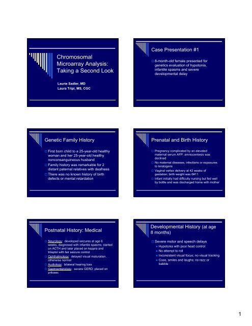
Chromosomal Microarray Analysis: Taking a ... - Kaleida Health
Chromosomal Microarray Analysis: Taking a ... - Kaleida Health
Chromosomal Microarray Analysis: Taking a ... - Kaleida Health

You also want an ePaper? Increase the reach of your titles
YUMPU automatically turns print PDFs into web optimized ePapers that Google loves.
<strong>Chromosomal</strong><br />
<strong>Microarray</strong> <strong>Analysis</strong>:<br />
<strong>Taking</strong> a Second Look<br />
Case Presentation #1<br />
8-month-old female presented for<br />
genetics evaluation of hypotonia,<br />
infantile spasms and severe<br />
developmental delay<br />
Laurie Sadler, MD<br />
Laura Tripi, MS, CGC<br />
Genetic Family History<br />
Prenatal and Birth History<br />
First born child to a 25-year-old healthy<br />
woman and her 25-year-old healthy<br />
nonconsanguineous husband<br />
Family history was remarkable for 2<br />
distant paternal relatives with deafness<br />
There was no known history of birth<br />
defects or mental retardation<br />
Pregnancy complicated by an elevated<br />
maternal serum AFP; amniocentesis was<br />
declined<br />
No maternal diseases, infections or exposures<br />
to teratogens<br />
Vaginal vertex delivery at 42 weeks of<br />
gestation; birth weight was 6#11<br />
Infant initially had difficulty nursing but fed well<br />
by bottle and was discharged home with mother<br />
Postnatal History: Medical<br />
Neurology: developed seizures at age 6<br />
weeks, diagnosed with infantile spasms; started<br />
on ACTH and later placed on keppra and<br />
trileptol with fair seizure control<br />
Ophthalmology: delayed visual maturation.<br />
otherwise normal<br />
Audiology: bilateral hearing loss<br />
Gastroenterology: severe GERD; placed on<br />
prilosec<br />
Developmental History (at age<br />
8 months)<br />
Severe motor and speech delays<br />
• Hypotonia with poor head control<br />
• No attempt to roll<br />
• Inconsistent visual focus; no visual tracking<br />
• Coos, smiles and laughs; no razz or<br />
babble<br />
1
Diagnostic studies prior to age<br />
8 months<br />
EEG: hypsarrythmia<br />
Echocardiogram: normal<br />
CT brain: normal<br />
MRI brain: normal<br />
CT middle and inner ear: narrowed<br />
external canals and partial fusion of<br />
incus to posterior wall on the left<br />
Routine karyotype: normal<br />
Physical Examination<br />
Growth<br />
• Height: 71 cm (75th-90th centile)<br />
• Weight: 8.2 kg (50th-75th centile)<br />
• OFC: 41.5 cm (5th centile)<br />
Neurologic examination: severe<br />
hypotonia with head lag; unable to lift<br />
head in prone; inconsistent visual focus<br />
Physical Examination<br />
Physical examination<br />
Craniofacial examination<br />
• Bitemporal narrowing<br />
• Flattened occiput with decreased<br />
anteroposterior dimension of the head<br />
• Large anterior fontanelle<br />
• Deeply set eyes<br />
Extremities: short dorsiflexed halluces<br />
Remainder of physical examination was<br />
normal<br />
Assessment<br />
8-month-old female with hypotonia, mild<br />
microcephaly, minor malformations,<br />
seizure disorder and global<br />
developmental delays<br />
Diagnostic considerations<br />
• Angelman syndrome<br />
• Submicroscopic chromosomal abnormality<br />
2
Recommended diagnostic<br />
studies<br />
Methylation testing for AS/PWS: normal<br />
<strong>Microarray</strong> analysis: abnormal;<br />
deletion 1p36<br />
Case #2<br />
3-day-old female presented for genetics<br />
evaluation of hypotonia and a large<br />
anterior fontanelle<br />
Genetic Family History<br />
Prenatal and Birth History<br />
Third born child to a 23-year-old healthy<br />
woman and her 23-year-old healthy<br />
nonconsanguineous husband<br />
Family history was remarkable for a<br />
maternal uncle with a VSD; several<br />
maternal relatives also had learning<br />
disabilities<br />
Pregnancy was complicated by exposure to<br />
varicella at 6 months of pregnancy; mother did<br />
not develop chicken pox<br />
Vaginal vertex delivery at 40 weeks of<br />
gestation; birth weight was 5#5<br />
Infant required transfer to the NICU for transient<br />
respiratory distress and hypoglycemia;<br />
discharged at 4 days of age<br />
Postnatal History: Medical<br />
Cardiology: VSD diagnosed at age 4<br />
months; required surgical closure<br />
Neurology: parents suspected seizures<br />
beginning at age 4 months; negative<br />
work-up<br />
Ophthalmology: pseudostrabismus;<br />
otherwise normal<br />
Audiology: normal hearing<br />
Developmental History<br />
(at age 3 1 / 2 years)<br />
Severe motor and speech delays<br />
• Hypotonia during infancy<br />
• Walked independently at age 34 months<br />
• No true speech; 2-3 consistent signs<br />
3
Diagnostic Studies prior to<br />
age 3 1 / 2 years<br />
EEG: normal<br />
Echocardiogram: VSD and PDA<br />
MRI brain: normal<br />
Routine karyotype: normal<br />
Physical Examination<br />
Growth<br />
• Height: 88 cm (
Deletion 1p36: A Recognizable<br />
Pattern of Malformation<br />
Most common terminal chromosomal<br />
deletion, occurring in 1/5000 live births<br />
Accounts for ~1% of cases of<br />
unexplained mental retardation<br />
2-3% of the general population has<br />
mental retardation, 50% of which have<br />
no identifiable etiology (genetic or<br />
environmental)<br />
Deletion 1p36: Growth and<br />
Development<br />
Postnatal growth deficiency (85%)<br />
Feeding difficulties: oropharyngeal<br />
dysphagia with failure to thrive<br />
Neurologic examination<br />
• Hypotonia (95%)<br />
• Seizures (45%)<br />
• Developmental delay (100%)<br />
• Severe to profound mental retardation with<br />
absent or very little speech<br />
Deletion 1p36 Phenotype<br />
Deletion 1p36: Other findings<br />
Craniofacial findings<br />
• Microcephaly (95%)<br />
• Large anterior fontanelle<br />
• Decreased AP dimension of the head<br />
• Straight eyebrows<br />
• Deeply set eyes<br />
• Flat nasal bridge<br />
• Minor ear anomalies (structure or position)<br />
• Pointed chin<br />
Cardiac defects (70%)<br />
Hearing loss (28%)<br />
5
So What???<br />
Prognosis<br />
Recurrence Risk<br />
Importance of Making a<br />
Specific Diagnosis<br />
Prognosis: poor; majority of patients with<br />
deletion 1p36 have severe to profound mental<br />
retardation<br />
Recurrence Risk: majority are de novo<br />
(unrelated to parents' chromosomes); no<br />
increased risk for future pregnancies<br />
Parental studies indicated in all cases; 6% of<br />
parents will have a balanced rearrangement<br />
with associated increased risks of recurrence<br />
Methods of Detecting<br />
<strong>Chromosomal</strong><br />
Abnormalities<br />
History of the Routine Karyotype<br />
Routine Karyotype<br />
19 th century: chromosomes first seen and<br />
named<br />
1952: Hsu discovered by accident that<br />
treatment of cells with a hypotonic solution<br />
allowed for improved chromosome spreading &<br />
visualization<br />
1956: Tijo and Levan establish human<br />
chromosome number as 46<br />
6
History of the Routine Karyotype<br />
Routine Karyotype<br />
1959: Lejeune discovers that Down syndrome is<br />
due to trisomy 21<br />
1966: Steele & Breg report cultured cells from<br />
amniotic fluid can be used to look at fetal<br />
chromosomes<br />
1970: Caspersson discovers banding for<br />
chromosome identification<br />
Standardized arrangement of all the<br />
chromosomes of a cell<br />
Banding identifies each chromosome uniquely<br />
Can detect numerical and gross structural<br />
abnormalities (trisomy, monosomy, translocation,<br />
inversion, marker, ring)<br />
Resolution to ~5Mb<br />
Detection rate of ~3% for unexplained<br />
developmental delay/MR +/- dysmorphic features<br />
Structure of a Chromosome<br />
Fluorescence in situ<br />
hybridization (FISH)<br />
7
FISH<br />
Developed in the 1990s<br />
Useful for clinically identifiable<br />
microdeletion syndromes that cannot be<br />
detected using standard cytogenetic<br />
methods<br />
• Deletion 22q11.2 (velocardiofacial syndrome)<br />
• Williams syndrome<br />
Also for rapid detection of common<br />
aneuploidy in amniotic fluid samples<br />
Multiprobe<br />
Subtelomere FISH<br />
Multiprobe Subtelomere FISH<br />
FISH at rearrangement hot-spots<br />
Developed in late 1990s, commercially available<br />
since about 2000-2001<br />
Typically uses 1 FISH probe per region<br />
Detection rate of 7-10% for unexplained<br />
developmental delay/mental retardation +/-<br />
dysmorphic features<br />
8
<strong>Microarray</strong> <strong>Analysis</strong><br />
<strong>Microarray</strong> <strong>Analysis</strong><br />
Relatively new technique examining “hotspots”<br />
for chromosomal deletions and<br />
duplications throughout the genome<br />
Detects copy number variations at<br />
resolution of 1Mb or less<br />
Detection rate 10-20% for individuals with<br />
normal karyotype, unexplained<br />
developmental delay/MR +/- dysmorphic<br />
features<br />
<br />
<strong>Microarray</strong> <strong>Analysis</strong> - Benefits<br />
Higher resolution than any previously available<br />
cytogenetic technique<br />
If a specific genomic imbalance is detected, may<br />
be able to provide the family with prognostic<br />
information and recurrence risk information and<br />
MDs with possible medical complications<br />
May get a diagnosis for previously undiagnosed<br />
individual (parent support groups, information)<br />
<strong>Microarray</strong> <strong>Analysis</strong> -<br />
Limitations<br />
New technology; chance of detecting benign copy<br />
number changes is high. The clinical significance of<br />
copy number changes may be refined through parental<br />
analysis<br />
For de novo rearrangements, prognosis is sometimes<br />
unknown (may be novel)<br />
Will not detect balanced rearrangements (translocations<br />
or inversions) that may interrupt gene(s) causing<br />
abnormal phenotype<br />
Expensive; $1500-$2900<br />
No laboratory has NY state approval yet<br />
9