Pediatric Venous Thrombosis Venous Thrombosis ... - Kaleida Health
Pediatric Venous Thrombosis Venous Thrombosis ... - Kaleida Health
Pediatric Venous Thrombosis Venous Thrombosis ... - Kaleida Health

Create successful ePaper yourself
Turn your PDF publications into a flip-book with our unique Google optimized e-Paper software.
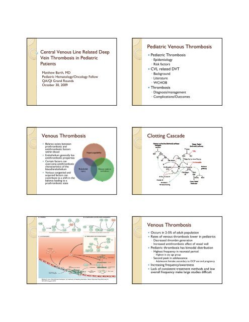
Central <strong>Venous</strong> Line Related Deep<br />
Vein <strong>Thrombosis</strong> in <strong>Pediatric</strong><br />
Patients<br />
Matthew Barth, MD<br />
<strong>Pediatric</strong> Hematology/Oncology Fellow<br />
QA/QI Grand Rounds<br />
October 30, 2009<br />
<strong>Pediatric</strong> <strong>Venous</strong> <strong>Thrombosis</strong><br />
• <strong>Pediatric</strong> <strong>Thrombosis</strong><br />
◦ Epidemiology<br />
◦ Risk factors<br />
• CVL related DVT<br />
◦ Background<br />
◦ Literature<br />
◦ WCHOB<br />
• <strong>Thrombosis</strong><br />
◦ Diagnosis/management<br />
◦ Complications/Outcomes<br />
<strong>Venous</strong> <strong>Thrombosis</strong><br />
Clotting Cascade<br />
• Balance exists between<br />
prothrombotic and<br />
antithrombotic factors<br />
within blood<br />
• Endothelium generally has<br />
antithrombotic properties<br />
• Certain factors can<br />
overcome antithrombotic<br />
characteristics of the<br />
blood/endothelium<br />
• Various congenital and<br />
acquired factors can<br />
contribute to a shift in the<br />
balance leading to a<br />
prothrombotic state<br />
<strong>Venous</strong> <strong>Thrombosis</strong><br />
• Occurs in 2-5% of adult population<br />
• Rates of venous thrombosis lower in pediatrics<br />
◦ Decreased thrombin generation<br />
◦ Increased antithrombotic effect of vessel wall<br />
• <strong>Pediatric</strong> thrombosis has bimodal distribution<br />
◦ Highest frequency in neonatal period<br />
• Highest in any age group<br />
◦ Second peak in adolescence<br />
• Adolescent females secondary to OCP use and pregnancy<br />
• Increasing frequency/awareness<br />
• Lack of consistent treatment methods and low<br />
overall frequency make large studies difficult<br />
Bishop P et al. Recombinant biologics for treatment of bleeding disorders. Nature Reviews Drug Discovery 3,<br />
684-694 (August 2004)
Frequency of <strong>Pediatric</strong> <strong>Thrombosis</strong><br />
• With increased awareness/frequency several registry<br />
studies attempted to determine overall rate<br />
• General pediatric population<br />
◦ van Ommen et al - Netherlands<br />
• 0.14 per 10,000 children<br />
◦ Monagle et al from Canadian registry<br />
• 0.07 per 10,000 children<br />
Hospitalized Patients<br />
• Increased risk in hospitalized patients<br />
◦ Canadian registry<br />
• 5.3 per 10,000 hospitalizations<br />
◦ Stein et al US National Hospital Discharge Survey<br />
• 4.9 per 10,000 childhood years<br />
• Highest risk noted in NICU patients<br />
◦ Up to 24 per 10,000 NICU admissions<br />
◦ van Ommen et al – Netherlands<br />
• 14.9/10,000<br />
• Most hospitalized children with VTE have at least<br />
one and often multiple identifiable risk factors<br />
Van Ommen et al. <strong>Venous</strong> thromboembolism in childhood: A<br />
prospective two-year registry in The Netherlands. J Pediatr 2001;139:676–81.<br />
Risk Factors for <strong>Thrombosis</strong><br />
• Congenital<br />
◦ Factor V mutations<br />
◦ Prothrombin gene<br />
mutation<br />
◦ Deficiency of protein<br />
C, protein S or<br />
antithrombin<br />
◦ Elevations of<br />
lipoprotein a,<br />
homocysteine, factor<br />
VII/VIII/IX/XI may also<br />
increase risk<br />
• Acquired<br />
◦ CVL<br />
• Most common risk<br />
• 60-90% of DVTs<br />
associated with CVL<br />
◦ Malignancy, trauma,<br />
surgery, hormone<br />
therapy, nephrotic<br />
syndrome/renal<br />
disease,<br />
antiphospholipid<br />
syndrome,<br />
medications,<br />
hemoglobinopathies,<br />
PNH<br />
Parasuraman S et al. <strong>Venous</strong> Thromboembolism in Children. Circulation.<br />
2006;113:e12-e16<br />
Central <strong>Venous</strong> Lines<br />
• Most common risk factor associated with<br />
development of DVT<br />
• Overall are the cause of majority of DVTs<br />
◦ Massicotte et al from Canadian registry<br />
◦ 244 cases of VTE<br />
◦ 60% associated with CVL<br />
◦ Schmidt et al<br />
◦ Neonates with non-renal vein thrombosis<br />
◦ 89% associated with CVL<br />
• With improved overall survival of critically and<br />
chronically ill children use of CVLs has become more<br />
frequent<br />
• Leads to increasing rate of DVT in pediatric patients<br />
Literature<br />
• Overall rate of DVT formation in the<br />
presence of a CVL varies from institution<br />
to institution and by diagnostic<br />
methodology/criteria
General <strong>Pediatric</strong> Population<br />
• Dubois et al<br />
◦ Review of 214 pediatric patients with PICC lines placed in<br />
their radiology dept<br />
◦ 9.3% of patients developed thrombus detected on<br />
screening at time line pulled<br />
• Only 1 symptomatic<br />
• Male et al<br />
◦ Prospective cohort study in general pediatric population<br />
including 158 children<br />
◦ Overall 13% developed thrombus on U/S or venogram<br />
◦ Femoral or subclavian lines were found to have statistically<br />
higher rate than brachial or jugular (32%/27% vs 12%/8%)<br />
◦ No statistically significant difference for type of line (PICC<br />
vs tunneled vs non-tunneled), size of line or duration of<br />
placement<br />
<strong>Pediatric</strong> ICU Patients<br />
• DeAngelis et al<br />
◦ 76 PICU patients screened with U/S<br />
◦ 4% with thrombus<br />
◦ All in femoral lines<br />
• Shefler et al<br />
◦ 56 PICU patients with femoral lines<br />
◦ screened with U/S of IVC<br />
◦ 11% with thrombus<br />
• One symptomatic<br />
<strong>Pediatric</strong> ICU Patients<br />
• Hanslik et al<br />
◦ Review of 90 pediatric patients with congenital<br />
heart disease with short term venous catheters<br />
◦ Predominantly jugular lines<br />
◦ Using combination of venography, U/S and Echo<br />
detected thrombus in 28% of patients<br />
• Sheridan et al<br />
◦ Review of 289 children in a burn ICU with 1056<br />
venous lines<br />
◦ Protocol includes rotating CVL sites weekly<br />
◦ Symptomatic DVT developed at site of previous<br />
cannulation in 0.6%<br />
Neonatal ICU<br />
• Butler-O’Hara et al<br />
◦ Review of 210 neonates with umbilical vein<br />
catheters<br />
◦ 20% of SGA neonates developed thrombus<br />
◦ 9% of AGA/LGA developed thrombus<br />
Oncology Patients<br />
• Male et al<br />
◦ Prospective cohort study of 85 children with ALL<br />
◦ 34% with thrombosis<br />
◦ Left sided, subclavian and percutaneously<br />
inserted catheters were independently associated<br />
with increased risk of thrombosis<br />
• Journeycake et al<br />
◦ 287 pediatric oncology patients<br />
◦ <strong>Thrombosis</strong> associated with line-related<br />
infections, repeated occlusions, need for multiple<br />
catheters<br />
CVL Related DVT<br />
• Many series report high rate of thrombus<br />
formation when screening using various<br />
methods to screen<br />
◦ When screening a group of pediatric patients<br />
with CVL regardless of symptoms >30% may<br />
have evidence of a thrombus
CVL Related DVT<br />
Outcomes with CVL Related DVT<br />
• Significantly smaller<br />
percentage of clots<br />
identified on screening<br />
are clinically<br />
symptomatic<br />
◦ Despite some even<br />
being occlusive clots<br />
• Long term outcome of<br />
the asymptomatic<br />
DVTs related to CVLs<br />
is unknown at this<br />
time<br />
Journeycake JM et al. Thrombotic complications<br />
of central venous catheters in children. Curr Opin<br />
Hematol 2003;10:369-374.<br />
• Outcomes often poorer<br />
◦ Likely related to seriousness of underlying<br />
disorders<br />
• Massicotte et al<br />
◦ Review of data from Canadian registry<br />
◦ All-cause mortality 23%<br />
◦ VTE-related mortality 4%<br />
◦ Recurrence in 6.5%<br />
◦ Post-thrombotic syndrome in 10%<br />
Prophylaxis with CVLs<br />
• Data thus far does not support<br />
prophylactic anticoagulation in children<br />
with CVLs<br />
• PROTEKT study<br />
◦ Multicenter, randomozed trial of prophylactic<br />
riviparin vs routine heparin flushes<br />
◦ Closed early with low enrollment<br />
◦ No major bleeding events<br />
◦ 14% developed a thrombus<br />
• Though only screened with venography<br />
QA/QI Analysis<br />
• Retrospective review of all patients admitted to<br />
the PICU at WCHOB from July 2008 to June<br />
2009 requiring placement of a CVL<br />
• Goals:<br />
◦ Determine rate of thrombotic complications of CVL<br />
placement<br />
◦ Identify risk factors for development of thrombosis<br />
◦ Review outcomes of patients with CVL related DVT<br />
Risk Factors<br />
• Based on historical data and theoretical risks will<br />
compare several groups of hypothetical risk<br />
factors<br />
◦ Patient age/weight<br />
◦ Admission diagnosis<br />
• Infectious , surgical, traumatic<br />
• Sepsis, malignancy<br />
◦ CVL duration<br />
◦ CVL size<br />
◦ CVL location<br />
◦ Evidence of low flow state<br />
• Hypotension requiring volume or pressor support<br />
◦ Immobility related to intubation and/or paralytics<br />
Subjects<br />
• Reviewed all available records for patients<br />
from January 2009 to June 2009<br />
◦ 637 total PICU admissions<br />
◦ 150 with CVL<br />
◦ 106 with available medical records/data<br />
• 2/106 (2%) dialysis catheters<br />
• 95/106 (90%) non-tunneled catheters<br />
• 67 (71%) femoral<br />
• 3 (3%) internal jugular<br />
• 1 (1%) subclavian<br />
• 24 (25%) PICC<br />
• 9/106 (8%) tunneled catheters<br />
• 2 Broviac<br />
• 7 Mediport
Characteristics<br />
• Evaluating individuals with non-tunneled,<br />
temporary CVLs<br />
◦ Femoral, jugular, subclavian lines<br />
• Mean age of 3.9 years<br />
• Diagnosis<br />
◦ 52/71 (73%) Infectious<br />
◦ 11/71 (15%) Other Medical<br />
◦ 4/71 (6%) Trauma<br />
◦ 6/71 (8%) Surgical<br />
<strong>Thrombosis</strong><br />
• Only 1/71 (1.5%) with symptomatic CVL<br />
related thrombus<br />
◦ Femoral line related clot in infant septic with<br />
meningococcemia<br />
◦ 2 other non-CVL-related DVTs<br />
• One related to local infection with osteomyelitis<br />
• One spontaneous DVT<br />
WCHOB PICU<br />
• Rate of thrombotic complications of<br />
CVLs comparable to reported data<br />
◦ Relatively low overall<br />
• Current numbers very small so at this<br />
point unable to assess other factors/risks<br />
<strong>Pediatric</strong> <strong>Venous</strong> <strong>Thrombosis</strong><br />
• Presentation<br />
• Diagnosis<br />
• Management<br />
• Long term<br />
◦ Complications<br />
◦ Prophylaxis?<br />
Presentation<br />
• Clinical presentation depends on site and<br />
extent of thrombus<br />
◦ Many are asymptomatic<br />
◦ Most commonly located in extremities<br />
• Swelling, pain, discoloration<br />
• SVC syndrome<br />
• Chylothorax/chylopericardium<br />
• CVL related clots often present with catheter<br />
dysfunction or catheter related sepsis<br />
• May present with thrombocytopenia from<br />
platelet consumption<br />
Diagnosis<br />
• Most commonly diagnosed with doppler<br />
ultrasound with compression<br />
◦ Non invasive<br />
◦ Sensitive and specific for diagnosing most<br />
lower extremity and many upper extremity<br />
DVTs<br />
• CT with contrast can be used for upper<br />
extremity, abdominal or pelvic clots<br />
• Venography not generally necessary but<br />
can be helpful in select situations
Acute Treatment<br />
• Anticoagulation<br />
◦ Initially with one form of heparin<br />
• Unfractionated (UFH)<br />
• Short half-life so can be turned off quickly<br />
• Low molecular weight<br />
• Decreased incidence of HIT<br />
• Requires adequate levels of antithrombin III which<br />
is normally low in neonates/infants<br />
• FFP prior to initiating therapy<br />
• Therapeutic monitoring<br />
◦ aPTT frequently does not correlate well with anti-<br />
FXa levels in pediatrics (particularly infants)<br />
Thrombolytics<br />
• Not generally used with few exceptions<br />
◦ Significant IVC thrombus or pulmonary<br />
embolism<br />
◦ Most DVTs related to CVLs are well<br />
organized by diagnosis<br />
Treatment<br />
• Beyond immediate acute treatment - two options<br />
◦ Warfarin<br />
• Oral<br />
• Affected by diet, absorption, other medications<br />
• Sensitivity to drug varies at different developmental stages<br />
• May require freq. INR monitoring to maintain therapeutic range<br />
• Long term use associated with risk of osteoporosis<br />
◦ LMWH<br />
• Daily subcutaneous injection<br />
• More predictable pharmacokinetics - less frequent monitoring<br />
• REVIVE trial<br />
◦ Randomized comparison of LMWH vs UFH/oral<br />
anticoagulant<br />
◦ Closed early due to poor accrual<br />
◦ Recurrence: 6% rivaparin vs 13% UFH/oral anticoag<br />
• Not significant though likely due to low power<br />
Evaluation of Initial Thrombus<br />
• Identify risk factors for recurrent<br />
thrombosis<br />
• Most pediatric cases have transient risk<br />
factors such as underlying disease state,<br />
CVL<br />
• Identify congenital, non-modifiable risk<br />
factors that may impact future treatment<br />
or counseling<br />
Risk Factors for <strong>Thrombosis</strong><br />
• Congenital<br />
◦ Factor V mutations<br />
◦ Prothrombin gene<br />
mutation<br />
◦ Deficiency of protein<br />
C, protein S or<br />
antithrombin<br />
◦ Elevations of<br />
lipoprotein a,<br />
homocysteine, factor<br />
VII/VIII/IX/XI may also<br />
increase risk<br />
• Acquired<br />
◦ CVL<br />
• Most common risk<br />
• 60-90% of DVTs<br />
associated with CVL<br />
◦ Malignancy, trauma,<br />
surgery, hormone<br />
therapy, nephrotic<br />
syndrome/renal<br />
disease,<br />
antiphospholipid<br />
syndrome,<br />
medications,<br />
hemoglobinopathies,<br />
PNH<br />
Risk Factors for <strong>Thrombosis</strong><br />
• Idiopathic venous thrombosis is rare in pediatrics<br />
• Almost always an underlying risk factor/disease<br />
• Andrew et al<br />
◦ Canadian registry<br />
◦ 50% with 3-4 identified risk factors<br />
◦
Initial Evaluation<br />
• Generally includes<br />
◦ Protein C Activity*<br />
◦ Protein S Levels* (Total/Free)<br />
◦ Antithrombin III*<br />
◦ LUPAC panel (Anticardiolipin Ab, dRVVT)<br />
◦ Activated protein C (APC) resistance<br />
• Potentially screen for factor V Leiden and other mutations<br />
◦ Homocysteine<br />
• Potentially screen for MTHFR mutations<br />
◦ Prothrombin 20210A mutation<br />
◦ Factor VIII<br />
◦ D-dimer<br />
◦ PT/PTT/Fibrinogen<br />
◦ ESR<br />
Congenital Risk Factors<br />
• Factor V Leiden<br />
◦ Mutation at cleavage site for activated protein<br />
C (APC)<br />
◦ Results in decreased sensitivity to natural<br />
anticoagulation effect of protein C and<br />
increased thrombin generation<br />
◦ Risk increased 7-fold for heterozygous and<br />
80-fold for homozygous<br />
◦ Around 5% of US population are carriers<br />
Congenital Risk Factors<br />
• Antithrombin deficiency<br />
◦ Leads to excess thrombin formation<br />
◦ Heterozygotes with 5-fold increased risk<br />
• Homozygous rare, likely not compatible with life<br />
• Protein C and Protein S deficiencies<br />
◦ Controls activity of factors Va and VIIIa<br />
◦ Heterozygotes with 5-10-fold risk<br />
◦ Homozygous generally present with purpura<br />
fulminans in infancy<br />
Congenital Risk Factors<br />
• Prothrombin 20210A mutation<br />
◦ Increased prothrombin levels<br />
◦ Heterozygotes with 3-fold increased risk<br />
• Hyperhomocysteinemia<br />
◦ Approximately 2.5-fold increased risk<br />
◦ Increases risk when associated with other<br />
known risk factors particularly factor V<br />
Leiden<br />
◦ Unknown mechanism<br />
◦ Related to mutation in MTHFR gene<br />
Acquired Risk Factors<br />
• Central venous line<br />
◦ Significant risk factor for venous thrombosis<br />
◦ Likely related to<br />
• <strong>Venous</strong> stasis<br />
• Turbulent blood flow around line<br />
• Endothelial cell damage<br />
• Thrombogenicity of catheter surface<br />
• Antiphospholipid Antibodies (Lupus anticoagulant)<br />
◦ Anticardiolipin, Antiphosphatidylserine, β2 glycoprotein Ab<br />
◦ Present in 6-8% of general population<br />
◦ Often associated with collagen vascular diseases or some<br />
medications<br />
◦ Mechanism not clear but may be related to impaired regulation<br />
of thrombin, acquired abnomalities in protein C-protein S<br />
system<br />
◦ Persistent presence increases risk around 20-fold i.e.<br />
antiphospholipid syndrome<br />
Acquired Risk Factors<br />
• Trauma<br />
◦ Significant risk factor for thrombosis in adult<br />
patients, but not in pediatrics<br />
◦ Azu et al<br />
• Retrospective review at Level 1 Trauma center<br />
• No thrombotic events in 2,320 patients under 13yo<br />
• Two PEs in 1,025 patients from 13-17yo<br />
• Both with high trauma scores<br />
• Most received prophylaxis in this group
Malignancy<br />
• Most common associated medical condition in CVL<br />
related VTE in Canadian Registry<br />
• Cancer itself induces hypercoagulable state<br />
• Some therapies have thrombotic risks<br />
• Highest rates in ALL<br />
◦ Reported rates from 1%-32%<br />
◦ Average rate about 3%<br />
◦ Higher risk related to frequent use of Asparaginase<br />
• Halts protein synthesis including those in the Protein C-Protein S system<br />
• Additional risks related to<br />
◦ Frequent long-term use of CVLs<br />
◦ Tumor compression of vessels<br />
Therapy Duration<br />
• No well established recommendations in<br />
pediatrics<br />
◦ Mostly extrapolated from adult data<br />
• Most uncomplicated clots treated 3-6 months<br />
• Individuals with clot progression, occlusive<br />
clots, persistently elevated inflammatory<br />
markers may be at increased risk of treatment<br />
failure<br />
◦ May benefit from longer period<br />
◦ Persistent elevation of Factor VIII or D-dimer may<br />
predict recurrence risk<br />
Complications<br />
• Thromboembolic events<br />
◦ Deep Vein <strong>Thrombosis</strong><br />
◦ Pulmonary embolism<br />
• Estimated to occur in 30-60% of patients w/documented DVT<br />
◦ Cerebrovascular events<br />
◦ Other (usually specific situations)<br />
• Portal Vein <strong>Thrombosis</strong><br />
• Renal Vein <strong>Thrombosis</strong><br />
• Superior Mesenteric Artery Syndrome<br />
• Treatment related complications<br />
◦ Bleeding though generally rare<br />
Long Term Clot Resolution<br />
• Up to 50% of clots fail to completely resolve<br />
following therapy<br />
• Recurrent <strong>Thrombosis</strong><br />
◦ Nowak-Gottl et al Childhood Thrombophilia Study<br />
Group registry<br />
• Registry of 301 neonates/children treated with 6m anticoag<br />
• 21% recurrence with mean f/u 7 years<br />
• Increasing data supporting importance of factor<br />
VIII and D-dimer in risk for recurrence<br />
◦ Goldenberg, NA et al<br />
• Elevated levels of either or both at diagnosis and at 3-6 months<br />
highly predictive of poor outcomes defined as incomplete clot<br />
resolution, recurrent thrombosis or post-thrombotic syndrome<br />
◦ Cosmi et al<br />
• D-dimer and factor VIII elevation 1 month following withdrawal<br />
of therapy are independent risk factors for recurrent VTE<br />
Post-Thrombotic Syndrome<br />
• Destruction of venous valves by thrombus or persistent<br />
thrombus<br />
◦ <strong>Venous</strong> hypertension<br />
◦ Increased hydrostatic pressure to soft tissue and skin<br />
• Most commonly in lower extremities<br />
• Pain, swelling, skin pigmentation, ulceration<br />
• Reported to affect up to 60% of patients<br />
◦ Less frequently in pediatrics<br />
• Monagle et al from Canadian pediatric registry<br />
◦ Over 12% diagnosed with PTS at mean f/u of 3y<br />
• No comment on severity of symptoms<br />
• Generally mild in children<br />
◦ Increased limb circumference, swelling, varicose veins, pain<br />
◦ Rarely ulcerations<br />
Mortality<br />
• Neonates<br />
◦ Schmidt et al from Canadian registry<br />
• All cause mortality 18%<br />
• Did not report VTE-specific mortality<br />
• Beyond Neonatal period<br />
◦ Monagle et al report from Canadian registry<br />
• Median f/u of 3 years in 356 patients<br />
• 2% VTE-related mortality<br />
• All deaths in CVL-related clots<br />
◦ Several other series with similar rates of 1-4%
Prophylaxis<br />
• Having genetic prothrombotic disorder does<br />
not necessarily require anticoagulation<br />
◦ Risks of long term anticoagulation often outweigh<br />
the benefit<br />
• Most asymptomatic individuals (no VTE<br />
history) with known prothrombotic<br />
disorder do not require prophylaxis<br />
◦ Screening asymptomatic individuals for<br />
prothrombotic disorders also generally not<br />
necessary<br />
Symptomatic Prophylaxis<br />
• Individuals with significant congenital risk factor or<br />
antiphospholipid syndrome may be considered for long term<br />
anticoagulation<br />
◦ Initial thrombotic event - often anticoagulated up to 6 months<br />
◦ After initial therapy need to decide on long term prophylaxis<br />
◦ Many would save long-term prophylaxis for individuals with<br />
recurrent thrombosis<br />
• Depends on age, presentation and severity of underlying<br />
disorder<br />
◦ Lifelong prophylaxis difficult when a diagnosis of thrombosis<br />
occurs in young age<br />
◦ Spontaneous clot<br />
◦ Significant/life-threatening clot<br />
Summary<br />
• <strong>Thrombosis</strong> is rare in kids<br />
◦ Most often seen with acquired risk factor<br />
◦ Most common risk is presence of a CVL<br />
• Overall still rare in patients with CVLs and<br />
prophylaxis likely not indicated<br />
• Underlying predisposition/thrombophilia<br />
adds to risk<br />
◦ May help guide therapeutic decisions<br />
◦ Despite added risk, screening in<br />
asymptomatic patients not necessary<br />
References<br />
• Goldenberg et al. Long-term outcomes of venous thrombosis in children. Current<br />
Opinion in Hematology 2005, 12:370—376.<br />
• Hagstrom et al. Prevalence of the factor V Leiden mutation in children and neonates with<br />
thrombotic disease. J Pediatr 1998, 133(6):777-781.<br />
• Massicotte et al. Central venous catheter related thrombosis in children: Analysis of the<br />
Canadian Registry of <strong>Venous</strong> Thromboembolic Complications. J Pediatr 1998,<br />
133(6):770-776.<br />
• Goldenberg N et al. Elevated plasma factor VIII and D-dimer levels as predictors of poor<br />
outcomes of thrombosis in children. N Engl J Med. 351(11):1081-1088.<br />
• Barnes C et al. Post-thrombotic syndrome. Arch Dis Child 2002;86:212-214.<br />
• Andrew M et al. <strong>Venous</strong> thrombotic complications (VTE) in children; first analysis of the<br />
Canadian registry of VTE. Blood 1994;83:1251-1257.<br />
• Monagle P et al. Outcome of pediatric thrombophilic disease: a report from the Canadian<br />
Childhood Thrombophilia Registry. Pedatr Res 2000;47:763-766.<br />
• Parsuraman S et al. <strong>Venous</strong> Thromboembolism in Children. Circulation 2006;113:e12-16.<br />
• Schmidt B et al. Neonatal thrombosis: report of a prospective Canadian and International<br />
Registry. <strong>Pediatric</strong>s 1995;96:939-943.<br />
• DuBois J et al. Incidence of deep vein thrombosis related to peripherally inserted central<br />
catheters in children and adolescents. CMAJ Can Med Ass J. 2007;177(10):1185-1190.<br />
More References<br />
• Sheridan RL et al. Mechanical and infectious complications of central venous cannulation<br />
in children: lessons learned from a 10-year experience placing more than 1000 catheters.<br />
Journal of Burn Care and Research. 2006;27(5):713-718.<br />
• Hanslik A et al. Incidence and diagnosis of thrombosis in children with short term central<br />
venous lines of the upper venous system. <strong>Pediatric</strong>s 2008;122(6):1284-1291.<br />
• Male C et al. Central venous line–related thrombosis in children: association with central<br />
venous line location and insertion technique. Blood 2003;101(11):4273-4278.<br />
• DeAngelis GA et al. Prevalence of deep venous thrombosis in the lower extremities of<br />
children in the intensice care unit. Pediatr Radiol 1996;26:821-824.<br />
• Sheffler A et al. Inferior vena cava thrombosis as a complication of femoral vein<br />
catheterization. Arch Dis Child 1995;72:343-345.<br />
• Van Ommen et al. <strong>Venous</strong> thromboembolism in childhood: A prospective two-year<br />
registry in The Netherlands. J Pediatr 2001;139:676–81<br />
• Azu CM. <strong>Venous</strong> Thromboembolic Events in <strong>Pediatric</strong> Trauma Patients: Is Prophylaxis<br />
Necessary? J Trauma. 2005;59:1345–1349.<br />
• Monagle P et al. Antithrombotic Therapy in Neonates and Children* American College of<br />
Chest Physicians Evidence-Based Clinical Practice Guidelines (8th Edition). CHEST 2008;<br />
133:887S–968S.