Long term prognosis of symptomatic occipital lobe epilepsy ... - sepeap
Long term prognosis of symptomatic occipital lobe epilepsy ... - sepeap
Long term prognosis of symptomatic occipital lobe epilepsy ... - sepeap

Create successful ePaper yourself
Turn your PDF publications into a flip-book with our unique Google optimized e-Paper software.
<strong>Long</strong>itudinal clinical course 97<br />
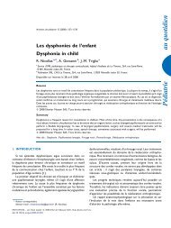
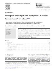
Figure 3 Sleep EEG recorded at the age <strong>of</strong> 5 years showing<br />
frequent sharp waves in the right <strong>occipital</strong> region.<br />
Figure 4 Sleep EEG recorded at the age <strong>of</strong> 5 years showing<br />
frequent sharp waves in the right <strong>occipital</strong> region.<br />
EEG and MRI findings (Table 4 and Figs. 3—5)<br />
Interictal EEGs showed <strong>occipital</strong> or parieto-<strong>occipital</strong> spikes<br />
in all patients (Figs. 3 and 4) during infancy and early childhood.<br />
Frequency and amplitude <strong>of</strong> spikes were gradually<br />
decreased and disappeared in 1 patient (Patient 3). Frontal<br />
spikes as well as <strong>occipital</strong> spikes were also seen in 1 patient<br />
(Patient 2). Frontal or central spikes were seen in late childhood<br />
or adolescence in 3 patients (Patients 2—4), while their<br />
clinical seizure symptoms went unchanged.<br />
MRI revealed cortical atrophy and T2 prolongation in the<br />
parieto-<strong>occipital</strong> region in 4 patients, <strong>of</strong> whom additional<br />
frontal lesions were present in 3 patients (Figs. 2, 3 and 5).<br />
One patient (Patient 6) showed hippocampal atrophy on both<br />
sides without temporal <strong>lobe</strong> seizures. MRI was unremarkable<br />
in 1 patient (Patient 4).<br />
Discussion<br />
It is well known that hypoglycemic brain injury predominantly<br />
involves the <strong>occipital</strong> <strong>lobe</strong> (Murakmi et al., 1999;<br />
Caraballo et al., 2004; Filan et al., 2006; Yalnizoglu and<br />
Halioglu, 2007). We reported on 6 patients who had a history<br />
<strong>of</strong> neonatal hypoglycemia and <strong>occipital</strong> <strong>lobe</strong> <strong>epilepsy</strong><br />
later on. Our patients had ictal symptoms compatible with<br />
<strong>occipital</strong> <strong>lobe</strong> <strong>epilepsy</strong>, such as eye deviation, eye blinking,<br />
staring, visual hallucinations, and ictal vomiting (Williamson<br />
et al., 1992). Interictal EEGs showed spike waves in the<br />
posterior region during the active periods <strong>of</strong> <strong>epilepsy</strong> in all<br />
patients. MRI also revealed posterior cerebral damage in 4<br />
patients. These findings indicate that epileptic foci <strong>of</strong> the<br />
patients could mainly be caused by neonatal hypoglycemia.<br />
Although 2 patients (Patients 4 and 6) could not be identified<br />
with posterior cerebral lesion on MRI, they showed<br />
typical clinical symptoms and EEG findings for <strong>occipital</strong><br />
<strong>lobe</strong> <strong>epilepsy</strong>. They could not be diagnosed as having idiopathic<br />
childhood <strong>occipital</strong> <strong>epilepsy</strong> coincidentally, because<br />
the onset and clinical course <strong>of</strong> seizures were not compatible<br />
with those <strong>of</strong> Panayiotopoulos syndrome or Gastaut type.<br />
They may have had very mild <strong>occipital</strong> lesions which could<br />
not be recognized on MRI.<br />
Perinatal hypoxic-ischemic encephalopathy in <strong>term</strong><br />
infants also causes parieto-<strong>occipital</strong> <strong>lobe</strong> <strong>epilepsy</strong> (Gil-Nagel<br />
et al., 2005; Oguni et al., 2008). It is sometimes difficult to<br />
distinguish between perinatal hypoxic-ischemic brain injury<br />
and neonatal hypoglycemic brain injury in clinical symptoms<br />
and neuroimaging findings. The perinatal hypoxic-ischemic<br />
brain injury in <strong>term</strong> infants usually involves the deeper sulcal<br />
portion <strong>of</strong> the convolutions and spares the crowns, so<br />
called ‘‘mushroom gyri’’ or ‘‘ulegyria’’. The brain lesions<br />
are located in parasagittal watershed vascular territories,<br />
such as frontal and parieto-<strong>occipital</strong> <strong>lobe</strong>s. It is usually more<br />
marked in the posterior regions at the watershed zone <strong>of</strong><br />
the 3 major cerebral arteries. Perinatal hypoxic-ischemic<br />
encephalopathy in <strong>term</strong> infants usually causes moderate to<br />
severe neurological deficit, including mental or developmental<br />
delay, motor palsy, visual impairment, and <strong>epilepsy</strong>,<br />
while milder cases have been reported (Oguni et al., 2008).<br />
Although EEG abnormalities and epileptic foci may be multifocal<br />
or widespread, parieto-<strong>occipital</strong> <strong>lobe</strong> <strong>epilepsy</strong> may be<br />
common. These two conditions sometimes coexist simultaneously<br />
and would enhance each other, creating more brain<br />
injury. In the series <strong>of</strong> Oguni et al. (2008), 3 <strong>of</strong> the 10<br />
patients with parieto-<strong>occipital</strong> <strong>lobe</strong> <strong>epilepsy</strong> as sequela <strong>of</strong><br />
perinatal asphyxia were reported to have hypoglycemia in<br />
the neonatal period. In the reports <strong>of</strong> neonatal hypoglycemic<br />
encephalopathy, other associated factors including perinatal<br />
asphyxia and fetal distress were highly present (Murakmi<br />
et al., 1999; Yalnizoglu and Halioglu, 2007; Montassir et<br />
al., 2009). In the present study, additional perinatal factors<br />
which were possibly associated with brain injury other than<br />
hypoglycemia were highly present; low birth weight for gestational<br />
age in 5, low Apgar score in 3, neonatal seizure in 5.<br />
MRI demonstrated abnormalities in the frontal <strong>lobe</strong> as well<br />
as the <strong>occipital</strong> <strong>lobe</strong> in 3 patients (Patients 2, 3, and 5). MRI<br />
<strong>of</strong> Patients 2 and 5 showed ‘‘mushroom gyri’’ or ‘‘ulegyria’’<br />
in the posterior regions. These findings indicate that not<br />
only hypoglycemia but also perinatal hypoxia or ischemia<br />
played an important role in the genesis <strong>of</strong> brain injury. The<br />
vulnerability <strong>of</strong> the brain to hypoglycemia is enhanced by<br />
concomitant other perinatal factors. In <strong>term</strong> infants, therefore,<br />
hypoglycemic encephalopathy and hypoxic-ischemic<br />
encephalopathy <strong>of</strong>ten overlap and sometimes cannot be distinguishable<br />
in practice. There may be some difference in<br />
response to treatment; epileptic patients with perinatal