Cladosporium leaf-blotch and stem rot of Paeonia spp. caused ... - Cbs
Cladosporium leaf-blotch and stem rot of Paeonia spp. caused ... - Cbs
Cladosporium leaf-blotch and stem rot of Paeonia spp. caused ... - Cbs

Create successful ePaper yourself
Turn your PDF publications into a flip-book with our unique Google optimized e-Paper software.
www.studiesinmycology.org<br />
10 changes<br />
Botryosphaeria ribis DQ316076<br />
Botryosphaeria stevensii AY343484<br />
100<br />
97<br />
70<br />
100<br />
100<br />
100<br />
97<br />
100<br />
100<br />
69<br />
<strong>Cladosporium</strong> tenellum CPC 12053<br />
<strong>Cladosporium</strong> variabile CPC 12753<br />
<strong>Cladosporium</strong> herbarum CPC 11600<br />
<strong>Cladosporium</strong> macrocarpum CBS 299.67<br />
<strong>Cladosporium</strong> bruhnei CPC 12211<br />
<strong>Cladosporium</strong> ossifragi CBS 842.91<br />
<strong>Cladosporium</strong> iridis CBS 138.40<br />
CPC 11383<br />
CPC 11969<br />
CBS 213.73<br />
CBS 100405<br />
100<br />
91<br />
Mycosphaerella punctiformis AY490763<br />
Passalora arachidicola AF297224<br />
Mycosphaerella aurantia AY509744<br />
Passalora fulva AF393701<br />
Cercospora apii AY840512<br />
Cercospora beticola AY840527<br />
Readeriella mirabilis AY725529<br />
Readeriella novaezel<strong>and</strong>iae DQ267603<br />
Teratosphaeria readeriellophora AY725577<br />
Teratosphaeria microspora AY260098<br />
Trimmatostroma abietis AY559362<br />
Trimmatostroma abietina AJ244267<br />
Trimmatostroma salicis AJ244264<br />
Dichocladosporium chlorocephalum<br />
DichoclaDosporium gen. nov.<br />
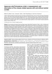
Fig. 1. One <strong>of</strong> two equally most parsimonious trees obtained from a heuristic search with 100 r<strong>and</strong>om taxon additions <strong>of</strong> the ITS sequence alignment. The scale bar shows<br />
10 changes, <strong>and</strong> bootstrap support values from 1 000 replicates are shown at the nodes. Thickened lines indicate the strict consensus branches. The tree was rooted to two<br />
Botryosphaeria species.<br />
Characters <strong>of</strong> the cladosporioid morph: Leaf-<strong>blotch</strong> symptoms on<br />
living leaves amphigenous, variable in shape <strong>and</strong> size, subcircularoval<br />
to irregular, broad, oblong to exp<strong>and</strong>ed, up to 30 mm long<br />
<strong>and</strong> 20 mm wide, at times covering the entire <strong>leaf</strong> surface, forming<br />
olivaceous-brown to blackish brown patches, rarely violet-brown,<br />
margin usually indefinite, attacked areas turning dry with age,<br />
also occurring on young, green <strong>stem</strong>s. Colonies amphigenous,<br />
punctiform to effuse, loose to dense, caespitose, brown, villose.<br />
Mycelium immersed, subcuticular to intraepidermal; hyphae<br />
sparingly branched, 4–7(–10) µm wide, septate, sometimes with<br />
swellings <strong>and</strong> constrictions, swollen cells up to 13 µm diam,<br />
subhyaline to pale brown, smooth, walls thickened, hyphae<br />
sometimes aggregated; in vitro mycelium at first mainly immersed,<br />
later also superficial, branched, 1–5(–7) µm wide, pluriseptate,<br />
<strong>of</strong>ten constricted at septa <strong>and</strong> with swellings <strong>and</strong> constrictions,<br />
therefore irregular in outline, smooth to verruculose or irregularly<br />
rough-walled, loosely verruculose with distinct large warts. Semimacronematous<br />
conidiophores formed on <strong>leaf</strong>-<strong>blotch</strong>es solitary<br />
or in small, loose groups, arising from internal hyphae or swollen<br />
hyphal cells, erumpent through the cuticle, occasionally emerging<br />
through stomata, erect, straight to somewhat flexuous, oblongcylindrical,<br />
usually unbranched or occasionally branched, 13–80<br />
(–120) × (4–)5–8(–10) µm, slightly attenuated towards the apex,<br />
septate, septa <strong>of</strong>ten dense, unconstricted, pale to medium brown,<br />
sometimes paler towards the apex, smooth, thick-walled, wall <strong>of</strong>ten<br />
with two distinct layers, <strong>of</strong>ten somewhat inflated at the very base,<br />
up to 14 µm diam, occasionally proliferating enteroblastically; in<br />
vitro conidiophores arising laterally from plagiotropous hyphae or<br />
terminally from ascending hyphae, the latter usually appearing<br />
more filiform than those arising laterally from plagiotropous hyphae,<br />
erect, straight to slightly flexuous, cylindrical-oblong, not geniculate,<br />
usually unbranched, rarely with a short lateral prolongation near<br />
the apex, 18–60(–100) × 3–6 µm, slightly attenuated towards<br />
the apex, septate, pale to medium brown or olivaceous-brown,<br />
smooth to asperulate, walls somewhat thickened. Conidiogenous<br />
cells integrated, terminal or intercalary, subcylindrical, 7–45 µm<br />
97