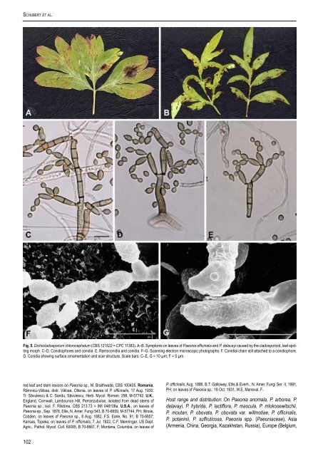
Schubert et al. Fig. 5. Dichocladosporium chlorocephalum (CBS 121522 = CPC 11383). A–B. Symptoms on leaves <strong>of</strong> <strong>Paeonia</strong> <strong>of</strong>ficinalis <strong>and</strong> P. delavayi <strong>caused</strong> by the cladosporioid, <strong>leaf</strong> spotting morph. C–D. Conidiophores <strong>and</strong> conidia. E. Ramoconidia <strong>and</strong> conidia. F–G. Scanning electron microscopic photographs. F. Conidial chain still attached to a conidiophore. G. Conidia showing surface ornamentation <strong>and</strong> scar structure. Scale bars: C–E, G = 10 µm; F = 5 µm. red <strong>leaf</strong> <strong>and</strong> <strong>stem</strong> lesions on <strong>Paeonia</strong> sp., M. Braithwaite, CBS 100405. romania, Râmnicu-Vâlcea, distr. Vâlcea, Oltenia, on leaves <strong>of</strong> P. <strong>of</strong>ficinalis, 17 Aug. 1930, Tr. Săvulescu & C. S<strong>and</strong>u, Săvulescu, Herb. Mycol. Roman. 298, M-57742. u.K., Engl<strong>and</strong>, Cornwall, Lambounce Hill, Perranzubuloe, isolated from dead <strong>stem</strong>s <strong>of</strong> <strong>Paeonia</strong> sp., isol. F. Rilstone, CBS 213.73 = IMI 048108a. u.s.A., on leaves <strong>of</strong> <strong>Paeonia</strong> sp., Sep. 1878, Ellis, N. Amer. Fungi 543, B 70-6659, M-57744, PH; Illinois, Cobden, on leaves <strong>of</strong> <strong>Paeonia</strong> sp., 8 Aug. 1882, F.S. Earle, No. 91, B 70-6657; Kansas, Topeka, on leaves <strong>of</strong> P. <strong>of</strong>ficinalis, 7 Jul. 1922, C.F. Menninger, US Dept. Agric., Pathol. Mycol. Coll. 60085, B 70-6661, F; Montana, Columbia, on leaves <strong>of</strong> 102 P. <strong>of</strong>ficinalis, Aug. 1886, B.T. Galloway, Ellis & Everh., N. Amer. Fungi Ser. II, 1991, PH; on leaves <strong>of</strong> <strong>Paeonia</strong> sp., 18 Oct. 1931, W.E. Maneval, F. Host range <strong>and</strong> distribution: On <strong>Paeonia</strong> anomala, P. arborea, P. delavayi, P. hybrida, P. lactiflora, P. mascula, P. mlokosewitschii, P. moutan, P. obovata, P. obovata var. willmotiae, P. <strong>of</strong>ficinalis, P. potaninii, P. suffruticosa, <strong>Paeonia</strong> <strong>spp</strong>. (<strong>Paeonia</strong>ceae), Asia (Armenia, China, Georgia, Kazakhstan, Russia), Europe (Belgium,
Czechoslovakia, Denmark, France, Germany, Italy, Latvia, Moldova, Pol<strong>and</strong>, Romania, Switzerl<strong>and</strong>, U.K., Ukraine), North America (Canada, U.S.A.), New Zeal<strong>and</strong>. Notes: Type material <strong>of</strong> Periconia chlorocephala is not preserved in the herbarium <strong>of</strong> G. Fresenius at FR (Forschungsinstitut Senkenberg, Frankfurt a. M., Germany). Hence, a new specimen collected in the Botanical Garden <strong>of</strong> the Martin-Luther-University Halle (Saale), Germany, is proposed to serve as neotype. A culture derived from this collection is deposited at the CBS, Utrecht, the Netherl<strong>and</strong>s as ex-neotype culture. A <strong>leaf</strong>-<strong>blotch</strong> sample, also collected in the Botanical Garden at Halle (Saale), from which we also derived a living culture, is designated as representative <strong>of</strong> the synanamorph, <strong>Cladosporium</strong> paeoniae. Both cultures have been used to generate DNA sequence data. The two stages (morphs) <strong>of</strong> this fungus are usually ecologically <strong>and</strong> seasonally separated, but sometimes conidiophores <strong>of</strong> the <strong>leaf</strong>-<strong>blotch</strong>ing (cladosporioid) morph also occur on dead <strong>stem</strong>s <strong>of</strong> peony intermixed with the macronematous conidiophores <strong>of</strong> the periconioid morph. In culture conidiophore <strong>and</strong> conidial width tends to be narrower than on the natural substratum, <strong>and</strong> the conidia are not as frequently septate. dIsCussIon Cultural studies by ourselves <strong>and</strong> McKemy & Morgan-Jones (1991), <strong>and</strong> molecular sequence analyses documented herein clearly demonstrate that <strong>Cladosporium</strong> chlorocephalum, occurring on nec<strong>rot</strong>ic <strong>stem</strong>s, <strong>and</strong> C. paeoniae, causing <strong>leaf</strong>-<strong>blotch</strong> symptoms on living leaves <strong>of</strong> <strong>Paeonia</strong> <strong>spp</strong>., are two synanamorphs <strong>of</strong> a single species, which has to be excluded from <strong>Cladosporium</strong> s. str. since the conidiogenous loci are quite distinct from the characteristically coronate scars in the latter genus <strong>and</strong> because ITS sequences indicate clear separation from <strong>Cladosporium</strong> s. str. Analysis <strong>of</strong> the morphology <strong>and</strong> conidiogenesis showed that the macronematous stage <strong>of</strong> this fungus (C. chlorocephalum, the periconioid morph) closely resembles Metulocladosporiella, recently introduced for the <strong>Cladosporium</strong> speckle disease <strong>of</strong> banana. There are, however, some differences. In Metulocladosporiella musae (E.W. Mason) Crous et al., the type species, micronematous conidiophores occur in vitro <strong>and</strong> in vivo, <strong>and</strong> macronematous conidiophores occur on <strong>leaf</strong>-spots, whereas in C. chlorocephalum the semi-macronematous conidiophores usually accompany <strong>leaf</strong>-<strong>blotch</strong> symptoms on living leaves <strong>and</strong> the macronematous conidiophores occur in saprobic growth on old nec<strong>rot</strong>ic <strong>stem</strong>s. Rhizoid hyphae arising from the swollen basal cells <strong>of</strong> the macronematous conidiophores are characteristic for M. musae, but lacking in C. chlorocephalum, <strong>and</strong> the conidia in the latter species are 0–5-septate, but only 0(–1)-septate in M. musae. The semimacronematous, <strong>leaf</strong>-<strong>blotch</strong>ing stage (the cladosporioid morph) is barely distinguishable from the present concept <strong>of</strong> Fusicladium, which includes species with catenate conidia (Schubert et al. 2003). However, the peony fungus does not cluster within the Venturiaceae. Since C. chlorocephalum clusters apart <strong>of</strong> the Chaetothyriales, the clade to which Metulocladosporiella belongs, the differences observed here seem to be sufficient to place this fungus in a new genus (also see Crous et al. 2007 – this volume). Crous et al. (2006a) discussed differences between Metulocladosporiella <strong>and</strong> allied dematiaceous hyphomycete genera <strong>and</strong> provided a key to the latter genus <strong>and</strong> morphologically similar genera. Using this key, attempts to determine the macronematous morph <strong>of</strong> <strong>Cladosporium</strong> www.studiesinmycology.org DichoclaDosporium gen. nov. chlorocephalum lead to Metulocladosporiella. Differences between morphologically similar genera have been discussed in the paper by Crous et al. (2006a) <strong>and</strong> are also valid for the new genus Dichocladosporium. Parapericoniella U. Braun, Heuchert & K. Schub., a fungicolous genus recently introduced to accommodate <strong>Cladosporium</strong> asterinae Deighton, is also morphologically similar in having apically, densely branched conidiophores <strong>and</strong> truncate, unthickened conidiogenous loci <strong>and</strong> hila, but is quite distinct in not having micronematous conidiophores (Heuchert et al. 2005). ACKnoWledGeMenTs We are much obliged to the curators <strong>of</strong> B, F, HBG, M, PH <strong>and</strong> SIENA for the loans <strong>of</strong> the collections studied. R. Kirschner <strong>and</strong> N. Ale-Agha are thanked for sending collections <strong>and</strong> cultures on <strong>Paeonia</strong> <strong>spp</strong>. We are very grateful to the Institute <strong>of</strong> Zoology <strong>of</strong> the Martin-Luther-University, above all to G. Tschuch, for providing access to SEM facilities. This work was supported in part by a grant <strong>of</strong> the “Graduiertenförderung des L<strong>and</strong>es Sachsen-Anhalt” <strong>and</strong> a grant <strong>of</strong> Synthesys (No. 2559) awarded to K.S. We thank M. Vermaas for preparing the photographic plates. reFerenCes Braun U, Crous PW, Dugan FM, Groenewald JZ, Hoog GS de (2003). Phylogeny <strong>and</strong> taxonomy <strong>of</strong> cladosporium-like hyphomycetes, including Davidiella gen. nov., the teleomorph <strong>of</strong> <strong>Cladosporium</strong> s.str. Mycological Progress 2: 3–18. Crous PW (1998). Mycosphaerella <strong>spp</strong>. <strong>and</strong> their anamorphs associated with <strong>leaf</strong> spot diseases <strong>of</strong> Eucalyptus. Mycologia Memoir 21: 1–170. Crous PW, Braun U, Schubert K, Groenewald JZ (2007). Delimiting <strong>Cladosporium</strong> from morphologically similar genera. Studies in Mycology 58: 33–56. Crous PW, Schroers HJ, Groenewald JZ, Braun U, Schubert K (2006a). Metulocladosporiella gen. nov. for the causal organism <strong>of</strong> <strong>Cladosporium</strong> speckle disease <strong>of</strong> banana. Mycological Research 110: 264–275. Crous PW, Slippers B, Wingfield MJ, Rheeder J, Marasas WFO, Phillips AJL, Alves A, Burgess T, Barber P, Groenewald JZ (2006b). Phylogenetic lineages in the Botryosphaeriaceae. Studies in Mycology 55: 235–253. David JC (1997). A contribution to the sy<strong>stem</strong>atics <strong>of</strong> <strong>Cladosporium</strong>. Revision <strong>of</strong> the fungi previously referred to Heterosporium. Mycological Papers 172: 1–157. Dhingra OD, Sinclair JB (1985). Basic plant pathology methods. CRC Press, Boca Raton, Florida. Fresenius JBGW (1850). Beiträge zur Mykologie 1. Heinrich Ludwig Brömmer Verlag, Frankfurt. Gams W, Verkley GJM, Crous PW (2007). CBS Course <strong>of</strong> Mycology. 5 th ed. CBS, Utrecht. Heuchert B, Braun U, Schubert K (2005). Morphotaxonomic revision <strong>of</strong> fungicolous <strong>Cladosporium</strong> species (hyphomycetes). Schlechtendalia 13: 1–78. Hoog GS de, Gerrits van den Ende AHG (1998). Molecular diagnostics <strong>of</strong> clinical strains <strong>of</strong> filamentous Basidiomycetes. Mycoses 41: 183–189. Kiffer E, Morelet M (1999). The Deuteromycetes. Mitosporic Fungi, Classification <strong>and</strong> Generic Key. Science Publishers, Enfield, NJ. Lee SB, Taylor JW (1990). Isolation <strong>of</strong> DNA from fungal mycelia <strong>and</strong> single spores. In: PCR P<strong>rot</strong>ocols: a guide to methods <strong>and</strong> applications (Innis MA, Gelf<strong>and</strong> DH, Sninsky JJ, White TJ, eds). Academic Press, San Diego, California: 282–287. Mason EW, Ellis MB (1953). British species <strong>of</strong> Periconia. Mycological Papers 56: 1–127. McKemy JM, Morgan-Jones G (1991). Studies in the genus <strong>Cladosporium</strong> sensu lato III. Concerning <strong>Cladosporium</strong> chlorocephalum <strong>and</strong> its synonym <strong>Cladosporium</strong> paeoniae, the causal organism <strong>of</strong> <strong>leaf</strong>-<strong>blotch</strong> <strong>of</strong> peony. Mycotaxon 41: 135– 146. Meuli LJ (1937). <strong>Cladosporium</strong> <strong>leaf</strong> <strong>blotch</strong> <strong>of</strong> peony. Phytopathology 27: 172–182. Moncalvo J-M, Rehner SA, Vilgalys R (1993). Sy<strong>stem</strong>atics <strong>of</strong> Lyophyllum section Difformia based on evidence from culture studies <strong>and</strong> ribosomal DNA sequences. Mycologia 85: 788–794. Passerini G (1876). <strong>Cladosporium</strong> paeoniae. Just’s Botanische Jahresberichte 4: 235. Rayner RW (1970). A mycological colour chart. CMI <strong>and</strong> British Mycological Society. Kew. Rehner SA, Samuels GJ (1994). Taxonomy <strong>and</strong> phylogeny <strong>of</strong> Gliocladium analysed from nuclear large subunit ribosomal DNA sequences. Mycological Research 98: 625–634. 103