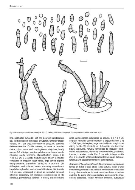
Schubert et al. Fig. 4. Dichocladosporium chlorocephalum (HAL 2011 F), cladosporioid, <strong>leaf</strong>-spotting morph. Conidiophores <strong>and</strong> conidia. Scale bar = 10 µm. long, proliferation sympodial, with one to several conidiogenous loci, subdenticulate or denticulate, p<strong>rot</strong>uberant, terminally broadly truncate, 1.5–3 µm wide, unthickened or almost so, somewhat darkened-refractive. Conidia catenate, in simple or branched chains, polymorphous, small conidia globose, subglobose, broadly obovoid, 3–9 × 3–5 µm, aseptate, pale to medium brown, smooth, intercalary conidia limoniform, ellipsoid-fusiform, oblong, 5–23 × 3.5–6.5 µm, 0–2-septate, medium brown, smooth to minutely verruculose or irregularly rough-walled, large conidia ellipsoid, oblong-cylindrical, ampulliform, 22–45(–52) × (4.5–)5–8 µm, 0–5-septate, medium brown, smooth to minutely verruculose or irregularly rough-walled, walls somewhat thickened, hila truncate, 1–3 µm wide, unthickened or almost so, somewhat darkenedrefractive; occasionally with microcyclic conidiogenesis; in vitro numerous, polymorphous, catenate, in loosely branched chains, 100 small conidia globose, subglobose, or obovoid, 3–8 × 3–4 µm, aseptate, intercalary conidia limoniform to ellipsoid-fusiform, 9–18 × 3.5–4.5 µm, 0–1-septate, large conidia ellipsoid to cylindricaloblong, 14–30(–38) × 3–6(–7) µm, 0–3-septate, pale to medium brown, asperulate, minutely verruculose to irregularly roughwalled, walls thickened, hila usually short denticle-like, p<strong>rot</strong>uberant, truncate, in smaller conidia 0.5–1.8 µm wide, in larger conidia (1.5–)2–3 µm wide, unthickened or almost so but usually darkenedrefractive; with occasional microcyclic conidiogenesis. Characters <strong>of</strong> the periconioid morph: Macronematous conidiophores formed on faded or dead <strong>stem</strong>s in late autumn, winter or after overwintering; colonies at first visible as reddish brown streaks, later turning olivaceous-brown to black, sometimes linear, sometimes encircling the <strong>stem</strong>s, <strong>of</strong>ten occupying large <strong>stem</strong> segments, effuse, densely caespitose, velvety. Mycelium immersed, subcuticular
to intraepidermal; hyphae at first sparsely branched, 3–7 µm wide, septate, not constricted at the septa, becoming swollen <strong>and</strong> wider, up to 11 µm wide, <strong>of</strong>ten branched, pale to medium olivaceous-brown, walls thickened, forming loose to dense hyphal aggregations; in vitro mycelium immersed to superficial, loosely branched, 2–6(–7) µm wide, pluriseptate, usually without swellings <strong>and</strong> constrictions, subhyaline to medium brown or olivaceousbrown, almost smooth to asperulate or irregularly rough-walled, in older colonies on PDA up to 10 µm wide, sometimes single hyphal cells distinctly swollen, up to 16(–20) µm wide, mainly at the base <strong>of</strong> conidiophores, sometimes covered by a slime coat or enveloped in a polysaccharide-like layer. Stromata well-developed, large <strong>and</strong> exp<strong>and</strong>ed, up to about 50–320 µm in length, 15–30 µm deep, composed <strong>of</strong> a single to several layers <strong>of</strong> swollen pale to medium brown stromatic cells, 5–18 µm diam, thick-walled. Conidiophores solitary or in loose groups, arising from swollen hyphal cells or stromata, erumpent through the cuticle, erect, straight, rigid to slightly flexuous, 150–680 µm long, composed <strong>of</strong> a subcylindrical stipe, 13–24 µm wide at the base, slightly attenuated towards the apex, 5–15 µm just below the branched head, pluriseptate, not constricted at the septa, young conidiophores pale medium olivaceous-brown, later medium to usually dark brown, sometimes slightly paler at the distal end, smooth or almost so, <strong>of</strong>ten appearing somewhat granular, roughened, walls distinctly thickened, 1.5–3(– 4) µm wide; apex with a roughly subglobose to ovoid head, about 35–70 µm diam, composed <strong>of</strong> dense branchlets <strong>and</strong> ramoconidia, primary branchlets close to the apex <strong>and</strong> below the first <strong>and</strong> sometimes second <strong>and</strong> third septa, solitary, in pairs or small verticils, appressed against the stipe or somewhat divergent, subcylindrical to ellipsoid-oval, aseptate, rarely 1-septate, pale olivaceous to dark brown, 10–20 × 5–8.5 µm; in vitro conidiophores initially micro- <strong>and</strong> semimacronematous, then progressively macronematous as colonies age, arising laterally from plagiotropous hyphae or terminally from ascending hyphae, sometimes also from swollen hyphal cells; micronematous conidiophores filiform, narrowly cylindrical-oblong, unbranched, up to 150 µm long, 2–3.5 µm wide, septate, septa <strong>of</strong>ten appear to be darkened, pale to pale medium olivaceous-brown, asperulate, walls slightly thickened; semimacronematous conidiophores <strong>of</strong>ten resembling those formed by the <strong>leaf</strong>-<strong>blotch</strong>ing (cladosporioid) morph on the natural host, subcylindrical to cylindrical-oblong, straight to slightly flexuous, unbranched, rarely branched, (10–)15–120 × 3–5(–6) µm, slightly attenuated towards the apex, septate, medium brown, minutely verruculose to irregularly rough-walled, walls more or less thickened; macronematous conidiophores formed in older cultures on SNA, PDA <strong>and</strong> also MEA (according to McKemy & Morgan-Jones 1991), but more prominent on PDA <strong>and</strong> MEA, resembling those formed by the <strong>stem</strong>-<strong>rot</strong>ting morph (i.e., the periconioid morph, in planta), consisting <strong>of</strong> a long unbranched stipe <strong>and</strong> a subglobose head, but in culture the heads are <strong>of</strong>ten more loosely branched than on the natural substratum, not always forming a compact head, up to 580 µm long, 5–13 µm wide, attenuated towards the apex, 4–8 µm just below the branched upper part, somewhat swollen at the base, septate, medium to very dark brown, minutely verruculose, walls distinctly thickened, two distinct wall layers visible, 1–2 µm thick. Conidiogenous cells holoblastic, integrated, terminal, intercalary or even discrete, ellipsoid to cylindrical or doliiform, subdenticulate, proliferation sympodial, multilocal, conidiogenous loci truncate, flat, unthickened, 1–3 µm wide, somewhat darkened-refractive; in culture conidiogenous loci appearing to be somewhat thickened <strong>and</strong> distinctly darkened-refractive, 1–2.5(–3) µm wide. Conidia catenate, in long, branched chains, straight, subglobose, aseptate, www.studiesinmycology.org DichoclaDosporium gen. nov. 3.5–7 µm diam, or ellipsoid-ovoid, 6–15 × 4–9 µm, 0(–1)-septate, pale olivaceous to olivaceous-brown, smooth to verruculose (under the light microscope), hila flat, truncate, unthickened, (0.5–)1– 2(–2.5) µm wide, not darkened, but somewhat refractive; in vitro conidia numerous, catenate, formed in long, branched chains, small conidia globose to subglobose, (2–)3–7 × (2–)3–4 µm, aseptate, intercalary ones ellipsoid-ovoid, 6–16 × 3.5–5 µm, 0(–1)-septate, secondary ramoconidia ellipsoid to cylindrical-oblong, (13–) 15–34(–47) × (3–)4–6(–7) µm, 0–2-septate, sometimes slightly constricted at the septa, medium olivaceous-brown, verruculose or irregularly rough-walled, walls slightly to distinctly thickened, hila more or less p<strong>rot</strong>uberant, subdenticulate to denticulate, in small <strong>and</strong> intercalary conidia 0.5–1(–1.5) µm, in secondary ramoconidia 1–2.5 (–3) µm, unthickened or somewhat thickened, darkened-refractive; occasional microcyclic conidiogenesis. Cultural characteristics: Colonies on PDA at first whitish or smoke grey, reverse smoke-grey to olivaceous-grey, with age smoke-grey to olivaceous or olivaceous-grey, sometimes even dark mouse-grey, reverse iron-grey to dark mouse-grey or black, felty; margin white to smoke-grey, narrow to more or less broad, regular to slightly undulate, glabrous to somewhat feathery; aerial mycelium at first mainly in the colony centre, with age abundantly formed, covering almost the whole colony, whitish, smoke-grey to olivaceous, felty; growth low convex to raised; numerous small exudates formed, sometimes becoming prominent; fertile. Specimens examined: Czechoslovakia, Bohemia, Turnau, on leaves <strong>of</strong> <strong>Paeonia</strong> arborea, 15 Sep. 1905, J.E. Kabát, Kabát & Bubák, Fungi Imperf. Exs. 396, B 70- 6669. France, on dead <strong>stem</strong>s <strong>of</strong> <strong>Paeonia</strong> sp., 1901, ex Herbario Musei Parisiensis, ex herb. Magnus, exs. Desmazières, Pl. Crypt. N. France, Ed. 2, Ser. 1, 1621, HBG, as “Periconia atra”; Chailly-en-Biere, Seine-et-Marne, Feuilleaubois, on <strong>stem</strong>s <strong>of</strong> P. <strong>of</strong>ficinalis, 27 Mar. 1881, Roumeguère, Fungi Sel. Gall. Exs. 1803, HBG, as “Periconia atra”. Germany, Baden-Würtemberg, Kreis Tübingen, Drusslingen, on leaves <strong>of</strong> P. <strong>of</strong>ficinalis, Jun. 1935, Raabe, B 70-6670; Bayern, Freising, on leaves <strong>of</strong> P. <strong>of</strong>ficinalis, Sep. 1918, Pr<strong>of</strong>. Dr. J.E. Weiß, Herbarium pathologicum, B 70- 6663; Br<strong>and</strong>enburg, Schloßpark zu Tamsel, on leaves <strong>of</strong> P. <strong>of</strong>ficinalis, 15 Aug. 1924, P. Vogel, Sydow, Mycoth. Germ. 2447, M-57751, PH; Triglitz, on leaves <strong>of</strong> P. <strong>of</strong>ficinalis, 3 Oct. 1909, Jaap, B 70-6668; Hessen, Frankfurt am Main, botanical garden, on leaves <strong>of</strong> P. potaninii, 7 Oct. 2004, R. Kirschner, HAL, RoKi 2222; Kreis Kassel, H<strong>of</strong>geismar, Garten von Pr<strong>of</strong>. Grupe, on leaves <strong>of</strong> P. <strong>of</strong>ficinalis, 3 Sep. 1947, Schulz, B 70-6658; Mecklenburg-Vorpommern, Rostock, neuer botanischer Garten, on leaves <strong>of</strong> P. corallina (= P. mascula), 27 Aug. 1950, Becker, B 70-6662; Nordrhein-Westfalen, Duisburg, Dinslake, private garden, on leaves <strong>of</strong> P. anomala, 9 Aug. 2005, N. Ale-Agha, HAL 2014 F; Hamborn, botanical garden, on leaves <strong>of</strong> P. obovata, 10 Aug. 2005, N. Ale-Agha, HAL 2017 F; Essen, botanical garden <strong>of</strong> the university <strong>of</strong> Essen, on leaves <strong>of</strong> P. mlokosewitschii, 10 Aug. 2005, N. Ale-Agha, HAL 2013 F; on leaves <strong>of</strong> P. <strong>of</strong>ficinalis <strong>and</strong> P. suffruticosa, 11 Aug. 2005, N. Ale- Agha, HAL 2016, 2017 F; Sachsen, Königstein, in Gärten, verbreitet, on leaves <strong>of</strong> P. <strong>of</strong>ficinalis, Aug. 1896, W. Krieger, Krieger, Fungi Saxon. Exs. 1545, M-57749; Aug., Sep. 1896, 1915, W. Krieger, Krieger, Schädliche Pilze, B 70-6666, 70-6667; Sachsen-Anhalt, Halle (Saale), Botanical Garden, on leaves <strong>of</strong> P. delavayi, 22 Jun. 2004, K. Schubert, HAL 2011 F, culture deposited at the CBS, CBS 121522 = CPC 11383; on leaves <strong>of</strong> P. <strong>of</strong>ficinalis, 22 Jun. 2004, K. Schubert, HAL 2012 F; on <strong>stem</strong>s <strong>of</strong> P. <strong>of</strong>ficinalis, 16 Mar. 2005, K. Schubert, neotype <strong>of</strong> Dichocladosporium chlorocephalum designated here HAL 1924 F, isoneotype CBS-H 19869, culture ex-neotype CBS 121523 = CPC 11969; on dead <strong>stem</strong>s <strong>of</strong> <strong>Paeonia</strong> sp., Jan. 1873, G. Winter, Rabenhorst, Fungi Eur. Exs. 1661, HBG, as “Periconia chlorocephala”; Thüringen, Fürstlicher Park zu Sondershausen, on leaves <strong>of</strong> P. arborea, 20 Aug. 1903, G. Oertel, Sydow, Mycoth. Germ. 196, PH. Italy, Thümen, Herb. Mycol. Oecon. 416, on living leaves <strong>of</strong> <strong>Paeonia</strong> lactiflora [= P. edulis] (M-57753), lectotype <strong>of</strong> “<strong>Cladosporium</strong> paeoniae” designated here; isolectotypes: Thümen, Herb. Mycol. Oecon. 416; Padova, on leaves <strong>of</strong> P. <strong>of</strong>ficinalis, Aug. 1902, P.A. Saccardo, Saccardo, Mycoth. Ital. 1186, B 70-6660, SIENA; Parma, on leaves <strong>of</strong> P. <strong>of</strong>ficinalis, Jul. 1876, Pr<strong>of</strong>. Passerini, Thümen, Mycoth. Univ. 670, B 70-6654, 70-6655, M- 57752; Pavia, botanical garden, on leaves <strong>of</strong> P. <strong>of</strong>ficinalis, summer 1889, Briosi & Cavara, Fung. Paras. Piante Colt. Utili Ess. 78, M-57748; F. Cavara, Roumeguère, Fungi Sel. Gall. Exs. 5193, mixed infection with <strong>Cladosporium</strong> herbarum, B 70- 6656; Siena, Hort. Bot., on leaves <strong>of</strong> <strong>Paeonia</strong> sp., Nov. 1899, SIENA. latvia, prov. Vidzeme, Kreis Riga, Riga, in a garden, on leaves <strong>of</strong> P. foemina [= P. <strong>of</strong>ficinalis], 28 Aug. 1936, J. Smarods, Fungi Lat. Exs. 799, M-57747. new Zeal<strong>and</strong>, isolated from 101