Chapter 1: The Characeae Plant
Chapter 1: The Characeae Plant
Chapter 1: The Characeae Plant

You also want an ePaper? Increase the reach of your titles
YUMPU automatically turns print PDFs into web optimized ePapers that Google loves.
11 <br />
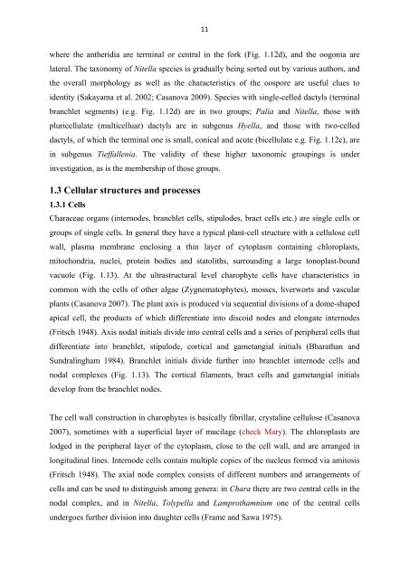
where the antheridia are terminal or central in the fork (Fig. 1.12d), and the oogonia are<br />
lateral. <strong>The</strong> taxonomy of Nitella species is gradually being sorted out by various authors, and<br />
the overall morphology as well as the characteristics of the oospore are useful clues to<br />
identity (Sakayama et al. 2002; Casanova 2009). Species with single-celled dactyls (terminal<br />
branchlet segments) (e.g. Fig. 1.12d) are in two groups; Palia and Nitella, those with<br />
pluricellulate (multicelluar) dactyls are in subgenus Hyella, and those with two-celled<br />
dactyls, of which the terminal one is small, conical and acute (bicellulate e.g. Fig. 1.12c), are<br />
in subgenus Tieffallenia. <strong>The</strong> validity of these higher taxonomic groupings is under<br />
investigation, as is the membership of those groups.<br />
1.3 Cellular structures and processes<br />
1.3.1 Cells<br />
<strong>Characeae</strong> organs (internodes, branchlet cells, stipulodes, bract cells etc.) are single cells or<br />
groups of single cells. In general they have a typical plant-cell structure with a cellulose cell<br />
wall, plasma membrane enclosing a thin layer of cytoplasm containing chloroplasts,<br />
mitochondria, nuclei, protein bodies and statoliths, surrounding a large tonoplast-bound<br />
vacuole (Fig. 1.13). At the ultrastructural level charophyte cells have characteristics in<br />
common with the cells of other algae (Zygnematophytes), mosses, liverworts and vascular<br />
plants (Casanova 2007). <strong>The</strong> plant axis is produced via sequential divisions of a dome-shaped<br />
apical cell, the products of which differentiate into discoid nodes and elongate internodes<br />
(Fritsch 1948). Axis nodal initials divide into central cells and a series of peripheral cells that<br />
differentiate into branchlet, stipulode, cortical and gametangial initials (Bharathan and<br />
Sundralingham 1984). Branchlet initials divide further into branchlet internode cells and<br />
nodal complexes (Fig. 1.13). <strong>The</strong> cortical filaments, bract cells and gametangial initials<br />
develop from the branchlet nodes.<br />
<strong>The</strong> cell wall construction in charophytes is basically fibrillar, crystaline cellulose (Casanova<br />
2007), sometimes with a superficial layer of mucilage (check Mary). <strong>The</strong> chloroplasts are<br />
lodged in the peripheral layer of the cytoplasm, close to the cell wall, and are arranged in<br />
longitudinal lines. Internode cells contain multiple copies of the nucleus formed via amitosis<br />
(Fritsch 1948). <strong>The</strong> axial node complex consists of different numbers and arrangements of<br />
cells and can be used to distinguish among genera: in Chara there are two central cells in the<br />
nodal complex, and in Nitella, Tolypella and Lamprothamnium one of the central cells<br />
undergoes further division into daughter cells (Frame and Sawa 1975).